Abstract
Partially cystic thyroid nodules (PCTNs) are common on ultrasound (US). However, there are insufficient data on the prevalence of thyroid carcinoma among such nodules. The purpose of this study was thus to evaluate the prevalence and differentiation of partially cystic thyroid cancers in US-guided fine needle aspiration (FNA).
A total of 1342 consecutive patients with 1360 thyroid nodules underwent prospective US diagnosis and FNA biopsy. In total, 281 nodules (20.7%) were partially cystic lesions. The nodules were prospectively analyzed based on US features (ie, solid portion positions, shapes, margins, and microcalcifications) and US diagnosis (benign, suspicious, or malignant).
Of the 281 partially cystic lesions, 22 nodules (8%) had inadequate FNA results, 14 nodules were diagnosed as malignant, 9 were suspicious for malignancy, and 236 were benign on FNA. Thirteen cancers were confirmed upon surgical histopathology examination or FNA, yielding a 4.6% rate of malignancy. Twelve of these cancers were papillary carcinomas, and 1 was an anaplastic carcinoma. The following individual sonographic characteristics had a statistically significant association with thyroid cancer: nodule composition (solid portion ≥50%, P = .000), eccentric solid portion (P = .001), irregular nodule shape (P = .000), microcalcification (P = .000), and intranodular vascularity (P = .001). The sensitivity, specificity, and accuracy of the US-based diagnoses were 84.6%, 84.0%, and 84.0%, respectively.
Fewer than 5% of the partially cystic nodules in this FNA series were malignant. Sonographic characteristics can be used to prioritize nodules for FNA biopsy.
Keywords: cystic, fine needle aspiration, malignancy, thyroid nodules, ultrasound-guided
1. Introduction
As an increasing number of patients are undergoing high-resolution thyroid ultrasound (US) studies for medical evaluations, progressively more thyroid nodules are being detected.[1] Partially cystic thyroid nodules (PCTNs) are common.[2–6] PCTNs are considered as a result of the cystic degeneration of either neoplastic or non-neoplastic nodules. Most of them can be managed conservatively. Recently, however, several studies have reported that the frequency of malignancy among cystic thyroid nodules varies from 5.2% to 17.6%, which is similar to what is observed for solid thyroid nodules.[4–9]
Previous studies have shown that a number of sonographic features are associated with an increased risk of cancer among PCTNs; these features differ for solid nodules. The features include an eccentric configuration with an acute angle, microcalcifications, macrolobulation or free-margin irregularities, perinodular infiltration, and intranodular vascularity.[5,7] Our goals were both to determine whether these sonographic features could help to differentiate malignant from benign nodules among partially cystic nodules and to assess the prevalence of malignancy in US-guided fine needle aspiration (FNA).
2. Materials and methods
2.1. Patients
Our institutional review board approved this study (Peking Union Medical College Hospital). From December 2004 to November 2012, 1342 patients with 1360 thyroid nodules underwent US-guided FNA at our hospital. Of the nodules, according to their composition on US, 1079 were classified as solid, and 281 were classified as mixed cystic. In total, 281 patients (44 male and 237 female, mean age 45.8 years) with 281 PCTNs were enrolled in this study. The guidelines we used to asses thyroid nodules were 2006 and 2009 American Thyroid Association management guidelines.[10,11] Before 2006, our indications for FNA were in concordance with the 2006 ATA guideline indications. The patients underwent US-FNA because of suspicious features (n = 54), the observation of substantial nodular growth at follow-up (n = 162), high risk factors for thyroid malignancy (n = 9) or their own request (n = 56). Suspicious features include microcalcifications; hypoechoic; increased nodular vascularity; infiltrative margins; taller than wide on transverse view. Definition of substantial nodular growth is a 20% increase in nodule diameter with a minimum increase in 2 or more dimensions of at least 2 mm. All the patients did not have a US-FNA performed previously. For each subject, we collected demographic, sonographic, and cytologic data, and subsequent management and outcome data.
2.2. Sonographic evaluation
Thyroid US was performed with an IU 22 scanner (Phillips, Bothell, WA) using a 5 to 12 MHz linear transducer. The US features of the thyroid nodules that underwent US-guided FNA were prospectively recorded by the radiologist who performed the US-guided FNA (YXJ). The reviewer was blinded to the FNA results when classifying the US results. Each nodule was evaluated for the following US findings: composition (a solid portion <50% vs a solid portion ≥50%, Fig. 1), nodule shape (regular vs irregular), position of the solid portion (eccentric vs non-eccentric), calcification (microcalcifications, macrocalcifications, or none), and nodule vascularity (peripheral, intranodular, or avascular). An eccentric nodule was a nodule with an internal solid portion that was not located in the center and that was abutted on only one side of the cyst wall (Fig. 2). The malignant US features included solid portion ≥50%, irregular shape, eccentric solid portion, microcalcification, and intranodular vascularity. The index nodule was diagnosed as suspicious for malignancy if more than one US-detected malignant feature was present in the nodule.
Figure 1.

An example of a predominantly cystic nodule with multiple septa inside the lesion in a 14-y-old boy. Longitudinal and transverse sonograms (A and B) showing an ovoid-shaped, smooth-margined cystic lesion with multiple septa inside the lesion. (C) Color Doppler flow image showing flow on the lesion wall. Papillary thyroid carcinoma was diagnosed by fine needle aspiration (FNA) and confirmed by surgical histopathology.
Figure 2.

An example of a mixed echoic nodule with an eccentric solid portion and microcalcifications in a 49-y-old man. Longitudinal and transverse sonograms (A and B) showing an eccentric configuration of the internal solid portion with multiple microcalcifications. (C) Color Doppler flow image showing intranodular flow inside the solid component. Papillary thyroid carcinoma was diagnosed by fine needle aspiration (FNA) and confirmed by surgical histopathology.
2.3. US-FNA technique
The US and US-FNA of the thyroid were performed by an experienced radiologist (YXJ) who has 30-year experience in US field using a 21-gauge needle with a 10 mL syringe. Once the needle was introduced into the solid part of the nodule, 3 to 5 mL of negative syringe pressure was applied. The cystic portion accounted for >50% of the nodule, the FNA was directed toward the solid portion or cyst wall. US and color-flow Doppler were used to direct sampling of the solid portion of the PCTNs that showed evidence of tissue perfusion. For each lesion, at least 4 smears of the aspirated material were performed. The smears were fixed in 95% ethyl alcohol and stained with Papanicolaou stain. There was no on-site cytopathologist assessment during the aspiration procedures.
2.4. Cytopathologic examination
The Bethesda classification was used to report the results of FNA cytology.[12] The cytologic results were categorized as nondiagnostic or adequate. A specimen was considered “adequate” if there was a minimum of 6 groupings of well-preserved thyroid cells consisting of at least 10 cells each. Adequate cytology results included benign, atypia of undetermined significance, follicular neoplasm or suspicious for a follicular neoplasm, suspicious for malignant, and malignant nodules. All nodules with a malignant FNA result were recommended to undergo surgery. Lesions with a benign FNA result were scheduled for clinical follow-up. In the group of patients with indeterminate or follicular lesions, surgery or clinical follow-up was recommended by the referring physicians from a quaternary referral center providing comprehensive care for patients with thyroid cancer, including dedicated thyroid endocrinology, surgery and nuclear medicine facilities. The decision of the referring physician was based on the patients’ history, high risk factors, symptom, physician examination, and US features. The patients with benign or indeterminate or follicular lesions cytology underwent US examination and clinical follow-up once a year.
2.5. Statistical analysis
The statistical analysis was performed using the SPSS software package (version 13.0 for Windows; SPSS, Chicago, IL). Univariate analysis was performed using Fisher exact test for categorical variables (US features), and an independent-sample t test was used to compare the means of the continuous normal data (patient age). Nodule size was not normally distributed, and thus, the Mann-Whitney U test was used. A P value of <.05 was considered to indicate a statistically significant difference.
3. Results
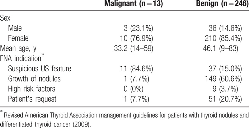
The study included 44 male and 237 female patients with a mean age of 45.8 years (range, 9–83 years). The mean nodule size was 2.5 ± 1.1 cm (range, 0.4–7.3 cm). Of the 281 patients, 9 patients had a personal history of thyroid carcinoma, and no patients reported childhood exposure of the head or neck to radiation. The baseline characteristics of the patients in malignant and benign thyroid nodules are shown in Table 1.
Table 1.
Baseline characteristics of the patients in malignant and benign thyroid nodules.

The results of FNA of 281 PCTNs are displayed in Figure 3. Of 281 PCTNs, 22 nodules (7.8%) had nondiagnostic cytology on FNA. Among the 259 nodules with adequate cytologic results, 234 were benign, 10 were atypia of undetermined significance, 4 were suspicious for malignant, and 11 were malignant. The correlations between FNA cytology and histological findings were demonstrated as following. Among the 234 benign lesions, surgery was performed on 9 nodules, which were all subsequently confirmed as benign on histopathology (8 nodular goiters and 1 lymphocytic thyroiditis). Of the 10 nodules that were atypia of undetermined significance, 4 underwent surgery and were confirmed as benign (3 nodular goiters and 1 adenoma with fibrosis). All of the 4 nodules that were suspicious for malignant underwent surgery, and these were confirmed as 2 papillary carcinoma and 2 nodular goiters. Finally, of the 11 nodules malignant on FNA, 9 were underwent surgery, and confirmed as cancerous on histopathology (8 papillary carcinomas and 1 anaplastic carcinoma). The remaining 2 were followed up clinically upon the patients’ request. Nodules without surgical histological results were clinically followed up as well and remained unchanged during the follow-up period (median 40 months, range from 24 months to 5 years). Among the 259 patients with adequate FNA samples, 13 nodules were confirmed as malignant upon surgical histopathology or FNA, yielding a 4.6% malignancy rate. The median size in benign and malignancy was not statistically different (2.5 vs 2.4 cm, P = .679).
Figure 3.

Results of FNA of 281 partially cystic thyroid nodules (PCTNs). AC = anaplastic carcinoma, AUS = atypia of undetermined significance, ND = nondiagnostic, PTC = papillary thyroid carcinoma.
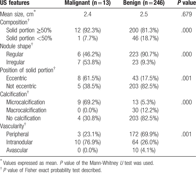
The following individual sonographic characteristics had a statistically significant association with thyroid cancer: nodule composition (solid portion ≥50%, P = .000), irregular shape (P = .000), eccentric solid portion (P = .001), microcalcification (P = .000), and intranodular vascularity (P = .001) (Table 2). Considering a nodule as suspicious if more than one malignant feature was present on US, the sensitivity, specificity, positive and negative predictive values, and accuracy of the US-based diagnoses in differentiating malignant from benign PCTNs were 84.6%, 84.0%, 20.4%, 99.1%, and 84.0%, respectively.
Table 2.
Sonographic findings for benign and malignant partially cystic thyroid nodules.

4. Discussion
PCTNs are common findings in thyroid imaging. Most cystic thyroid nodules result from a degenerative process arising in underlying lesions; the occurrence of a true epithelial cyst is rare. The appropriate recommendation for management depends on the prevalence of thyroid cancer in these cases. There is considerable variation among published studies regarding the risk of malignancy among mixed echoic thyroid nodules, ranging from 4.6% to 17.6%. In a review of 927 consecutive aspirations, García-Pascual[9] reported an 11.1% (4/36) malignancy rate among PCTNs with nondiagnostic FNA cytology (FNAC). Patients with echographic PCTNs and nondiagnostic FNAC who underwent surgery were included in that study. In a study of 119 patients with cystic thyroid nodules who underwent US-FNA biopsy and subsequently thyroidectomy, Bellantone observed carcinoma in 21 patients (17.6%).[6] The aforementioned studies were subjected to ascertainment bias in that a definitive pathology was identified only in a select subset of patients who underwent surgery, thus skewing the data toward those patients with the most suspicious nodules. Furthermore, these studies were based on small patient populations and therefore may have overestimated the malignancy rate among PCTNs. In fact, there are large amount of the PCTNs and the history of thyroid carcinoma is very long. So it is very difficult to follow up all the nodules in clinical. 2015 ATA guideline recommends PCTNs with suspicious US features or nodules with the greatest dimension >1.5 cm underwent FNA and follow-up. So we choose nodules in concordance with the ATA guideline indications for FNA and follow up these nodules in clinic. In our study the malignancy rate in PCTNs was 4.6%, with a relatively narrow range, which was comparable with the findings of a number of prior studies. For example, Frates determined the prevalence of thyroid cancer in patients with solitary or multiple thyroid nodules >10 mm in diameter and found a 7% (34/457) malignancy rate among PCTNs.[4] Among 1056 thyroid nodules submitted to US-FNA biopsy, Lee reported that 18 of the PCTNs were malignant, yielding a 5.4% malignancy rate.[5] These results support the notions that the frequency of cancer among PCTNs is low and that conservative management is most likely a valid recommendation.
Sonographic characteristics can be used to prioritize PCTNs for FNA.[4] As the prevalence of cancer among PCTNs is low and given that these are the most common type of lesion in the thyroid, it is desirable to be able to identify nodules with a high risk of malignancy based on sonographic features. Unselected nodules without any suspicious US feature have a low risk of malignancy (<2%).[13] The possibility of malignancy or benignancy for thyroid nodules is considered to be related to the number of malignant or benign US features.[14,15]
The US criteria used for assessing PCTNs are different from those used for solid nodules. In our study, nodule composition was an indicator of malignancy, with a predominantly solid nodule being more likely to be malignant. In a study of 3483, nodules measuring >10 mm in the largest dimension (found during physical exams or after discovering an “incidental” nodule on imaging), the prevalence of thyroid cancer among predominantly solid, mixed solid and cystic, and cystic nodules was 10.3%, 5.8%, and 2.3%, respectively.[4] Only one cancer in our study presented as mainly cystic. This large lesion (4 cm) was found in a 14-year-old boy. The lesion showed multiple septa inside and was diagnosed as most likely benign by US. However, based on FNA, the lesion was diagnosed as malignant, and it was later confirmed as a focal papillary carcinoma following surgical thyroidectomy. Current estimates suggest that up to 25% of thyroid nodules that occur during childhood are malignant, and thyroid carcinomas have larger tumor sizes, distant spread, and greater atypia.
In the present study, an eccentric solid portion and the presence of microcalcifications were also correlated with malignancy among PCTNs. The position of the solid portion is unique to PCTNs, indicating the presence of cancerous tissue protruding from one side of the cyst wall. Eccentric positioning of the solid component has been reported as an indicator of malignancy, although with low sensitivity (44.4%) and a low positive predictive value (13.1%).[5] Thus, the shape of the internal solid portion should be further evaluated. Non-smooth margins for the internal solid portion are indicative of malignancy and can be explained by the histologic tendency of malignancies to grow unevenly and in an infiltrative manner, without pseudocapsule formation.[16] A recent study[7] that subdivided the eccentric configuration based on either an acute angle or a blunt angle to the adjacent cyst wall found that only the cases with an acute angle were associated with malignancy. This observation emphasizes the importance of evaluating the shape of the internal solid portions of PCTNs. Meanwhile, the presence of microcalcifications is highly specific for papillary thyroid cancer.[15] Microcalcifications in a predominantly solid nodule are associated with an approximately 3-fold increase in cancer risk. Most notably, the presence of microcalcifications inside the solid components of PCTNs is associated with a high prevalence of malignancy. Color Doppler US has also been evaluated as a diagnostic tool for predicting thyroid cancer, with the hypothesis that flow that is predominantly at the periphery of the nodule is suggestive of a benign nodule, while flow predominantly in the central portion of the nodule is suggestive of malignancy. The result of these studies are mixed, with some reporting that Doppler US is helpful[17,18] and others reporting that Doppler US did not improve diagnostic accuracy.[19] In our study, intranodular vascularity was seen in a higher percentage of malignant nodules than benign nodules (76.9% vs 26.0%) among PCTNs.
Our study had several limitations. First of all, the malignancy rate in our study may have been affected by some degree of selection bias. The patients underwent US-FNA because of suspicious sonographic features or substantial nodular growth would probably tend to cause us to overestimate the incidence of cancer in PCTNs. On the other hand, the patients that requested biopsy presumably might have the opposite effect. Second, the sample size was small. Third, there were limited pathologic follow-up for inadequate findings (n = 22) and cytologically benign or malignant nodules without pathologic confirmation.
In conclusion, we noted a relatively low malignancy rate (4.6%) in our series of PCTNs and found that a nodule with predominantly solid components, an eccentric solid portion, and microcalcification is highly suggestive of malignancy. The application of these findings may aid in prioritizing nodules for FNA with high accuracy.
Footnotes
Abbreviations: FNA = fine needle aspiration, PCTNs = partially cystic thyroid nodules, US = ultrasound.
This work was supported by the National Natural Science Foundation of China (81171354).
The authors report no conflicts of interest.
References
- [1].Frates MC, Benson CB, Charboneau JW, et al. Management of thyroid nodules detected at US: Society of Radiologists in Ultrasound consensus conference statement. Radiology 2005;237:794–800. [DOI] [PubMed] [Google Scholar]
- [2].Hammer M, Wortsman J, Folse R. Cancer in cystic lesions of the thyroid. Arch Surg 1982;117:1020–3. [DOI] [PubMed] [Google Scholar]
- [3].McHenry CR, Slusarczyk SJ, Khiyami A. Recommendations for management of cystic thyroid disease. Surgery 1999;126:1167–71. [DOI] [PubMed] [Google Scholar]
- [4].Frates MC, Benson CB, Doubilet PM, et al. Prevalence and Distribution of Carcinoma in Patients with Solitary and Multiple Thyroid Nodules on Sonography. J Clin Endocrinol Metab 2006;91:3411–7. [DOI] [PubMed] [Google Scholar]
- [5].Lee MJ, Kim EK, Kwak JY, et al. Partially cystic thyroid nodules on ultrasound: probability of malignancy and sonographic differentiation. Thyroid 2009;19:341–6. [DOI] [PubMed] [Google Scholar]
- [6].Bellantone R, Lombardi CP, Raffaelli M, et al. Management of cystic or predominantly cystic thyroid nodules: the role of ultrasound-guided fine-needle aspiration biopsy. Thyroid 2004;14:43–7. [DOI] [PubMed] [Google Scholar]
- [7].Kim DW, Lee EJ, In HS, et al. Sonographic differentiation of partially cystic thyroid nodules: a prospective study. Am J Neuroradiol 2010;31:1961–6. [DOI] [PMC free article] [PubMed] [Google Scholar]
- [8].Choi KU, Kim JY, Park DY, et al. Recommendations for the management of cystic thyroid nodules. ANZ J Surg 2005;75:537–41. [DOI] [PubMed] [Google Scholar]
- [9].García-Pascual L, Barahona MJ, Balsells M, et al. Complex thyroid nodules with nondiagnostic fine needle aspiration cytology: histopathologic outcomes and comparison of the cytologic variants (cystic vs. acellular). Endocrine 2011;39:33–40. [DOI] [PubMed] [Google Scholar]
- [10].Cooper DS, Doherty GM, Haugen BR, et al. Management guidelines for patients with thyroid nodules and differentiated thyroid cancer. Thyroid 2006;16:109–42. [DOI] [PubMed] [Google Scholar]
- [11].Cooper DS, Doherty GM, Haugen BR, et al. American Thyroid Association (ATA) Guidelines Taskforce on Thyroid Nodules and Differentiated Thyroid Cancer. Revised American Thyroid Association management guidelines for patients with thyroid nodules and differentiated thyroid cancer. Thyroid 2009;19:1167–214. [DOI] [PubMed] [Google Scholar]
- [12].Cibas ES, Ali SZ. The Bethesda system for reporting thyroid cytopathology. Am J Clin Pathol 2009;132:658–65. [DOI] [PubMed] [Google Scholar]
- [13].Bastin S, Bolland MJ, Croxson MS. Role of ultrasound in the assessment of nodular thyroid disease. J Med Imaging Radiat Oncol 2009;53:177–87. [DOI] [PubMed] [Google Scholar]
- [14].Kwak JY, Han KH, Yoon JH, et al. Thyroid imaging reporting and data system for US features of nodules: a step in establishing better stratification of cancer risk. Radiology 2011;260:892–9. [DOI] [PubMed] [Google Scholar]
- [15].Horvath E, Majlis S, Rossi R, et al. An ultrasonogram reporting system for thyroid nodules stratifying cancer risk for clinical management. J Clin Endocrinol Metab 2009;94:1748–51. [DOI] [PubMed] [Google Scholar]
- [16].Park JM, Choi Y, Kwag HJ. Partially cystic thyroid nodules: ultrasound findings of malignancy. Korean J Radiol 2012;13:530–5. [DOI] [PMC free article] [PubMed] [Google Scholar]
- [17].Bakhshaee M, Davoudi Y, Mehrabi M, et al. Vascular pattern and spectral parameters of power Doppler ultrasound as predictors of malignancy risk in thyroid nodules. Laryngoscope 2008;118:2182–6. [DOI] [PubMed] [Google Scholar]
- [18].Frates MC, Benson CB, Doubilet PM, et al. Can color Doppler sonography aid in the prediction of malignancy of thyroid nodules? J Ultrasound Med 2003;22:127–31. [DOI] [PubMed] [Google Scholar]
- [19].Moon HJ, Kwak JY, Kim MJ, et al. Can vascularity at power Doppler US help predict thyroid malignancy? Radiology 2010;255:260–9. [DOI] [PubMed] [Google Scholar]


