Summary
Adenosine 5′-triphosphate (ATP) mediates a variety of biological functions following nerve-evoked release, via activation of either G-protein-coupled P2Y- or ligand-gated P2X receptors. In smooth muscle, ATP, acting via P2Y receptors (P2YR), may act as an inhibitory neurotransmitter. The underlying mechanism(s) remain unclear, but have been proposed to involve the production of inositol 1,4,5-trisphosphate [Ins(1,4,5)P3] by phospholipase C (PLC), to evoke Ca2+ release from the internal store and stimulation of Ca2+-activated potassium (KCa) channels to cause membrane hyperpolarization. This mechanism requires Ca2+ release from the store. However, in the present study, ATP evoked transient Ca2+ increases in only ~10% of voltage-clamped single smooth muscle cells. These results do not support activation of KCa as the major mechanism underlying inhibition of smooth muscle activity. Interestingly, ATP inhibited Ins(1,4,5)P3-evoked Ca2+ release in cells that did not show a Ca2+ rise in response to purinergic activation. The reduction in Ins(1,4,5)P3-evoked Ca2+ release was not mimicked by adenosine and therefore, cannot be explained by hydrolysis of ATP to adenosine. The reduction in Ins(1,4,5)P3-evoked Ca2+ release was, however, also observed with its primary metabolite, ADP, and blocked by the P2Y1R antagonist, MRS2179, and the G protein inhibitor, GDPβS, but not by PLC inhibition. The present study demonstrates a novel inhibitory effect of P2Y1R activation on Ins(1,4,5)P3-evoked Ca2+ release, such that purinergic stimulation acts to prevent Ins(1,4,5)P3-mediated increases in excitability in smooth muscle and promote relaxation.
Keywords: ATP; Smooth muscle; Ins(1,4,5)P3; Calcium
Introduction
The purinergic agonist, adenosine 5′-triphosphate (ATP), is an important and ubiquitous extracellular signalling molecule that mediates diverse physiological effects in numerous cell types and is pivotal in regulating smooth muscle activity, ranging from gastrointestinal motility to vascular tone (Abbracchio et al., 2006; Burnstock, 2006; De Man et al., 2003; Gallego et al., 2006). Alterations in ATP signalling are implicated in several pathological conditions of smooth muscle, including inflammatory bowel disease, partial bladder outlet obstruction and hypertension (Burnstock, 2006; Calvert et al., 2001; Neshat et al., 2009) and there is an emerging role of purinergic receptors as therapeutic targets in such conditions. A major regulator of smooth muscle function is the cytoplasmic Ca2+ concentration [Ca2+]c (Berridge et al., 2003; Himpens et al., 1995). Ca2+ release from the intracellular Ca2+ store, the sarcoplasmic reticulum (SR), provides a significant mechanism by which agonists such as ATP may regulate [Ca2+]c and thus, smooth muscle activity. Release occurs via two ligand-gated channel–receptor complexes, the inositol 1,4,5-trisphosphate receptor [Ins(1,4,5)P3R] and the ryanodine receptor (RyR) (Bootman et al., 2001; McCarron et al., 2004). In several cell types, such as smooth muscle, Ins(1,4,5)P3R is the predominant Ca2+ release mechanism and is activated by Ins(1,4,5)P3, generated via G-protein- or tyrosine kinase-linked receptor-dependent activation of phospholipase C (PLC) (Bootman et al., 2001; Iacovou et al., 1990; Marks, 1992).
Smooth muscle cells express ligand-gated P2X receptors (P2XR) and G-protein-coupled P2Y receptors (P2YR). Many of the physiological effects of neuronally released ATP in smooth muscle are influenced by the relaxant actions of P2YR (Abbracchio et al., 2006; Burnstock, 2009; Gallego et al., 2006; King et al., 1998), which are largely coupled to Gαq proteins and thus to the activation of PLC. Indeed, the direct inhibitory response to ATP on smooth muscle has been proposed to involve PLC-mediated phosphoinositide hydrolysis and the subsequent ATP-dependent production of Ins(1,4,5)P3 to evoke local Ca2+ release near the plasma membrane via Ins(1,4,5)P3Rs. The [Ca2+]c rise, it is proposed, may activate Ca2+-activated K+ (KCa) channels to hyperpolarize the plasma membrane and decrease bulk average [Ca2+]c (Bakhramov et al., 1996; Burnstock, 1990; Gallego et al., 2006; Koh et al., 1997; Strøbaek et al., 1996a; Zizzo et al., 2006). Yet, the frequency with which ATP-induced [Ca2+]c rises are observed varies substantially. For example, a recent study reported that [Ca2+]c rises to ATP were not observed in colonic smooth muscle cells (Kurahashi et al., 2011) and in other preparations approximately one-third of cells failed to respond to ATP with a rise in [Ca2+]c (Dockrell et al., 2001; Friel and Bean, 1988; Liu et al., 2002) or ATP-induced Ca2+ responses may be either limited or not observed at all (Bardoni et al., 1997; Fukushi, 1999; Kimelberg et al., 1997; Oshimi et al., 1999). These observations suggest that activation of KCa channels, following Ca2+ store release, may not be a universal mechanism of P2YR-mediated signal transduction.
In the present investigation, the effect of ATP on Ins(1,4,5)P3R-mediated Ca2+ release was initially examined to characterise the release of Ca2+ from the SR by ATP. Freshly isolated single colonic smooth muscle cells were selected, which provide a robust model for studying Ins(1,4,5)P3R activity. The use of flash photolysis of caged Ins(1,4,5)P3 minimised the activation of second messenger systems, to give a clearer understanding of the control of Ca2+ release via Ins(1,4,5)P3R. The study shows that ATP failed to evoke a rise in [Ca2+]c in most cells, but instead inhibited Ins(1,4,5)P3-evoked Ca2+ release. This effect was dependent on G-protein-coupled P2Y1R activation, but independent of PLC. Thus we propose here, a novel mechanism by which ATP regulates Ins(1,4,5)P3-evoked Ca2+ release in smooth muscle.
Results
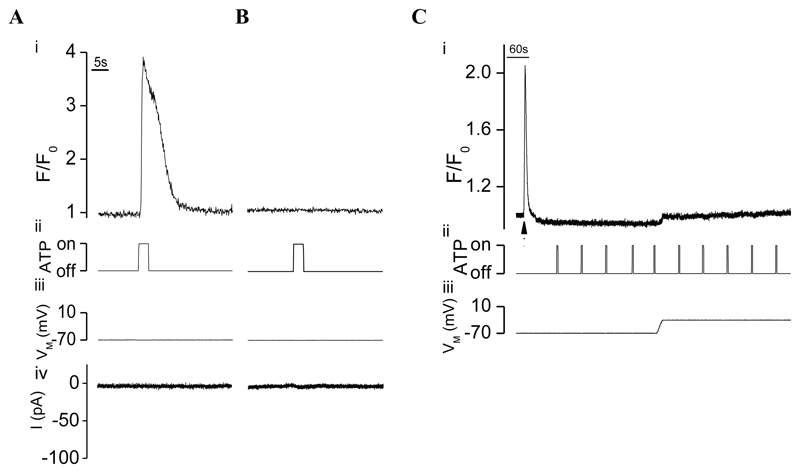
Initial experiments were designed to characterise the release of store Ca2+ by ATP. Unexpectedly, however, ATP (1 mM) transiently increased [Ca2+]c (ΔF/F0=2.0±0.4) in only ~10% of single smooth muscle cells (Fig. 1A, 9 of 87 cells) voltage-clamped at −70 mV, but did not evoke any resolvable current responses in these cells (Fig. 1A). ATP failed to evoke Ca2+ release in the remaining cells (Fig. 1B). The absence of a Ca2+ rise in response to ATP cannot be explained by clamping the cells at −70 mV since the agonist also failed to evoke Ca2+ release at −30 mV (Fig. 1C, n=11).
Fig. 1. ATP evoked [Ca2+]c rises in only a minority of single voltage-clamped myocytes.
(A,B) ATP (1 mM by pressure ejection; ii) evoked transient [Ca2+]c increases in only ~10% of cells voltage-clamped at –70 mV (iii) as indicated by F/F0 (Ai) and did not elicit any discernible current responses in these cells (iv). ATP failed to evoke Ca2+ release in the remaining cells (Bi). (C) Although photolysed caged Ins(1,4,5)P3 (▲) increased [Ca2+]c (i), ATP (1 mM by pressure ejection; ii), however, failed to evoke [Ca2+]c rises as indicated by F/F0 (i) in cells voltage clamped at –30 mV (iii).
ATP inhibits Ins(1,4,5)P3R-mediated Ca2+ release
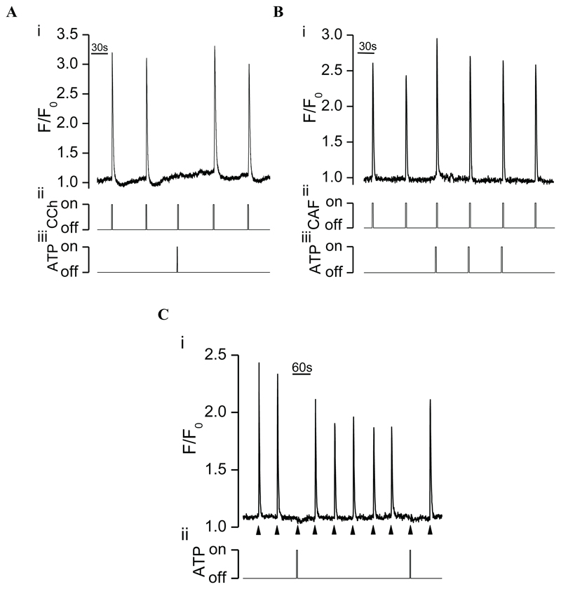
We hypothesised that ATP may inhibit Ins(1,4,5)P3R-mediated Ca2+ signalling in cells that did not display an increase in [Ca2+]c in response to purinergic stimulation. Therefore, the effect of ATP on Ins(1,4,5)P3R-mediated Ca2+ release from the store was examined. In these experiments, the Ins(1,4,5)P3 generating agonist, carbachol (CCh), was applied in a Ca2+-free bath solution to prevent Ca2+ influx and record purely store Ca2+ release. CCh (100 μM) elicited reproducible transient [Ca2+]c increases in single myocytes (Fig. 2A) which is routinely observed in ~50% of cells. ATP (1 mM), applied 2 seconds before CCh, failed to release Ca2+ from the store, but significantly (P<0.05) inhibited CCh-evoked [Ca2+]c increases (ΔF/F0) by 76±7% (from 2.28±0.2 to 0.61±0.1, n=6, Fig. 2A). Since ATP did not release Ca2+, neither activation of KCa channels nor depletion of the store of Ca2+ can explain these results. In fact, if the reduction in Ins(1,4,5)P3R-mediated Ca2+ release arose by depletion of the store of Ca2+, then ATP would be expected to inhibit RyR-mediated Ca2+ release, because both Ins(1,4,5)P3R and RyR access a single common Ca2+ store in this smooth muscle preparation (McCarron and Olson, 2008). Hence, SR Ca2+ release was evoked by the RyR activator, caffeine (10 mM), which reproducibly increased [Ca2+]c (Fig. 2B). ATP (1 mM), applied 2 seconds before caffeine, did not alter caffeine-evoked [Ca2+]c increases [(ΔF/F0) from 2.01±0.4 (control) compared with 2.04±0.3 (following ATP), n=4]. Together, the results suggest that ATP inhibits Ins(1,4,5)P3R-mediated Ca2+ signalling.
Fig. 2. ATP inhibited Ins(1,4,5)P3R- but not RyR-mediated [Ca2+]c increases in voltage-clamped single myocytes.
(A) The Ins(1,4,5)P3-generating agonist, CCh (100–250 µM by pressure ejection; ii), increased [Ca2+]c (i) as indicated by F/F0. CCh was applied in a Ca2+-free bath solution to ensure that [Ca2+]c rises arose by Ca2+ release from the SR. ATP (1 mM), applied by pressure ejection 2 s before CCh (iii), decreased CCh-evoked [Ca2+]c increases. (B) Ca2+ release was evoked using the RyR activator, caffeine. Caffeine (CAF, 10 mM by pressure ejection; ii) increased [Ca2+]c (i) as indicated by F/F0. ATP (1 mM), applied by pressure ejection 2 s before caffeine (iii), did not alter caffeine-evoked [Ca2+]c increases. (C) Photo release of caged Ins(1,4,5)P3 (▲), which activated Ins(1,4,5)P3R directly, increased [Ca2+]c (i) as indicated by F/F0. ATP (1 mM), applied by pressure ejection 2 s before Ins(1,4,5)P3 (ii), decreased Ins(1,4,5)P3-evoked [Ca2+]c increases (VM −70 mV).
The inhibition of CCh-evoked Ca2+ release by ATP may arise from either inhibition of Ins(1,4,5)P3 synthesis or alternatively, Ins(1,4,5)P3R-mediated Ca2+ release. To distinguish between these possibilities, Ins(1,4,5)P3R were activated directly using caged Ins(1,4,5)P3 which obviates the synthesis of Ins(1,4,5)P3. In these experiments, photo release of caged Ins(1,4,5)P3 elicited reproducible transient [Ca2+]c elevations in voltage-clamped single myocytes (Fig. 2C). Again, ATP (1 mM), applied 2 seconds before Ins(1,4,5)P3, significantly (P<0.05) decreased Ins(1,4,5)P3-evoked [Ca2+]c increases (ΔF/F0) by 87±5% (from 1.17±0.04 to 0.15±0.01, n=5, Fig. 2C). A similar inhibition was observed when ATP was applied at up to 10-fold lower concentrations. Since the [Ca2+]c increase evoked by photolysis of caged Ins(1,4,5)P3 does not require Ins(1,4,5)P3 synthesis, these results suggest that ATP inhibited Ins(1,4,5)P3R-mediated Ca2+ release.
ATP inhibits Ins(1,4,5)P3R-mediated Ca2+ release via P2Y1R
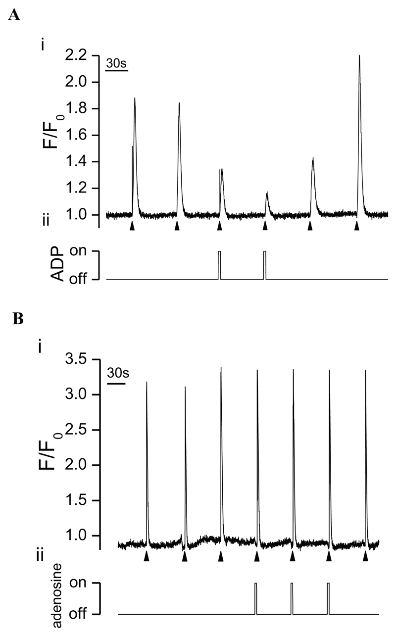
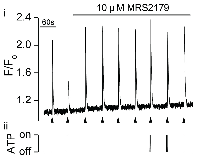
A structural analogue of ATP, adenosine 5′-diphosphate (ADP, 1 mM), also significantly decreased Ins(1,4,5)P3-evoked [Ca2+]c increases (ΔF/F0) by 76±7% (from 1.76±0.3 to 0.40±0.1, n=3, P<0.05, Fig. 3A) indicating that the purinergic inhibitory response may be mediated by P2YR. Again, a similar inhibition was observed when ADP was applied at up to 10-fold lower concentrations. In contrast, adenosine (1 mM), applied 2 seconds before Ins(1,4,5)P3, did not however, alter Ins(1,4,5)P3-evoked [Ca2+]c rises (ΔF/F0 from 1.95±0.2 to 2.02±0.2, n=4, Fig. 3B), excluding a role for adenosine receptors. Consistent with these data, the inhibitory effect of ATP on Ins(1,4,5)P3-evoked Ca2+ release was blocked by the selective P2Y1R antagonist, MRS2179 (Boyer et al., 1998). ATP (1 mM), applied 2 seconds before Ins(1,4,5)P3, significantly (P<0.05) decreased Ins(1,4,5)P3-evoked Ca2+ release (ΔF/F0 from 1.12±0.04 to 0.38±0.09, n=5, Fig. 4). The inhibitory response to ATP was abolished by MRS2179 (10 μM) (ΔF/F0 1.17±0.04, n=5, P>0.05 compared to control, Fig. 4), confirming a role of P2Y1R in this response.
Fig. 3. The inhibitory effect of ATP on Ins(1,4,5)P3-evoked [Ca2+]c increases was mimicked by ADP, but not by adenosine in voltage-clamped single myocytes.
At –70 mV photolysed caged Ins(1,4,5)P3 (▲) increased [Ca2+]c (i) as indicated by F/F0. ADP (1 mM; A) and adenosine (1 mM; B) were each applied by pressure ejection 2 s before Ins(1,4,5)P3 (ii). ADP decreased, whereas adenosine did not alter Ins(1,4,5)P3-evoked [Ca2+]c increases.
Fig. 4. The inhibitory response to ATP was blocked by the P2Y1R antagonist, MRS2179, in voltage-clamped single myocytes.
At –70 mV photolysed caged Ins(1,4,5)P3 (▲) increased [Ca2+]c (i) as indicated by F/F0. ATP (1 mM), applied by pressure ejection 2 s before Ins(1,4,5)P3 (ii), decreased Ins(1,4,5)P3-evoked [Ca2+]c increases. The inhibitory effect of ATP on Ins(1,4,5)P3-evoked Ca2+ release was blocked by MRS2179 (10 μM).MRS2179 was perfused into the solution bathing the cells.
Role of G proteins in ATP-evoked inhibition of Ins(1,4,5)P3R-mediated Ca2+ release
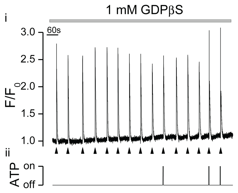
Although P2Y1R classically couple to Gαq/11 G proteins (Abbracchio et al., 2006), agonist stimulation of P2Y1R can lead, in some instances, to physiological responses that are independent of G protein activation (Fam et al., 2005; Hall et al., 1998; Lee et al., 2003; O’Grady et al., 1996). The potential role of G proteins in the ATP-mediated inhibition of Ins(1,4,5)P3-evoked Ca2+ release was therefore, examined using the membrane-impermeable G protein inhibitor, GDP 5′-O-(2-thio-diphosphate) (GDPβS), introduced into the cell via the patch electrode (Fig. 5). GDPβS (10 μM) abolished the inhibitory effect of ATP on Ins(1,4,5)P3-evoked Ca2+ release [ΔF/F0 from 1.79±0.1 (control) compared with 1.85±0.1 (following ATP), n=8], indicating that the inhibitory response to ATP requires G protein activation.
Fig. 5. The G protein inhibitor, GDPβS, blocked the inhibitory effect of ATP on Ins(1,4,5)P3-evoked [Ca2+]c increases in voltage-clamped single myocytes.
Photolysed caged Ins(1,4,5)P3 (▲) increased [Ca2+]c (i) as indicated by F/F0. GDPβS (1 mM, introduced via the patch pipette; pretreated for 7–15 mins) blocked the inhibitory effect of ATP (1 mM by pressure ejection, applied 2 s before Ins(1,4,5)P3; ii) on Ins(1,4,5)P3-evoked Ca2+ release (VM –70 mV).
ATP-evoked inhibition of Ins(1,4,5)P3R-mediated Ca2+ release does not require PLC
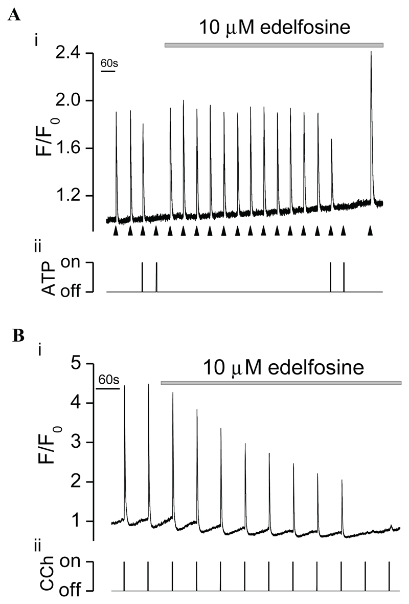
G-protein-coupled P2YR-mediated effects are mainly mediated by activation of PLC (Abbracchio et al., 2006). Thus, to investigate the role of PLC in mediating the ATP-dependent inhibition of Ins(1,4,5)P3-evoked Ca2+ release, we examined the effects of the PLC inhibitor, edelfosine. ATP (1 mM), applied 2 seconds before Ins(1,4,5)P3, significantly (P<0.05) decreased Ins(1,4,5)P3-evoked [Ca2+]c increases (ΔF/F0 from 2±0.39 to 0.14±0.01, n=4, Fig. 6A) and remained effective in decreasing Ins(1,4,5)P3-evoked [Ca2+]c rises in the presence of the PLC inhibitor, edelfosine (10 μM) (ΔF/F0 from 2±0.39 to 0.17±0.01, n=4, P<0.05, Fig. 6A). On the other hand, edelfosine significantly inhibited Ca2+ release evoked by the Ins(1,4,5)P3 generating agonist, CCh, (ΔF/F0) by 93±2% (from 1.88±0.3 to 0.12±0.03, n=6, P<0.05; Fig. 6B), a result that is consistent with the proposed mechanism of action of the drug (Powis et al., 1992).
Fig. 6. The PLC inhibitor, edelfosine, did not alter the ATP-mediated reduction in Ins(1,4,5)P3-evoked Ca2+ release in voltage-clamped single myocytes.
(A) Photolysed caged Ins(1,4,5)P3 (▲) increased [Ca2+]c (i) as indicated by F/F0. ATP (1 mM by pressure ejection, applied 2 s before Ins(1,4,5)P3; ii) decreased Ins(1,4,5)P3-evoked [Ca2+]c increases. Edelfosine (10 μM, n=4) did not alter the inhibitory effect of ATP on Ins(1,4,5)P3-evoked Ca2+ release (VM –70 mV). (B) The Ins(1,4,5)P3-generating agonist, CCh (100–250 μM by pressure ejection; ii), increased [Ca2+]c (i) as indicated by F/F0. Edelfosine (10 μM) decreased CCh-evoked [Ca2+]c increases (i), which is consistent with the proposed mechanism of action of the drug. Edelfosine was perfused into the solution bathing the cells.
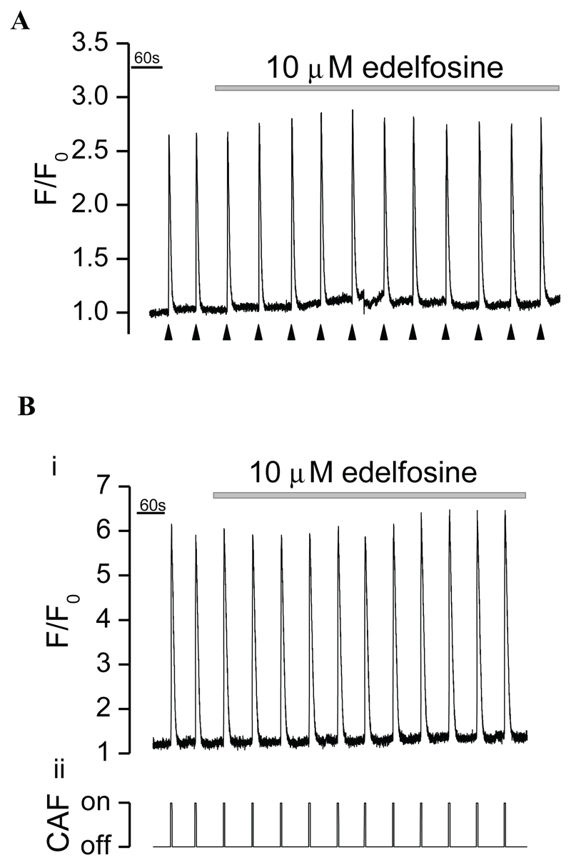
Having previously demonstrated that the commonly used PLC inhibitor, U-73122, exerts non-selective effects on SR Ca2+ pumps in this tissue (MacMillan and McCarron, 2010), it was necessary to confirm the selectivity of edelfosine as a PLC inhibitor. Edelfosine affected neither Ins(1,4,5)P3- [ΔF/F0 from 1.70±0.3 (control) to 1.73±0.3 (edelfosine), n=4, Fig. 7A] or RyR- [ΔF/F0 from 2.84±0.1 (control) to 3.06±0.2 (edelfosine), n=4, Fig. 7B] evoked Ca2+ release nor the rate of Ca2+ removal from the cytoplasm following release. The 80–20% decay interval following Ins(1,4,5)P3- and caffeine-evoked Ca2+ release was 3.1±0.4 s and 3.6±0.5 s in controls and 3.2±0.5 s and 3.4±0.5 s in edelfosine (n=4 and 4), respectively.
Fig. 7. The PLC inhibitor, edelfosine, neither altered Ins(1,4,5)P3R- nor RyR-mediated [Ca2+]c increases in voltage-clamped single myocytes.
Photolysed caged Ins(1,4,5)P3 (▲; A) and the RyR activator, caffeine (CAF, 10 mM by pressure ejection; Bii), each increased [Ca2+]c as indicated by F/F0. Edelfosine (10 μM) neither altered Ins(1,4,5)P3-evoked [Ca2+]c increases (A) nor caffeine-evoked [Ca2+]c increases (Bi; VM –70 mV). Edelfosine was perfused into the solution bathing the cells.
In conclusion, these results suggest that P2Y1R inhibition of Ins(1,4,5)P3-evoked Ca2+ release is G-protein dependent. A product of the PLC-mediated signalling pathway is not responsible for ATP-dependent inhibition of Ca2+ release.
Discussion
At present the mechanism by which ATP mediates smooth muscle relaxation is contentious. The present study demonstrates that ATP induced Ca2+ release in only a minority of smooth muscle cells. Instead, in cells that did not show a [Ca2+]c rise in response to purinergic stimulation, ATP inhibited Ins(1,4,5)P3-evoked Ca2+ release. This response was mediated by P2Y1R and was G protein dependent, but did not involve activation of PLC. Thus we propose a novel mechanism for smooth muscle relaxation whereby stimulation of a G-protein-coupled receptor inhibits Ins(1,4,5)P3-evoked Ca2+ release to promote relaxation.
Our initial experiments found that ATP increased [Ca2+]c in only ~10% of voltage-clamped single smooth muscle cells. The absence of an increase in [Ca2+]c in response to ATP was not influenced by changes in membrane potential and so, cannot be accounted for by voltage control of Ins(1,4,5)P3 production or Ins(1,4,5)P3-dependent Ca2+ release (Billups et al., 2006; Ganitkevich and Isenberg, 1993; Itoh et al., 1992; Mahaut-Smith et al., 1999; Martinez-Pinna et al., 2004; Mason et al., 2000). This is intriguing because it is generally believed that purinergic activation leads to the mobilization of Ins(1,4,5)P3-sensitive Ca2+ stores and hyperpolarization of smooth muscle (Gallego et al., 2006; Hata et al., 2000; Koh et al., 1997; Lecci et al., 2002; Strøbaek et al., 1996a; Strøbaek et al., 1996b; Zizzo et al., 2006). Evidently, a requirement for this mechanism is Ca2+ release from the internal store. However, recent evidence from another group, although having previously been in support of this model, also suggest that ATP failed to elicit increased responses in isolated smooth muscle cells (Kurahashi et al., 2011), while the relatively small purinergic KCa current evoked by ATP or the inability to elicit KCa currents in smooth muscle (Kurahashi et al., 2011) is inconsistent with previous studies supporting the concept of purinergic signalling via intracellular Ca2+ release and subsequent activation of KCa channels in smooth muscle. Thus, it appears that only a sub-population of smooth muscle cells may be organised to hyperpolarize in response to ATP.
Our results provide an additional mechanism by which ATP may evoke smooth muscle relaxation, that is by modulation of Ins(1,4,5)P3R-mediated Ca2+ signalling. ATP suppressed [Ca2+]c increases evoked by CCh (Ins(1,4,5)P3 generating agonist) and by photolysis of a caged form of the inositide, to release Ins(1,4,5)P3, in all cells that failed to respond to purinergic activation with an increase in [Ca2+]c. Since ATP did not evoke Ca2+ release from the store, neither KCa channel activation nor Ca2+ store depletion can explain the inhibitory response to ATP. Neither can the inhibitory response to ATP be attributed to inhibition of Ins(1,4,5)P3 synthesis since Ins(1,4,5)P3 was released by photolysis and so synthesis was not required. Consistent with our data, ATP exerted an inhibitory effect on muscarinic-mediated increases in intracellular Ca2+ release in platelets and acinar cells (Fukushi, 1999; Hurley et al., 1993; Jørgensen et al., 1995; Métioui et al., 1996; Soslau et al., 1995). These studies also concluded that the mechanism underlying the ATP-dependent inhibition of Ins(1,4,5)P3R-mediated Ca2+ release appears to extend beyond the plasma membrane, but is not due to depletion of the store. ATP, at a concentration which fully inhibits Ca2+ store release, affected neither the interaction of agonists with their respective cell surface receptors nor Ca2+ store content (Hurley et al., 1993; Jørgensen et al., 1995).
In the present study the ATP-mediated inhibitory response was mimicked by ADP and blocked by the P2Y1R antagonist, MRS2179, in accordance with previous studies which demonstrate that ATP mediates its relaxant actions through the activation of P2Y1R (Brizzolara and Burnstock, 1991; Gallego et al., 2006; Gallego et al., 2008; Mathieson and Burnstock, 1985). Furthermore, ATP was not being hydrolysed to adenosine by ectonucleotidases (Guibert et al., 1998; Kadowaki et al., 2000; Liu et al., 1989; Moody et al., 1984), as demonstrated by the lack of effect of the nucleoside on Ins(1,4,5)P3-evoked Ca2+ release. The inability of ATP to elicit inward currents in these cells also excludes a role of P2XR. Thus, it appears that the inhibitory response to ATP may be ascribed to the inhibition of Ins(1,4,5)P3-evoked Ca2+ release via P2Y1R activation.
The inhibitory effects of ATP in these experiments were abolished by the G protein inhibitor, GDPβS, indicating that activation of P2Y1R led to downstream stimulation of G proteins. This is important as in some cases P2Y1R stimulation can evoke responses that are G protein independent (Fam et al., 2005; Hall et al., 1998; Lee et al., 2003; O’Grady et al., 1996). However, P2Y1R characteristically couple to the Gαq/11 G proteins (Abbracchio et al., 2006), which in turn stimulate PLC. Thus, we focused on a role of PLC in this inhibition. However, the actions of ATP were unaffected by the PLC inhibitor, edelfosine, at a concentration that inhibited CCh-evoked [Ca2+]c increases. This is consistent with our conclusion that ATP did not act by inhibiting Ins(1,4,5)P3 synthesis, as has been proposed in glandular cells (Jørgensen et al., 1995; Métioui et al., 1996). We have also demonstrated that the PLC inhibitor used in the former study to confirm a contribution of PLC attenuates Ins(1,4,5)P3-evoked Ca2+ release by depleting the store of Ca2+, a mechanism unrelated to inhibition of PLC activity (MacMillan and McCarron, 2010).
A likely candidate is activation of the cAMP second messenger pathway since P2Y1R can couple to cAMP pathways in addition to phosphoinositide pathways (del Puerto et al., 2012). However, studies carried out previously by us (Flynn et al., 2001) have demonstrated that cAMP does not regulate Ins(1,4,5)P3R activity in these cells. Elevation of intracellular cAMP levels by either forskolin (which stimulates adenylate cyclase) or IBMX (which inhibits phosphodiesterase) did not significantly affect Ins(1,4,5)P3-evoked Ca2+ release. We also examined the involvement of kinase activation, since Ins(1,4,5)P3R can be greatly affected by phosphorylation. However, Ins(1,4,5)P3-evoked Ca2+ release was not altered by the broad spectrum kinase inhibitor, H7 (100 μM; data not shown), and ATP remained effective in decreasing Ins(1,4,5)P3-evoked Ca2+ release in the presence of the broad spectrum PI3K (phosphoinositide 3-kinase) inhibitor, wortmannin (10 μM; data not shown), which is consistent with our previous work confirming that neither kinase (i.e. PKA, PKG and PKC) inhibition nor activation modulated Ins(1,4,5)P3-mediated Ca2+ release (McCarron et al., 2002; McCarron et al., 2004).
The intracellular signalling cascade downstream of G protein receptor-coupled (GPRC) activation clearly exhibits greater diversity than previously appreciated. Purinergic signalling is further complicated by the fact that P2Y1R may couple directly to more than one G protein isoform (Hermans, 2003; Ostrom and Insel, 2004; Rashid et al., 2004) and each G protein isoform may also activate multiple signalling pathways (Maudsley et al., 2005; Ostrom, 2002), resulting in a wide range of cellular responses (Boeynaems et al., 2000; Communi et al., 1997; Murthy and Makhlouf, 1998; Neary et al., 1999; Qi et al., 2001). This may also be influenced by the physical proximity of the P2Y1R and its signalling components, which can be partitioned into specialised membrane microdomains. For example, GPCR (Norambuena et al., 2008), G proteins (Oh and Schnitzer, 2001) and G protein effector enzymes (Ostrom et al., 2002) localise to these structures. Moreover, membrane organisation may be critically important for ATP to release store Ca2+ in a subpopulation of cells, despite being a potent inhibitor of Ins(1,4,5)P3R-mediated Ca2+ release. Thus, the microenvironment may permit selective cellular responses (Delmas et al., 2002; Haley et al., 2000; Segawa et al., 2002; Takemura and Horio, 2005) by providing a mechanism for achieving specificity of the receptor-mediated Ins(1,4,5)P3 pathway.
The complexity of purinergic signalling pathways constitute a formidable obstacle to the complete understanding of the molecular mechanism of this inhibition. Equally perplexing is the very rapid speed of onset of the inhibition of Ca2+ release (i.e. 2 seconds) which likely excludes signalling through slower second messenger systems. We speculate that a such a rapid time course of inhibition may more likely be explained by the modulation of proteins which interact directly with the Ins(1,4,5)P3R such as IRBIT [Ins(1,4,5)P3R binding protein released with Ins(1,4,5)P3] or Ca2+ binding proteins (CaBP), and which can inhibit the Ca2+ release activity of the Ins(1,4,5)P3R channel. Further studies are necessary to characterise the precise molecular mechanism underlying inhibition of Ins(1,4,5)P3R activity by ATP, but may prove challenging.
In conclusion, purinergic relaxation of smooth muscle has largely been attributed to the mobilization of Ins(1,4,5)P3-sensitive Ca2+ stores to hyperpolarize the cell, but we propose quite the reverse, that P2Y1R activation at the plasma membrane inhibits direct activation of Ins(1,4,5)P3R in the SR by Ins(1,4,5)P3 to inhibit smooth muscle activity. Smooth muscle may be under dual, inhibitory control by purinergic nerves. Thus, we report here for the first time that ATP can regulate the mobilization of Ins(1,4,5)P3-sensitive Ca2+ stores by two diametrically opposed mechanisms which are mediated by the same receptor. Although the biological significance of this divergence in ATP signalling is not yet clear, it is tempting to speculate that this novel mode of regulation may reflect complementary mechanisms of smooth muscle relaxation which have developed to acquire the ability of coordinated contraction and relaxation under a variety of circumstances and in response to a variety of stimuli. The existence of multiple regulatory mechanisms represents an advantageous strategy that allows impairment of one or more of its components. It is therefore understandable that several complementary and cooperating mechanisms are in place to control smooth muscle relaxation. Nevertheless, this study highlights the differential ability of ATP to regulate Ins(1,4,5)P3R-mediated mobilization of intracellular Ca2+ which may provide the molecular basis for such heterogeneous responses to ATP which are recognised in a variety of cells.
Materials and Methods
Cell isolation
All animal care and experimental procedures complied with the Animal (Scientific Procedures) Act UK 1986. Male guinea pigs (500–700 g) were sacrificed by cervical dislocation and immediate exsanguination. A segment of distal colon was immediately removed and transferred to an oxygenated (95% O2, 5% CO2) physiological saline solution of the following composition (mM): NaCl 118.4, NaHCO3 25, KCl 4.7, NaH2PO4 1.13, MgCl2 1.3, CaCl2 2.7 and glucose 11 (pH 7.4). Following the removal of the mucosa from this tissue, single smooth muscle cells, from circular muscle, were isolated using a two-step enzymatic dissociation protocol (McCarron and Muir, 1999), stored at 4°C and used the same day. All experiments and loading of cells with fluorescent dyes were conducted at room temperature (20±2°C).
Electrophysiological experiments
Cells were voltage clamped using conventional tight seal whole-cell recording (MacMillan and McCarron, 2010; Rainbow et al., 2009). The composition of the extracellular solution was (mM): Na glutamate 80, NaCl 40, tetraethylammonium chloride 20, MgCl2 1.1, CaCl2 3, HEPES 10 and glucose 30 (pH 7.4 adjusted with NaOH 1 M). The Ca2+-free extracellular solution additionally contained (mM): MgCl2, 3 (substituted for Ca2+); and EGTA, 1. The pipette solution contained (mM): Cs2SO4 85, CsCl 20, MgCl2 1, HEPES 30, pyruvic acid 2.5, malic acid 2.5, KH2PO4 1, MgATP 3, creatine phosphate 5, guanosine triphosphate 0.5, fluo-3 penta-ammonium salt 0.1 and caged Ins(1,4,5)P3-trisodium salt 0.025 (pH 7.2 adjusted with CsOH 1 M). Whole cell currents were amplified by an Axopatch amplifier (Axon instruments, Union City, CA, USA), low pass filtered at 500 Hz (8-pole bessel filter; Frequency Devices, Haverhill, MA, USA), and digitally sampled at 1.5 kHz using a Digidata interface, pCLAMP software (version 6.0.1, Axon Instruments) and stored on a personal computer for analysis.
[Ca2+]c measurement
[Ca2+]c was measured as fluorescence using either the membrane-impermeable dye fluo-3 (penta-ammonium salt), introduced into the cell via the patch pipette (MacMillan and McCarron, 2009), or the membrane-permeable dye, fluo-3 acetoxymethylester (AM) (McCarron et al., 2004; Rainbow et al., 2009). Where [Ca2+]c measurements were made using the AM dye, cells were loaded with fluo-3 AM (10 μM) in the presence of wortmannin (10 μM; to prevent contraction) for 30 minutes prior to the beginning of the experiment (n=10). Fluorescence was quantified using a microfluorimeter which consisted of an inverted microscope (Nikon, Surrey, UK) and a photomultiplier tube with a bi-alkali photo cathode. Fluo-3 was excited at 488 nm (bandpass 9 nm) by a PTI Delta Scan (Photon Technology International Inc., London, UK) through the epi-illumination port of the microscope (using one arm of a bifurcated quartz fibre optic bundle). Excitation light was passed through a field stop diaphragm to reduce background fluorescence and reflected off a 505 nm long-pass dichroic mirror. Emitted light was guided through a 535 nm barrier filter (bandpass 35 nm) to a photomultiplier in photon counting mode (Photon Technology International Inc., London, UK). Interference filters and dichroic mirrors were obtained from Glen Spectra (London, UK).
Caged Ins(1,4,5)P3 was photolysed to the uncaged compound by ultraviolet light, which was selected by passing the output of a xenon flash lamp (Rapp Optoelektronik, Hamburg, Germany) through a UG-5 filter to select UV light and merging into the excitation light path of the microfluorimeter using a quartz bifurcated fibre optic bundle. The nominal flash lamp energy was 57 mJ, measured at the output of the fibre optic bundle and the flash duration was ~1 ms.
Statistical analysis
Changes in [Ca2+]c were expressed as the ratio (F/F0) of fluorescence counts (F) relative to baseline (control) values (taken as 1) before stimulation (F0). Summarised data are expressed as mean ± s.e.m. for n cells. Student’s paired t-tests were applied to test and control conditions; a value of P<0.05 was considered significant.
Materials
Caged Ins(1,4,5)P3-trisodium salt and fluo-3 AM were each purchased from Invitrogen (Paisley, UK). Fluo-3 penta-ammonium salt was purchased from TEF labs (Austin, Texas, USA). Edelfosine and MRS2179 were purchased from Tocris Bioscience (Bristol, UK). Papain and collagenase were purchased from Worthington Biochemical Corporation (Lakewood, NJ, USA). All other reagents were purchased from Sigma (Poole, Dorset, UK). Ins(1,4,5)P3 was released from its caged compound by flash photolysis. ATP (100 μM–1 mM), ADP (100 μM–1 mM), adenosine (1 mM), carbachol (100–250 μM) and caffeine (10 mM) were each applied by hydrostatic pressure ejection using a pneumatic pump (PicoPump PV 820, World Precision Instruments, Stevenage, Herts, UK). With pressure ejection, the concentration of the ejected drug at the cell is unknown, but will be significantly lower than that in the pipette owing to dilution in the bathing solution. Possible ejection artefacts were excluded by pressure ejection of the vehicle solution alone. The concentration of GDPβS and caged, non-photolysed Ins(1,4,5)P3 refers to that in the pipette. ATP, ADP, adenosine, carbachol and caffeine were each dissolved in extracellular bathing solution. Edelfosine was dissolved in water and GDPβS was dissolved in pipette solution. MRS2179 was dissolved in DMSO (final bath concentration of the solvent, 0.05%, was by itself ineffective). MRS2179 (10 μM) and edelfosine (10 μM) were each perfused into the solution bathing the cells (~5 ml per min).
Funding
This work was supported by the British Heart Foundation [grant number PG/10/79/28603 to D.M., J.G.M, C.K.]; and the Wellcome Trust [grant number 092292/Z/10/Z to J.G.M.]. Deposited in PMC for immediate release.
References
- Abbracchio MP, Burnstock G, Boeynaems JM, Barnard EA, Boyer JL, Kennedy C, Knight GE, Fumagalli M, Gachet C, Jacobson KA, et al. International Union of Pharmacology LVIII: update on the P2Y G protein-coupled nucleotide receptors: from molecular mechanisms and pathophysiology to therapy. Pharmacol Rev. 2006;58:281–341. doi: 10.1124/pr.58.3.3. [DOI] [PMC free article] [PubMed] [Google Scholar]
- Bakhramov A, Hartley SA, Salter KJ, Kozlowski RZ. Contractile agonists preferentially activate CL- over K+ currents in arterial myocytes. Biochem Biophys Res Commun. 1996;227:168–175. doi: 10.1006/bbrc.1996.1484. [DOI] [PubMed] [Google Scholar]
- Bardoni R, Goldstein PA, Lee CJ, Gu JG, MacDermott AB. ATP P2X receptors mediate fast synaptic transmission in the dorsal horn of the rat spinal cord. J Neurosci. 1997;17:5297–5304. doi: 10.1523/JNEUROSCI.17-14-05297.1997. [DOI] [PMC free article] [PubMed] [Google Scholar]
- Berridge MJ, Bootman MD, Roderick HL. Calcium signalling: dynamics, homeostasis and remodelling. Nat Rev Mol Cell Biol. 2003;4:517–529. doi: 10.1038/nrm1155. [DOI] [PubMed] [Google Scholar]
- Billups D, Billups B, Challiss RA, Nahorski SR. Modulation of Gq-protein-coupled inositol trisphosphate and Ca2+ signaling by the membrane potential. J Neurosci. 2006;26:9983–9995. doi: 10.1523/JNEUROSCI.2773-06.2006. [DOI] [PMC free article] [PubMed] [Google Scholar]
- Boeynaems JM, Communi D, Savi P, Herbert JM. P2Y receptors: in the middle of the road. Trends Pharmacol Sci. 2000;21:1–3. doi: 10.1016/s0165-6147(99)01415-7. [DOI] [PubMed] [Google Scholar]
- Bootman MD, Collins TJ, Peppiatt CM, Prothero LS, MacKenzie L, De Smet P, Travers M, Tovey SC, Seo JT, Berridge MJ, et al. Calcium signalling—an overview. Semin Cell Dev Biol. 2001;12:3–10. doi: 10.1006/scdb.2000.0211. [DOI] [PubMed] [Google Scholar]
- Boyer JL, Mohanram A, Camaioni E, Jacobson KA, Harden TK. Competitive and selective antagonism of P2Y1 receptors by N6-methyl 2′-deoxyadenosine 3′,5′-bisphosphate. Br J Pharmacol. 1998;124:1–3. doi: 10.1038/sj.bjp.0701837. [DOI] [PMC free article] [PubMed] [Google Scholar]
- Brizzolara AL, Burnstock G. Endothelium-dependent and endothelium-independent vasodilatation of the hepatic artery of the rabbit. Br J Pharmacol. 1991;103:1206–1212. doi: 10.1111/j.1476-5381.1991.tb12325.x. [DOI] [PMC free article] [PubMed] [Google Scholar]
- Burnstock G. Dual control of local blood flow by purines. Ann N Y Acad Sci. 1990;603:31–44. doi: 10.1111/j.1749-6632.1990.tb37659.x. discussion 44-35. [DOI] [PubMed] [Google Scholar]
- Burnstock G. Pathophysiology and therapeutic potential of purinergic signaling. Pharmacol Rev. 2006;58:58–86. doi: 10.1124/pr.58.1.5. [DOI] [PubMed] [Google Scholar]
- Burnstock G. Purinergic regulation of vascular tone and remodelling. Auton Autacoid Pharmacol. 2009;29:63–72. doi: 10.1111/j.1474-8673.2009.00435.x. [DOI] [PubMed] [Google Scholar]
- Calvert RC, Thompson CS, Khan MA, Mikhailidis DP, Morgan RJ, Burnstock G. Alterations in cholinergic and purinergic signaling in a model of the obstructed bladder. J Urol. 2001;166:1530–1533. [PubMed] [Google Scholar]
- Communi D, Govaerts C, Parmentier M, Boeynaems JM. Cloning of a human purinergic P2Y receptor coupled to phospholipase C and adenylyl cyclase. J Biol Chem. 1997;272:31969–31973. doi: 10.1074/jbc.272.51.31969. [DOI] [PubMed] [Google Scholar]
- De Man JG, De Winter BY, Seerden TC, De Schepper HU, Herman AG, Pelckmans PA. Functional evidence that ATP or a related purine is an inhibitory NANC neurotransmitter in the mouse jejunum: study on the identity of P2X and P2Y purinoceptors involved. Br J Pharmacol. 2003;140:1108–1116. doi: 10.1038/sj.bjp.0705536. [DOI] [PMC free article] [PubMed] [Google Scholar]
- del Puerto A, Díaz-Hernández JI, Tapia M, Gomez-Villafuertes R, Benitez MJ, Zhang J, Miras-Portugal MT, Wandosell F, Díaz-Hernández M, Garrido JJ. Adenylate cyclase 5 coordinates the action of ADP P2Y1, P2Y13 and ATP-gated P2X7 receptors on axonal elongation. J Cell Sci. 2012;125:176–188. doi: 10.1242/jcs.091736. [DOI] [PubMed] [Google Scholar]
- Delmas P, Wanaverbecq N, Abogadie FC, Mistry M, Brown DA. Signaling microdomains define the specificity of receptor-mediated InsP(3) pathways in neurons. Neuron. 2002;34:209–220. doi: 10.1016/s0896-6273(02)00641-4. [DOI] [PubMed] [Google Scholar]
- Dockrell ME, Noor MI, James AF, Hendry BM. Heterogeneous calcium responses to extracellular ATP in cultured rat renal tubule cells. Clin Chim Act. 2001;303:133–138. doi: 10.1016/s0009-8981(00)00391-0. [DOI] [PubMed] [Google Scholar]
- Fam SR, Paquet M, Castleberry AM, Oller H, Lee CJ, Traynelis SF, Smith Y, Yun CC, Hall RA. P2Y1 receptor signaling is controlled by interaction with the PDZ scaffold NHERF-2. Proc Natl Acad Sci USA. 2005;102:8042–8047. doi: 10.1073/pnas.0408818102. [DOI] [PMC free article] [PubMed] [Google Scholar]
- Flynn ER, Bradley KN, Muir TC, McCarron JG. Functionally separate intracellular Ca2+ stores in smooth muscle. J Biol Chem. 2001;276:36411–36418. doi: 10.1074/jbc.M104308200. [DOI] [PubMed] [Google Scholar]
- Friel DD, Bean BP. Two ATP-activated conductances in bullfrog atrial cells. J Gen Physiol. 1988;91:1–27. doi: 10.1085/jgp.91.1.1. [DOI] [PMC free article] [PubMed] [Google Scholar]
- Fukushi Y. Heterologous desensitization of muscarinic receptors by P2Z purinoceptors in rat parotid acinar cells. Eur J Pharmacol. 1999;364:55–64. doi: 10.1016/s0014-2999(98)00824-3. [DOI] [PubMed] [Google Scholar]
- Gallego D, Hernández P, Clavé P, Jiménez M. P2Y1 receptors mediate inhibitory purinergic neuromuscular transmission in the human colon. Am J Physiol Gastrointest Liver Physiol. 2006;291:G584–G594. doi: 10.1152/ajpgi.00474.2005. [DOI] [PubMed] [Google Scholar]
- Gallego D, Vanden Berghe P, Farré R, Tack J, Jiménez M. P2Y1 receptors mediate inhibitory neuromuscular transmission and enteric neuronal activation in small intestine. Neurogastroenterol Motil. 2008;20:159–168. doi: 10.1111/j.1365-2982.2007.01004.x. [DOI] [PubMed] [Google Scholar]
- Ganitkevich VYa, Isenberg G. Membrane potential modulates inositol 1,4,5-trisphosphate-mediated Ca2+ transients in guinea-pig coronary myocytes. J Physiol. 1993;470:35–44. doi: 10.1113/jphysiol.1993.sp019845. [DOI] [PMC free article] [PubMed] [Google Scholar]
- Guibert C, Loirand G, Vigne P, Savineau JP, Pacaud P. Dependence of P2-nucleotide receptor agonist-mediated endothelium-independent relaxation on ectonucleotidase activity and A2A-receptors in rat portal vein. Br J Pharmacol. 1998;123:1732–1740. doi: 10.1038/sj.bjp.0701773. [DOI] [PMC free article] [PubMed] [Google Scholar]
- Haley JE, Abogadie FC, Fernandez-Fernandez JM, Dayrell M, Vallis Y, Buckley NJ, Brown DA. Bradykinin, but not muscarinic, inhibition of M-current in rat sympathetic ganglion neurons involves phospholipase C-beta 4. J Neurosci. 2000;20:RC105. doi: 10.1523/JNEUROSCI.20-21-j0003.2000. [DOI] [PMC free article] [PubMed] [Google Scholar]
- Hall RA, Ostedgaard LS, Premont RT, Blitzer JT, Rahman N, Welsh MJ, Lefkowitz RJ. A C-terminal motif found in the beta2-adrenergic receptor, P2Y1 receptor and cystic fibrosis transmembrane conductance regulator determines binding to the Na+/H+ exchanger regulatory factor family of PDZ proteins. Proc Natl Acad Sci US. 1998;95:8496–8501. doi: 10.1073/pnas.95.15.8496. [DOI] [PMC free article] [PubMed] [Google Scholar]
- Hata F, Takeuchi T, Nishio H, Fujita A. Mediators and intracellular mechanisms of NANC relaxation of smooth muscle in the gastrointestinal tract. J Smooth Muscle Res. 2000;36:181–204. doi: 10.1540/jsmr.36.181. [DOI] [PubMed] [Google Scholar]
- Hermans E. Biochemical and pharmacological control of the multiplicity of coupling at G-protein-coupled receptors. Pharmacol Ther. 2003;99:25–44. doi: 10.1016/s0163-7258(03)00051-2. [DOI] [PubMed] [Google Scholar]
- Himpens B, Missiaen L, Casteels R. Ca2+ homeostasis in vascular smooth muscle. J Vasc Res. 1995;32:207–219. doi: 10.1159/000159095. [DOI] [PubMed] [Google Scholar]
- Hurley TW, Shoemaker DD, Ryan MP. Extracellular ATP prevents the release of stored Ca2+ by autonomic agonists in rat submandibular gland acini. Am J Physiol. 1993;265:C1472–C1478. doi: 10.1152/ajpcell.1993.265.6.C1472. [DOI] [PubMed] [Google Scholar]
- Iacovou JW, Hill SJ, Birmingham AT. Agonist-induced contraction and accumulation of inositol phosphates in the guinea-pig detrusor: evidence that muscarinic and purinergic receptors raise intracellular calcium by different mechanisms. J Urol. 1990;144:775–779. doi: 10.1016/s0022-5347(17)39590-3. [DOI] [PubMed] [Google Scholar]
- Itoh T, Seki N, Suzuki S, Ito S, Kajikuri J, Kuriyama H. Membrane hyperpolarization inhibits agonist-induced synthesis of inositol 1,4,5-trisphosphate in rabbit mesenteric artery. J Physiol. 1992;451:307–328. doi: 10.1113/jphysiol.1992.sp019166. [DOI] [PMC free article] [PubMed] [Google Scholar]
- Jørgensen TD, Gromada J, Tritsaris K, Nauntofte B, Dissing S. Activation of P2z purinoceptors diminishes the muscarinic cholinergic-induced release of inositol 1,4,5-trisphosphate and stored calcium in rat parotid acini. ATP as a co-transmitter in the stimulus-secretion coupling. Biochem J. 1995;312:457–464. doi: 10.1042/bj3120457. [DOI] [PMC free article] [PubMed] [Google Scholar]
- Kadowaki M, Takeda M, Tokita K, Hanaoka K, Tomoi M. Molecular identification and pharmacological characterization of adenosine receptors in the guinea-pig colon. Br J Pharmacol. 2000;129:871–876. doi: 10.1038/sj.bjp.0703123. [DOI] [PMC free article] [PubMed] [Google Scholar]
- Kimelberg HK, Cai Z, Rastogi P, Charniga CJ, Goderie S, Dave V, Jalonen TO. Transmitter-induced calcium responses differ in astrocytes acutely isolated from rat brain and in culture. J Neurochem. 1997;68:1088–1098. doi: 10.1046/j.1471-4159.1997.68031088.x. [DOI] [PubMed] [Google Scholar]
- King BF, Townsend-Nicholson A, Burnstock G. Metabotropic receptors for ATP and UTP exploring the correspondence between native and recombinant nucleotide receptors. Trends Pharmacol Sci. 1998;19:506–514. doi: 10.1016/s0165-6147(98)01271-1. [DOI] [PubMed] [Google Scholar]
- Koh SD, Dick GM, Sanders KM. Small-conductance Ca(2+)- dependent K+ channels activated by ATP in murine colonic smooth muscle. Am J Physiol. 1997;273:C2010–C2021. doi: 10.1152/ajpcell.1997.273.6.C2010. [DOI] [PubMed] [Google Scholar]
- Kurahashi M, Zheng H, Dwyer L, Ward SM, Don Koh S, Sanders KM. A functional role for the ‘fibroblast-like cells’ in gastrointestinal smooth muscles. J Physiol. 2011;589:697–710. doi: 10.1113/jphysiol.2010.201129. [DOI] [PMC free article] [PubMed] [Google Scholar]
- Lecci A, Santicioli P, Maggi CA. Pharmacology of transmission to gastrointestinal muscle. Curr Opin Pharmacol. 2002;2:630–641. doi: 10.1016/s1471-4892(02)00225-4. [DOI] [PubMed] [Google Scholar]
- Lee SY, Wolff SC, Nicholas RA, O’Grady SM. P2Y receptors modulate ion channel function through interactions involving the C-terminal domain. Mol Pharmacol. 2003;63:878–885. doi: 10.1124/mol.63.4.878. [DOI] [PubMed] [Google Scholar]
- Liu SF, McCormack DG, Evans TW, Barnes PJ. Characterization and distribution of P2-purinoceptor subtypes in rat pulmonary vessels. J Pharmacol Exp Ther. 1989;251:1204–1210. [PubMed] [Google Scholar]
- Liu R, Bell PD, Peti-Peterdi J, Kovacs G, Johansson A, Persson AE. Purinergic receptor signaling at the basolateral membrane of macula densa cells. J Am Soc Nephrol. 2002;13:1145–1151. doi: 10.1097/01.asn.0000014827.71910.39. [DOI] [PubMed] [Google Scholar]
- MacMillan D, McCarron JG. Regulation by FK506 and rapamycin of Ca2+ release from the sarcoplasmic reticulum in vascular smooth muscle: the role of FK506 binding proteins and mTOR. Br J Pharmacol. 2009;158:1112–1120. doi: 10.1111/j.1476-5381.2009.00369.x. [DOI] [PMC free article] [PubMed] [Google Scholar]
- MacMillan D, McCarron JG. The phospholipase C inhibitor U-73122 inhibits Ca(2+) release from the intracellular sarcoplasmic reticulum Ca(2+) store by inhibiting Ca(2+) pumps in smooth muscle. Br J Pharmacol. 2010;160:1295–1301. doi: 10.1111/j.1476-5381.2010.00771.x. [DOI] [PMC free article] [PubMed] [Google Scholar]
- Mahaut-Smith MP, Hussain JF, Mason MJ. Depolarization-evoked Ca2+ release in a non-excitable cell, the rat megakaryocyte. J Physiol. 1999;515:385–390. doi: 10.1111/j.1469-7793.1999.385ac.x. [DOI] [PMC free article] [PubMed] [Google Scholar]
- Marks AR. Calcium channels expressed in vascular smooth muscle. Circulation. 1992;6(Suppl.):III61–III67. [PubMed] [Google Scholar]
- Martinez-Pinna J, Tolhurst G, Gurung IS, Vandenberg JI, Mahaut-Smith MP. Sensitivity limits for voltage control of P2Y receptor-evoked Ca2+ mobilization in the rat megakaryocyte. J Physiol. 2004;555:61–70. doi: 10.1113/jphysiol.2003.056846. [DOI] [PMC free article] [PubMed] [Google Scholar]
- Mason MJ, Hussain JF, Mahaut-Smith MP. A novel role for membrane potential in the modulation of intracellular Ca2+ oscillations in rat megakaryocytes. J Physiol. 2000;524:437–446. doi: 10.1111/j.1469-7793.2000.00437.x. [DOI] [PMC free article] [PubMed] [Google Scholar]
- Mathieson JJ, Burnstock G. Purine-mediated relaxation and constriction of isolated rabbit mesenteric artery are not endothelium-dependent. Eur J Pharmacol. 1985;118:221–229. doi: 10.1016/0014-2999(85)90132-3. [DOI] [PubMed] [Google Scholar]
- Maudsley S, Martin B, Luttrell LM. The origins of diversity and specificity in G protein-coupled receptor signaling. J Pharmacol Exp Ther. 2005;314:485–494. doi: 10.1124/jpet.105.083121. [DOI] [PMC free article] [PubMed] [Google Scholar]
- McCarron JG, Muir TC. Mitochondrial regulation of the cytosolic Ca2+ concentration and the InsP3-sensitive Ca2+ store in guinea-pig colonic smooth muscle. J Physiol. 1999;516:149–161. doi: 10.1111/j.1469-7793.1999.149aa.x. [DOI] [PMC free article] [PubMed] [Google Scholar]
- McCarron JG, Olson ML. A single luminally continuous sarcoplasmic reticulum with apparently separate Ca2+ stores in smooth muscle. J Biol Chem. 2008;283:7206–7218. doi: 10.1074/jbc.M708923200. [DOI] [PubMed] [Google Scholar]
- McCarron JG, Craig JW, Bradley KN, Muir TC. Agonist-induced phasic and tonic responses in smooth muscle are mediated by InsP(3) J Cell Sci. 2002;115:2207–2218. doi: 10.1242/jcs.115.10.2207. [DOI] [PubMed] [Google Scholar]
- McCarron JG, MacMillan D, Bradley KN, Chalmers S, Muir TC. Origin and mechanisms of Ca2+ waves in smooth muscle as revealed by localized photolysis of caged inositol 1,4,5-trisphosphate. J Biol Chem. 2004;279:8417–8427. doi: 10.1074/jbc.M311797200. [DOI] [PubMed] [Google Scholar]
- Mètioui M, Amsallem H, Alzola E, Chaib N, Elyamani A, Moran A, Marino A, Dehaye JP. Low affinity purinergic receptor modulates the response of rat submandibular glands to carbachol and substance P. J Cell Physiol. 1996;168:462–475. doi: 10.1002/(SICI)1097-4652(199608)168:2<462::AID-JCP25>3.0.CO;2-3. [DOI] [PubMed] [Google Scholar]
- Moody CJ, Meghji P, Burnstock G. Stimulation of P1-purinoceptors by ATP depends partly on its conversion to AMP and adenosine and partly on direct action. Eur J Pharmacol. 1984;97:47–54. doi: 10.1016/0014-2999(84)90511-9. [DOI] [PubMed] [Google Scholar]
- Murthy KS, Makhlouf GM. Coexpression of ligand-gated P2X and G protein-coupled P2Y receptors in smooth muscle. Preferential activation of P2Y receptors coupled to phospholipase C (PLC)-beta1 via Galphaq/11 and to PLC-beta3 via Gbetagammai3. J Biol Chem. 1998;273:4695–4704. doi: 10.1074/jbc.273.8.4695. [DOI] [PubMed] [Google Scholar]
- Neary JT, Kang Y, Bu Y, Yu E, Akong K, Peters CM. Mitogenic signaling by ATP/P2Y purinergic receptors in astrocytes: involvement of a calcium-independent protein kinase C, extracellular signal-regulated protein kinase pathway distinct from the phosphatidylinositol-specific phospholipase C/calcium pathway. J Neurosci. 1999;19:4211–4220. doi: 10.1523/JNEUROSCI.19-11-04211.1999. [DOI] [PMC free article] [PubMed] [Google Scholar]
- Neshat S, deVries M, Barajas-Espinosa AR, Skeith L, Chisholm SP, Lomax AE. Loss of purinergic vascular regulation in the colon during colitis is associated with upregulation of CD39. Am J Physiol Gastrointest Liver Physiol. 2009;296:G399–G405. doi: 10.1152/ajpgi.90450.2008. [DOI] [PubMed] [Google Scholar]
- Norambuena A, Poblete MI, Donoso MV, Espinoza CS, González A, Huidobro-Toro JP. P2Y1 receptor activation elicits its partition out of membrane rafts and its rapid internalization from human blood vessels: implications for receptor signaling. Mol Pharmacol. 2008;74:1666–1677. doi: 10.1124/mol.108.048496. [DOI] [PubMed] [Google Scholar]
- O’Grady SM, Elmquist E, Filtz TM, Nicholas RA, Harden TK. A guanine nucleotide-independent inwardly rectifying cation permeability is associated with P2Y1 receptor expression in Xenopus oocytes. J Biol Chem. 1996;271:29080–29087. doi: 10.1074/jbc.271.46.29080. [DOI] [PubMed] [Google Scholar]
- Oh P, Schnitzer JE. Segregation of heterotrimeric G proteins in cell surface microdomains. G(q) binds caveolin to concentrate in caveolae, whereas G(i) and G(s) target lipid rafts by default. Mol Biol Cell. 2001;12:685–698. doi: 10.1091/mbc.12.3.685. [DOI] [PMC free article] [PubMed] [Google Scholar]
- Oshimi Y, Miyazaki S, Oda S. SATP-induced Ca2+ response mediated by P2U and P2Y purinoceptors in human macrophages: signalling from dying cells to macrophages. lmmunology. 1999;98:220–227. doi: 10.1046/j.1365-2567.1999.00858.x. [DOI] [PMC free article] [PubMed] [Google Scholar]
- Ostrom RS. New determinants of receptor-effector coupling: trafficking and compartmentation in membrane microdomains. Mol Pharmacol. 2002;61:473–476. doi: 10.1124/mol.61.3.473. [DOI] [PubMed] [Google Scholar]
- Ostrom RS, Insel PA. The evolving role of lipid rafts and caveolae in G protein-coupled receptor signaling: implications for molecular pharmacology. Br J Pharmacol. 2004;143:235–245. doi: 10.1038/sj.bjp.0705930. [DOI] [PMC free article] [PubMed] [Google Scholar]
- Ostrom RS, Liu X, Head BP, Gregorian C, Seasholtz TM, Insel PA. Localization of adenylyl cyclase isoforms and G protein-coupled receptors in vascular smooth muscle cells: expression in caveolin-rich and noncaveolin domains. Mol Pharmacol. 2002;6:983–992. doi: 10.1124/mol.62.5.983. [DOI] [PubMed] [Google Scholar]
- Powis G, Seewald MJ, Gratas C, Melder D, Riebow J, Modest EJ. Selective inhibition of phosphatidylinositol phospholipase C by cytotoxic ether lipid analogues. Cancer Res. 1992;52:2835–2840. [PubMed] [Google Scholar]
- Qi AD, Kennedy C, Harden TK, Nicholas RA. Differential coupling of the human P2Y(11) receptor to phospholipase C and adenylyl cyclase. Br J Pharmacol. 2001;132:318–326. doi: 10.1038/sj.bjp.0703788. [DOI] [PMC free article] [PubMed] [Google Scholar]
- Rainbow RD, Macmillan D, McCarron JG. The sarcoplasmic reticulum Ca2+ store arrangement in vascular smooth muscle. Cell Calcium. 2009;46:313–322. doi: 10.1016/j.ceca.2009.09.001. [DOI] [PubMed] [Google Scholar]
- Rashid AJ, O’Dowd BF, George SR. Minireview: Diversity and complexity of signaling through peptidergic G protein-coupled receptors. Endocrinology. 2004;145:2645–2652. doi: 10.1210/en.2004-0052. [DOI] [PubMed] [Google Scholar]
- Segawa A, Takemura H, Yamashina S. Calcium signalling in tissue: diversity and domain-specific integration of individual cell response in salivary glands. J Cell Sci. 2002;115:1869–1876. doi: 10.1242/jcs.115.9.1869. [DOI] [PubMed] [Google Scholar]
- Soslau G, McKenzie RJ, Brodsky I, Devlin TM. Extracellular ATP inhibits agonist-induced mobilization of internal calcium in human platelets. Biochim Biophys Acta. 1995;1268:73–80. doi: 10.1016/0167-4889(95)00051-s. [DOI] [PubMed] [Google Scholar]
- Strøbaek D, Christophersen P, Dissing S, Olesen SP. ATP activates K and Cl channels via purinoceptor-mediated release of Ca2+ in human coronary artery smooth muscle. Am J Physiol. 1996a;271:C1463–C1471. doi: 10.1152/ajpcell.1996.271.5.C1463. [DOI] [PubMed] [Google Scholar]
- Strøbaek D, Olesen SP, Christophersen P, Dissing S. P2-purinoceptor-mediated formation of inositol phosphates and intracellular Ca2+ transients in human coronary artery smooth muscle cells. Br J Pharmacol. 1996b;118:1645–1652. doi: 10.1111/j.1476-5381.1996.tb15587.x. [DOI] [PMC free article] [PubMed] [Google Scholar]
- Takemura H, Horio Y. Spatial microenvironment defines Ca2+ entry and Ca2+ release in salivary gland cells. Biochem Biophys Res Commun. 2005;336:223–231. doi: 10.1016/j.bbrc.2005.07.200. [DOI] [PubMed] [Google Scholar]
- Zizzo MG, Mulè F, Serio R. Mechanisms underlying hyperpolarization evoked by P2Y receptor activation in mouse distal colon. Eur J Pharmacol. 2006;544:174–180. doi: 10.1016/j.ejphar.2006.06.030. [DOI] [PubMed] [Google Scholar]


