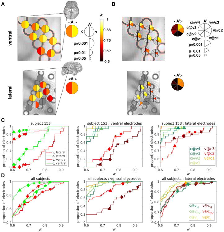
Figure 6. Subjective visibility can be decoded better than physical contrast.

(A) Decoding accuracies A′ for a selected group of electrodes from an example subject are shown color-coded on the ventral (top) and lateral (bottom) brain images. A′c are indicated in the left half of the bisected disks, A′v in the right half. Only decoding accuracies that are significant at p<0.05 (uncorrected for multiple comparisons) are shown, with larger symbol size indicating higher significance. Brain images show the areas that are enlarged in the main panels. Summary disks indicate the average A′ among the face-responsive electrodes in the selected area. (B) As in A, but with A′c@v indicated in the left sectors of the pie charts, A′v@c in the right sectors, as shown in the legend. (C), (D): Cumulative probability density functions of decoding accuracy over the populations of ventral and lateral electrodes for an example subject (C), and pooling over all subjects (D). Only results from decoding analyses with at least 10 trials in the least populated class are shown. Symbols indicate threshold A′ at several significance levels for each classification considered, obtained by permutation. Symbols are only shown if there is at least one electrode that is significant for the corresponding decoding analysis and significance level. Triangles: p=0.01; Diamonds: p=0.001; Stars: p=0.0001; Hexagons: p=0.00001. The FDR-corrected (over the number of tested electrodes and decoding analyses) p-value thresholds for these 4 levels of significance are the following: C, left panel: 0.023, 0.003, 0.0003, 0.00003; C, middle and right panels: 0.08 (n.s.), 0.012, 0.002, 0.0003; D, left panel: 0.036, 0.007, 0.001, 0.0002; D, middle and right panels: 0.2 (n.s.), 0.051, 0.014, 0.003. Single-subject cumulative probability density functions of decoding accuracy for the remaining subjects are shown in Fig. S6.
