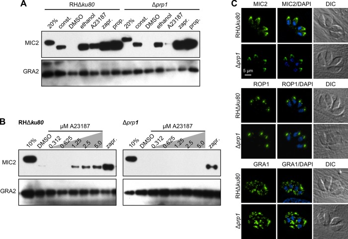
FIG 3 .
Absence of PRP1 disrupts high-Ca2+-trigger-induced microneme secretion in tachyzoites. (A) Representative Western blot image of the microneme secretion assay performed with Δprp1 and parent parasites. The secretion of the microneme protein MIC2 was used as the marker. The 20% lane contains the nonsecreted total protein lysate from 20% of the total parasites used in the secretion assay. Extracellular tachyzoites were treated with 0.25% (vol/vol) ethanol, 1.25 μM A23187, 500 μM zaprinast (zapr.), 500 μM propranolol (prop.) or DMSO as the control for 5 min at 37°C. For constitutive (const.) secretion, extracellular tachyzoites were allowed to release the protein for 1 h in the absence of a pharmacological trigger. Proteolytic processing of the secreted microneme protein can be seen as a shift in the MIC2 band. Dense granule protein GRA2 was used as a control for microneme- and Ca2+-independent secretion. (B) Titration of the Ca2+ ionophore A23187 used for triggering microneme secretion in Δprp1 and RHΔku80 parent parasites. Extracellular tachyzoites were treated with the indicated concentrations of A23187 and zaprinast as the control for 5 min at 37°C. The 10% lane contains the nonsecreted total protein lysate from 10% of the total parasites used in the secretion assay. (C) Immunofluorescence displaying morphology of the secretory organelles in Δprp1 and RHΔku80 parent parasites. Micronemes are shown in MIC2 panels, rhoptries are shown in ROP1 panels, and GRA1 dense granules are shown in green. DAPI marks DNA. DIC, differential interference contrast.

