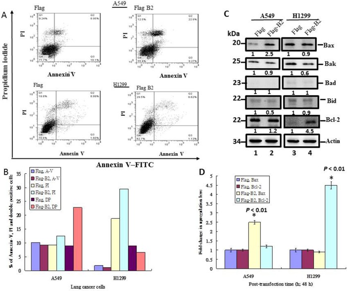
Figure 3. B2 protein induces Bax-mediated apoptosis in A549 cells, but induces RIP3-mediated necroptosis in H1299 cells.
(A) Representative flow cytometry results at 48 h post-transfection. Fluorescence of Annexin-V and PI were measured in 10,000 cells. Annexin-V-FITC+ cells indicate early apoptosis and PI+ cells indicate late apoptotic/secondary necrosis. (B) Quantitation of the percentage of viable cells (Annexin-V-FITC+ and PI+) from flow cytometry experiments. (C) Immunoblot analysis of A549 and H1299 cells using monoclonal antibodies against pro-apoptotic and anti-apoptotic proteins shows the expression of various forms of Bax, Bak, Bad, Bid and Bcl-2. ß-actin was a loading control. (D) Quantitative analysis of the pro-apoptotic and anti-apoptotic proteins from Figure 3C. Error bars represent the SEM of 3 independent experiments. All data were analyzed using a paired or unpaired Student’s t-test, as appropriate. *P < 0.01 significantly different from the control.

