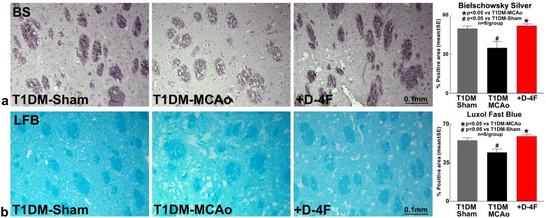
Figure 2. D-4F treatment of stroke in T1DM rats decreases white matter damage.
Stroke in T1DM rats significantly decreases axon (Bielschowsky silver staining) and myelin density (Luxol fast blue staining) compared to T1DM-sham control rats (#p<0.05; n=8/group). D-4F treatment of stroke in T1DM rats significantly increases (a) axon density (Bielschowsky silver staining) and (b) myelin density (Luxol fast blue staining) in the ischemic border zone compared to PBS treated T1DM stroke control rats (*p<0.05, n=8/group) at 48 hours after stroke. Data are represented as mean ± SE.

