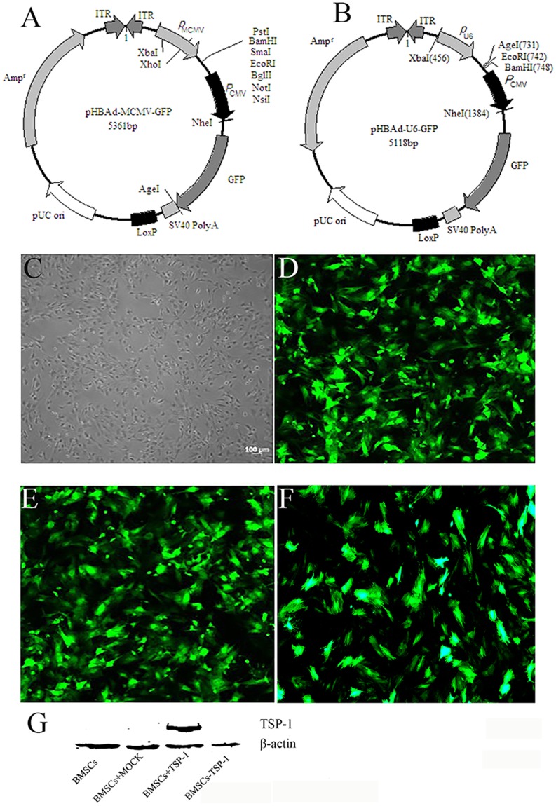
Figure 1. TheTSP-1 and TSP-1 siRNA plasmid construction and expression.
(A) Maps of pHBAd-MCMV-GFP. (B) Maps of pHBAd-U6-GFP. (C) Morphology characterization of cultured BMSCs (Light microscopy). (D) GFP expression in BMSCs 48 h after transduction of empty vector into BMSCs. (E) GFP expression in BMSCs 48 h after transduction of Adv-TSP-1 into BMSCs. (F) GFP expression in BMSCs 48 h after transduction of Adv-TSP-1-shRNA into BMSCs. Scale bar:100μm. (G) Western blotting detection of TSP-1 protein expression.

