Abstract
Background
Water serves as a potential reservoir for Campylobacter, the leading cause of bacterial gastroenteritis in humans. However, little is understood about the mechanisms underlying variations in survival characteristics between different strains of C. jejuni in natural environments, including water.
Results
We identified three Campylobacter jejuni strains that exhibited variability in their ability to retain culturability after suspension in tap water at two different temperatures (4°C and 25°C). Of the three, strains C. jejuni M1 exhibited the most rapid loss of culturability whilst retaining viability. Using RNAseq transcriptomics, we characterised C. jejuni M1 gene expression in response to suspension in water by analyzing bacterial suspensions recovered immediately after introduction into water (Time 0), and from two sampling time/temperature combinations where considerable loss of culturability was evident, namely (i) after 24 h at 25°C, and (ii) after 72 h at 4°C. Transcript data were compared with a culture-grown control. Some gene expression characteristics were shared amongst the three populations recovered from water, with more genes being up-regulated than down. Many of the up-regulated genes were identified in the Time 0 sample, whereas the majority of down-regulated genes occurred in the 25°C (24 h) sample.
Conclusions
Variations in expression were found amongst genes associated with oxygen tolerance, starvation and osmotic stress. However, we also found upregulation of flagellar assembly genes, accompanied by down-regulation of genes involved in chemotaxis. Our data also suggested a switch from secretion via the sec system to via the tat system, and that the quorum sensing gene luxS may be implicated in the survival of strain M1 in water. Variations in gene expression also occurred in accessory genome regions. Our data suggest that despite the loss of culturability, C. jejuni M1 remains viable and adapts via specific changes in gene expression.
Introduction
Campylobacter jejuni is the most common bacterial cause of gastroenteritis in Europe and the USA [1–3]. Although campylobacteriosis is primarily considered to be a zoonotic infection, mostly transmitted to humans through consumption of contaminated poultry [4], it is clear that the environment can play a role in transmission either directly, for example via unchlorinated drinking water [5], or indirectly, via farm animals that acquire the pathogen from the environment [4, 6]. Indeed, contaminated groundwater is considered to be a potential source of transmission of Campylobacter to poultry flocks and to humans directly [7–15].
The survival mechanisms employed by Campylobacter outside of the host are not well understood. Genomic studies suggest that the ability of C. jejuni to regulate gene expression in response to environmental stresses may be limited because of the lack of many of the stress response mechanisms possessed by other bacterial species [6, 16, 17]. However, despite its apparent limited ability to respond to stress, Campylobacter can survive in the environment and remain infectious [18]. It has been suggested that Campylobacter spp. can survive in natural water by entering a viable but non culturable (VBNC) state [19]. During the VBNC state, Campylobacter form coccoid-shaped cells with an intact cell membrane, which remain viable according to various measures of metabolic activity, but are unable to grow on routine culture media [20]. Bacteria in this state are capable of causing infections or colonising a host and can be returned to a state of culturability [21].
Previous studies have shown that survival of Campylobacter in natural water samples or ground water is highly dependent upon temperature and strain origin [22–24]. Low temperatures (around 4°C) enhance the survival of Campylobacter, whereas at increased temperatures (20–25°C) viability declines rapidly [22, 25]. Survival times for different C. jejuni isolates in water can vary from a few days at ambient temperatures to four months at 4°C [26].
Many factors could contribute to the variations observed between Campylobacter strains with respect to survival in water [18]. These include the ability to metabolize available nutrients and to deal with oxidative and osmotic stress [25, 27]. Previous epidemiological studies have suggested that some strain types, defined using schemes such as Multi Locus Sequence Typing (MLST), are more commonly found in the environment [28–30]
There are significant gaps in our knowledge concerning variations in the survival of different C. jejuni strain types in the environment. These could be due to differences either in gene content or in gene expression. Variations in the gene content of C. jejuni strains, linked to virulence potential, have been described previously [31]. These include variations in surface structures, such as glycosylation patterns of flagellin or the structures of lipooligosaccharides, but also include metabolic traits [32]. Genes supporting oxygen-independent respiration and the catabolism of amino acids and peptides are particularly over-represented in strains that are robust colonisers of poultry, compared to strains that do not colonise well [30, 31, 33–36]. Little is known about how variations in gene expression contribute to the ability to survive in the environment. A better understanding of the occurrence and behaviour of Campylobacter in water, and the mechanisms underlying variations, has implications for food safety and public health [37].
In this study we demonstrate how strains of C. jejuni adjust differently to an aquatic lifestyle by comparing the culturability of C. jejuni strains after prolonged exposure to water. We show that C. jejuni strain M1, though rapidly losing culturability on standard laboratory media, retains viability in water and, using strand-specific Illumina RNA Seq analysis, we identify gene expression changes occurring during the survival process.
Materials and methods
Bacterial growth conditions
The C. jejuni strains used in this study were strain M1, associated with transmission from poultry to a human, strain 1336, isolated from a wild bird and strain 414, isolated from a bank vole [30, 38]. Campylobacter strains were stored on storage beads in glycerol broth (M Lab) at -80°C. C. jejuni strains were grown on sterile Columbia Blood Agar Base (CBA, Oxoid) with 5% (v/v) defibrinated horse blood (Oxoid) at 37°C for 48 h under microaerobic conditions (85% [v/v] N2, 5% [v/v] O2, and 10% [v/v] CO2), in a Whitley VA500 Workstation incubator (Don Whitley Scientific Ltd).
Preparation of cell suspensions for testing survival in sterile water
For survival experiments, bacteria were sub-cultured on blood agar for 24 h at 37°C under microaerobic conditions. A 5 μL loop of cultured cells was taken, and suspended in 5 mL of Muller-Hinton Broth (MHB, Oxoid) supplemented with Campylobacter growth supplement (LAB M). Suspended bacterial samples were adjusted to a final optical density of 0.05 at 600 nm (OD600) [3.8×10⁷–3.5×10⁸ Colony Forming Unit (CFU)/mL] (Spectronic Biomate 5).
Preparation and inoculation of sterile water sample
Filtered-tap water (PUR1TE SELECT) was collected, and autoclaved at 121°C for 15 min. Aliquots of 99 mL (pH 6.5) of autoclaved water samples were transferred into 250 mL sterile borosilicate glass bottles with screw caps (Schott, Duran, Germany) in triplicate. These were inoculated with 1 mL MHB bacterial suspensions to a final concentration of approximately 8×10⁵–3.7×10⁶ cells/mL. The inoculated samples were kept in the dark at 25°C (for 24h) or 4°C (for 72 h). An uninoculated sterile distilled water sample for each temperature was used as a control for the presence of contamination. A further control was used whereby the water was inoculated with 1 mL of bacterial suspension and the bacteria were collected immediately (time 0). All experiments were conducted using three independent technical replicates and three biological replicates (strains M1, 1336 and 414).
Enumeration of colony forming units (CFU)
At time 0, 25°C (24 h), and 4°C (72 h), a 100 μL sample was taken and 10-fold dilutions were made in MHB supplemented with Campylobacter growth supplement. A 10 μL spot assay of appropriate dilutions was carried out on CBA plates in triplicate. The plates were incubated for 48 h at 37°C under microaerophilic conditions, and the survival was then determined by enumerating the CFU/ mL.
Cell survival by LIVE/DEAD staining
Inoculated water samples were prepared as described above. At time 0, 25°C (24 h), and 4°C (72 h), inoculated water samples C. jejuni strains M1, 414 and 1336 were concentrated by centrifugation at 3893 × g for 20 min (3-16PK—SIGMA) in Falcon tubes (Corning, Appleton Woods). Supernatant was removed and approximately 1 mL remained at the bottom of each Falcon tube and was subsequently transferred into a 1.5 mL Eppendorf tube. Cells were pelleted by centrifugation at 5000 × g for 20 min. The pellet was re-suspended in 1 mL of sterile distilled water. Using the LIVE/DEAD BacLight, Invitrogen kit, 3 μL of the mixture (SYTO 9 green-fluorescent and propidium iodide red-fluorescent) were added for each 1 mL of bacterial suspension and this mixture was incubated for 15 min at room temperature. 5 μL of the cell suspension was then placed on a microscope slide, covered with a 22 × 22 mm cover slip and sealed.
The LIVE/DEAD BacLight kit (Invitrogen) contains two nucleic acid stains, SYTO 9 dye, which penetrates live cells (intact membranes) causing the cells to stain fluorescent green, and propidium iodide dye that cannot cross the cell membrane and therefore only stains cells red if the membranes are damaged and the cell is therefore presumed dead.
Enumeration of viable cells was carried out under a fluorescence microscope (Nikon ECLIPSE 80i). For each sample, three fields were enumerated at an average of 90–180 cells in each field. The percentage of viable cells was calculated as follows: % viable cells = [viable cell count (green cells)/total cell count (green cells + red cells)] × 100. The experiment was conducted in three independent replicates.
RNA extraction from water survival experiments
100 mL of inoculated water samples were prepared for C. jejuni M1 in triplicate at time 0, 25°C (24 h) and 4°C (72 h). Cells were concentrated by centrifugation at 3893 × g (3–16 pk -SIGMA) for 20 min at the corresponding temperature in 50ml Falcon tubes (Corning, Appleton Woods). Supernatants were removed but, for each sample, approximately 1 ml was retained at the bottom of each Falcon tube and subsequently combined per sample and transferred into a 1.5 mL Eppendorf tube. Cells were pelleted by centrifugation at 5000 × g for 10 min. A negative control was included using only sterile distilled water. Additionally a control of C. jejuni M1 grown in Mueller Hinton Broth was prepared. Cells were grown to a density of 1x10⁷ in a 25 cm2 cell culture flask with gas exchange lid (Corning) at 37°C microaerophilic conditions. Cells were collected by centrifugation at 3000xg in a microcentrifuge (Eppendorf). Once collected, cells were immediately resuspended in TRIzol solution (3 times TRIzol volume to 1 volume of cells)(Ambion) and stored at -80°C until further processing.
TRIzol samples were allowed to reach room temperature and cells were disrupted using vigorous vortexing. The samples were then incubated at room temperature for 5 min. RNA was extracted using the Direct-zol RNA MiniPrep Kit (Zymo Research), following the manufacturer’s instructions. Quality of RNA was confirmed by Agilent 2100 Bioanalyzer (S1 Fig).
Illumina library construction and sequencing
116 ng of total RNA was depleted using the Illumina Ribo-zero rRNA Removal Kit (Bacteria) and purified with Ampure XP beads. Successful depletion was confirmed using Qubit and Agilent 2100 Bioanalyzer. All of the depleted RNA was used as input material for the ScriptSeq v2 RNA-Seq Library Preparation protocol. Following 15 cycles of amplification the libraries were purified using Ampure XP beads. Each library was quantified using Qubit and the size distribution assessed using the Agilent 2100 Bioanalyzer.
The final libraries were pooled in equimolar amounts using the Qubit and Bioanalyzer data. The quantity and quality of each pool was assessed by Bioanalyzer and subsequently by qPCR using the Illumina Library Quantification Kit from Kapa on a Roche Light Cycler LC480II according to manufacturer's instructions. Sequencing was performed at the Centre for Genomic Research, University of Liverpool, on one lane of the Illumina HiSeq 2500 2x125 bp using v4 chemistry (Illumina).
RNA Seq data analysis
The raw FASTQ data files were trimmed for the presence of Illumina adapter sequences using Cutadapt (v1.2.1.) [39], using the–O 3 option. The reads were further trimmed using Sickle (v1.200) (https://github.com/najoshi/sickle) with a minimum window quality score of 20. Reads shorter than 10bp after trimming were removed.
Sense and antisense overlaps between the annotation and mapped reads were counted using the HTSEQ package [40], using the stranded and union options. Read counts were then normalised and Differential Expression calculated using EdgeR implemented in R [version 3.1.2 (2014-10-31)], using Loess-style weighting to estimate the trended dispersion values, heatmaps were constructed using heatmap2 in R.
For pairwise Differential Expression analysis between samples, the data were re-mapped to the C. jejuni M1 genome [38] using Bowtie2 [41], and parsed using the BitSeq (Bayesian Inference of Transcripts from Sequencing data) pipeline [42]. BitSeq takes into account biological replicates and technical noise, and thereby calculates a posterior distribution of differential expression between samples.
Statistical Significance of BitSeq results was visualized in Artemis [43]. Regions of Difference between the genomes of M1 (CP001900) [38], 1336 (ADGL00000000), 414 (ADGM00000000) [30] and NCTC11168 (AL111168) [44, 45] were derived through pairwise genome comparisons in ACT [46]. Putative / Hypothetical genes were selected in Artemis and a putative function was derived from searches using BLASTX (http://blast.ncbi.nlm.nih.gov/blast/Blast.cgi?PROGRAM=blastx&PAGE_TYPE=BlastSearch&LINK_LOC=blasthome).
Results and discussion
Loss of culturability during survival in water
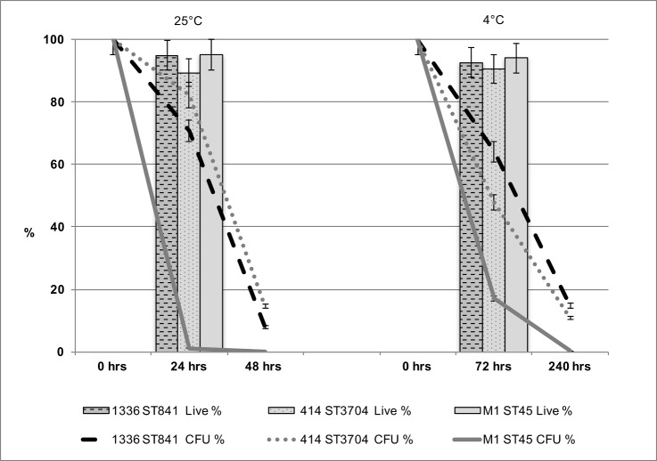
In this study, it was necessary to choose a water system (tap water) that would allow excellent sample reproducibility. Although it could be argued that tap water is not representative of environmental water, farm animals are exposed to tap water for drinking. We focused on three genome-sequenced C. jejuni strains exhibiting differences in their ability to retain culturability during incubation in water at specific time points and temperatures (24 h at 25°C and 72 h at 4°C). The ability to retain culturability in water was tested using three biological replicates for each of the three strains of C. jejuni: M1 (ST137, clonal complex ST45, associated with severe human infection) [38], 1336 (ST841, a representative of the water/wild-life clade)[30], and strain 414 (ST3704, associated with bank voles) [47]. Clear variations were observed between the three strains with respect to the ability to retain culturability on Columbia Blood Agar (CBA) containing 5% (v/v) defibrinated horse blood after exposure to sterile distilled water at two temperatures: 25°C and 4°C (Fig 1). At 4°C (72 h), whereas only approximately 17% of strain M1 cells were still culturable, for strains 1336 and 414 much higher levels (64% and 48% respectively) remained culturable. At 25°C (24 h), whereas only approximately 1.2% of strain M1 cells remained culturable, for strains 1336 and 414 approximately 71% and 82% respectively remained culturable. At these two sampling points, the survival of strain M1, based on culturability on CBA media, was significantly lower (p < 0.01; 2-tailed Student’s t-test) than for either of the other two strains.
Fig 1. Culturability of C. jejuni strains during survival periods in sterile distilled water.
The percentage of the original inoculum retaining culturability is shown at 0 h, 72 h and 240 h at 4°C and at 0 h, 24 h and 48 h at 25°C. The percentage of viable cells at 72 h at 4°C and 24 h at 25°C (indicated by the line graphs) was determined by LIVE/DEAD staining (BacLight, Invitrogen) for C. jejuni. The error bars represent a 5% error, three independent biological replicates were performed.
We further investigated viability of cells using LIVE/DEAD staining (BacLight, Invitrogen) at Time 0, 4°C (72 h) and 25°C (24 h). Whereas the prevalence of culturable cells on CBA media declined rapidly, especially for strain M1, at both 4°C and 25°C, the percentage of viable cells according to LIVE/DEAD staining remained high throughout the experiment (Fig 1). Hence, although strain M1 rapidly loses culturability on CBA media during exposure to water, the staining method used suggests that the majority of cells remain viable.
Overview of differential gene expression during survival of C. jejuni M1 in water
In order to better understand the process by which strain M1 remains viable but loses culturability, we analysed the transcriptome of cells recovered from water survival experiments. C. jejuni M1 gene expression in water was investigated, using Illumina RNA Seq under two key experimental conditions: i) 25°C for 24h, ii) 4°C for 72 h, selected because of the high viability but low culturability exhibited by strain M1 at these sampling points (Fig 1). In addition, two controls were included in the study. The first control involved suspending an inoculum of C. jejuni M1 in 100 ml of sterile distilled water and recovering the cells immediately; this control will be referred to as the “Time 0” sample. It should be noted that the bacterial cells in the Time 0 sample were pre-exposed to water at room temperature for approximately 20 min due to the sample processing time, prior to RNA extraction. The second control consisted of cells cultured in Mueller Hinton Broth (MHB) for 24 h at 37°C under microaerophilic conditions; this transcriptome will be referred to as the “MHB Control”. All initial inocula were taken from the same starter culture.
The transcriptomics data were analysed using two separate approaches to determine differential expression of genes: (i) based on counts per million (cpm) and (ii) using BitSeq analysis.
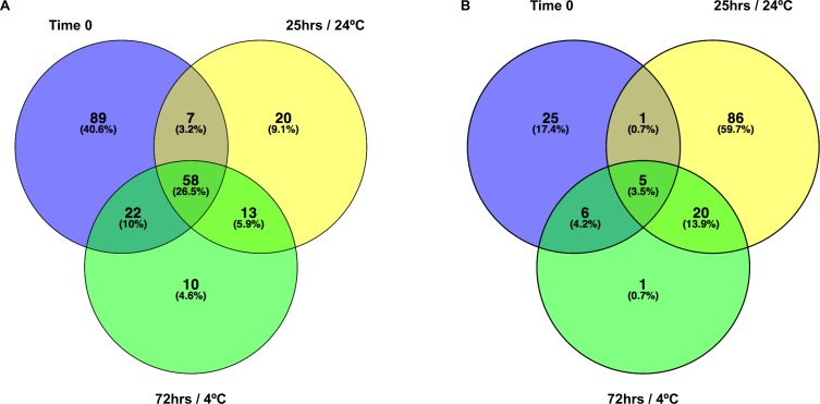
A summary of all M1 RNA Seq data in counts per million (cpm) is shown in S1 Table. It is apparent from these data that significant transcriptome changes occurred during the preparation of the Time 0 sample compared to the MHB Control, indicating that C. jejuni gene expression changes in response to suspension in water occur rapidly. Fig 2 shows comparisons of genes up (Fig 2A) or down (Fig 2B) regulated 2-fold or more relative to the MHB Control during the different experimental conditions. S2 Table summarizes genes that are up- or downregulated 2-fold or more across all three test conditions compared to the MHB control.
Fig 2. Summary of gene expression changes.
(A) Venn diagram of Genes upregulated ≥ 2-fold (log counts per million) compared to the MHB control. (B) Venn diagram of Genes downregulated ≥ 2-fold (log counts per million) compared to the MHB control.
Fig 2 clearly shows that more genes were upregulated (58 genes) across the three time-point/temperature combinations [Time 0, 25°C (24 h) and 4°C (72 h)], than downregulated (5 genes). The biggest response in terms of upregulation in gene expression was observed during the early exposure to water at Time 0. The biggest response in terms of downregulation occurred at 25°C (24 h) (Fig 2).
Among genes upregulated across all three conditions were several flagellar genes (flgBCDEGHFJ and flaG), as well as the flmA (pseB) gene, which is also involved in flagellar assembly and glycosylation; flmA mutants are non-motile and accumulate intracellular flagellin [48]. Four of the genes upregulated across all three conditions were hypothetical ones with their function currently unknown (S2 Table). The list of genes downregulated 2-fold included three members of the nap-operon.
The results of the Bayesian Inference of Transcripts from Sequencing data (BitSeq) [42] analysis are summarized in S3 Table. 546 genes were statistically significantly upregulated at Time 0, compared to the MHB Control, whereas 204 genes were downregulated; 872 genes showed no significant change. In the survival experiments, at 25°C after 24 h in sterile distilled water, 161 genes were upregulated, 557 genes were downregulated and 904 showed no significant change, compared to the MHB Control. At 4°C after 72 h, 202 genes were upregulated, 301 were downregulated and 1119 showed no significant change in expression compared to the MHB Control.
Differential gene expression between the 25°C (24 h) and 4°C (72 h) samples and the Time 0 sample
At 4°C after 72 h in sterile distilled water, 91 genes were upregulated, 589 genes were downregulated and 942 showed no significant change, compared to Time 0.
140 genes were statistically significantly upregulated at 25°C (24 h), compared to Time 0; 531 genes showed no significant change, whereas 951 genes were downregulated at 25°C (24 h) compared to Time 0 (S3 Table).
Genes downregulated at 25°C (24 h) compared to Time 0 included several genes involved in biosynthesis pathways including glycosylation (pgl genes), lipoologosaccharide production (waa genes), and peptidoglycan synthesis (mur genes). Other genes down-regulated were involved in metabolism or iron uptake (chu genes) (S3 Table). A number of flagellar genes (for example, flgE, flgM, flhF, flaD) were down-regulated in the 25°C (24 h) or 4°C (72 h) samples compared to time zero, suggesting that for some flagellar genes at least the up-regulation in response to water was transient (S3 Table).
Differential expression of known stress response genes
Because most gene expression changes occurred rapidly and could already be observed in the Time 0 sample, we subsequently focus on comparing all three water samples [Time 0, 25°C (24 h) and 4°C (72 h)] against the MHB control.
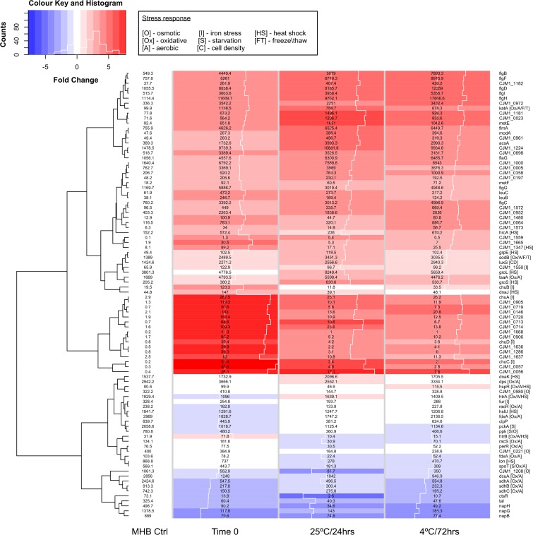
Despite its lack of many of the conventional pathways possessed by other enteropathogenic bacteria, Campylobacter does adapt quickly to environmental stressors [49, 50]. During adaptation to water, C. jejuni M1 must potentially counter oxidative and short term aerobic stress, hypo-osmotic stress, starvation and temperature shock in the different experimental conditions tested. S4 Table summarises the expression profiles of a number of previously characterized stress response genes compared to the MHB Control, across the experimental conditions tested. Fig 3 highlights the expression of known stress response genes and also includes all of the genes that showed significant up- or down-regulation (≥2-fold) compared to MHB Control (S2 Table).
Fig 3. Expression changes in stress-related genes.
The heatmap shows log fold change (logFC) calculated in edgeR compared to the MHB Control (blue = negative, red = positive, the white traceline in the columns indicates the size of the logFC measurement), and displays normalized counts per million (CPM) in numbers. It includes known stress response genes and genes up- or down-regulated ≥2-fold compared to the MHB control based on normalized CPM (numbers displayed in the heatmap matrix) for MHB Control, Time 0, 24 h (25°C) and 72 h(4°C). Square brackets display the type of stress response the gene is involved in (if known). The Histogram in the Colour key indicates the distribution of the data included in the heatmap.
Oxidative, short term aerobic and temperature stress responses
Catalase (katA) and superoxide dismutase (sodB) genes were significantly upregulated in all three experimental conditions compared to the MHB control (Fig 3, S4 Table). These genes are involved in both oxidative stress and freeze-thaw stress response [51]. Despite an apparent lack of cold shock proteins, Campylobacter maintains its metabolic rate at low temperatures (4°C) and appears to survive better at 4°C than at 25°C [52, 53]. In addition, an ankyrin-containing protein CJM1_1347 (Cj1386) has recently been identified in C. jejuni, encoded by a gene based directly downstream of the katA gene, and this is thought to be involved in the same detoxification pathway as catalase [54]; our data indicated significant upregulation of this gene as an early response to exposure to water in the Time 0 sample (Fig 3).
Previous studies have suggested that expression of the catalase (katA) gene, increased after exposure to oxidative stress, but not the superoxide dismutase (sodB) gene, the main antioxidant defence of most organisms [55]. It is essential for C. jejuni to counter atmospheric oxygen tensions and the resulting damage to nucleic acid through the toxicity of reactive oxygen species (ROS). Unlike the three classified types of superoxide dismutases (SODs) present in Escherichia coli, C. jejuni only possess one (SodB) [56, 57]. It has been suggested that both SodB and catalase may play an important role in intracellular survival of C. jejuni [58, 59].
The htrA gene was significantly downregulated during the early response to exposure to water (Time 0). However, downregulation was not evident at the other time points. HtrA is important for stress tolerance and survival of Gram-negative bacteria generally [60, 61] and is required for both heat and oxygen tolerance [62]. It has been reported previously that htrA is downregulated in C. jejuni in response to low nutrient and especially oxidative stress [63]. HtrA, a periplasmic serine protease, displays both chaperone and protease properties, both implicated in the ability to tolerate stress, though the chaperone activity was identified as more important for resistance to oxidative stress [64].
Campylobacter cells show signs of heat stress at temperatures of 46°C and above; these conditions accelerate the transition of spiral cells to coccoid shaped ones. Arguably our experimental conditions tested for cold shock conditions, encountered by Campylobacter in the natural environment outside the host, with test temperatures below the ideal growth temperatures of 37 to 42°C. We did, however, observe changes in the expression levels of several heat shock proteins at 25°C including the chaperones groLS and dnaK the encoding genes are summarized in S4 Table.
Osmotic stress
While a number of studies have contributed to our understanding of hyper-osmotic stress responses [65–67], mainly related to food preservation and in vivo environments, hypo-osmotic stress responses are still poorly understood. Introducing the bacteria into water (Time 0) leads to a marked influx of water into the cells due to the osmotic gradient. Hence, C. jejuni needs to react quickly to prevent cell lysis. Genes implicated in responding to hyper-osmotic conditions include htrB, ppk and a sensor histidine kinase (CJM1_1208). Both CJM1_1208 and ppk were downregulated at all three time points (Fig 3, S4 Table).
The obvious response to changes in osmolarity, exhibited by many bacteria, is to pump out both water and solutes. Three stretch-activated mechano-sensitive (Msc) channels have been described in E. coli: MscM (mini), MscS (small) and MscL (large). A homologue for MscL has been described in H. pylori [68–70]. Kakuda et al. recently identified two putative mechanosensitive channels in the strain 81–176 (Cjj0263 and Cjj1025), corresponding to CJM1_0221 (mechanosensitive ion channel family protein) and CJM1_0980 (putative membrane protein) respectively in M1 [71]. Both genes were upregulated during the early response at Time 0, compared to the MHB control, with CJM1_0980 showing statistically significant upregulation (PPRL = 0.965). Interestingly, both genes were downregulated at 4°C (72 h), suggesting that C. jejuni M1 had adjusted to the hypo-osmotic conditions after 24 h (Fig 3).
Iron acquisition
Iron acquisition is a vital process for bacterial survival and persistence. Due to the toxic potential of free iron, storage and uptake are tightly regulated.
The putative hemin uptake gene cluster chuABCD and Cj1613c (CJM1_1550) are regulated by the ferric uptake repressor (Fur), which in turn is governed by the availability of free iron [72]. In this study, the chuABCD genes were upregulated >2 fold across all three experimental conditions tested, compared to the MHB control; chuA and chuC were statistically significantly upregulated over all three conditions, whereas chuB, chuD and the heme oxygenase gene CJM1_1550 (Cj1613c) were only upregulated at Time 0. These findings strongly support the importance of iron regulation during water survival. The fur gene was statistically significantly downregulated at 25°C (24 h) compared to the MHB control, but did not vary significantly between the non-control samples.
Starvation response
The stringent response is rapidly induced during starvation and stationary phase. Classically, RpoS is the global regulator for the stationary phase in bacteria. In lacking the rpoS gene, C. jejuni presents an RpoS-independent response to starvation and stationary phase. Inorganic polyphosphate (poly-P) is a linear polymer of phosphate residues linked by phosphoanhydride bonds which provide a high energy to the cell [73]. Poly-P, a source of energy and an essential molecule for survival during starvation, is synthesized by mediating the key enzyme polyphosphate kinase 1 (ppk) [74]; we observed that ppk was down-regulated in response to water (Fig 3; S4 Table). Interestingly, it has been reported that a mutant C. jejuni (Δppk) showed decreased ability to enter a VBNC state due to lack in poly-P synthesis [16, 75]. Our observations of down-regulation appear to contradict this notion and may be indicative of variations between strains.
Even if glucose was available, Campylobacter spp. are incapable of using glucose as an energy source and have a very restricted carbohydrate catabolism (non-saccharolytic), a characteristic that distinguishes them greatly from other gastrointestinal pathogens. Phosphoenolpyruvate carboxykinase (PCK)(CJM1_0407), an essential enzyme in gluconeogenesis, was significantly upregulated in the early response (Time 0) compared to the MHB Control (S2 Table).
Quorum sensing
The transfer from an exponentially growing culture into water suddenly presents C. jejuni M1 cells with very low cell density conditions. Bacterial communities can communicate, in a density-dependent manner, through sensing autoinducers, extracellular signal molecules, produced by members of the community. The LuxS product autoinducer 2 (AI-2) is found in over 55 species, and is common to both Gram positive and Gram negative bacteria [76, 77]. Compared to the MHB control, we observed a significant upregulation of luxS expression at Time 0, and at both 25°C (24 h) and 4°C (72 h) (S2 Table). The gene encoding CosR (CJM1_0334), a known positive regulator of luxS [78], was significantly upregulated at 4°C (72 h) compared to 25°C (24 h), but not found to be upregulated otherwise.
Protein translocation and secretion
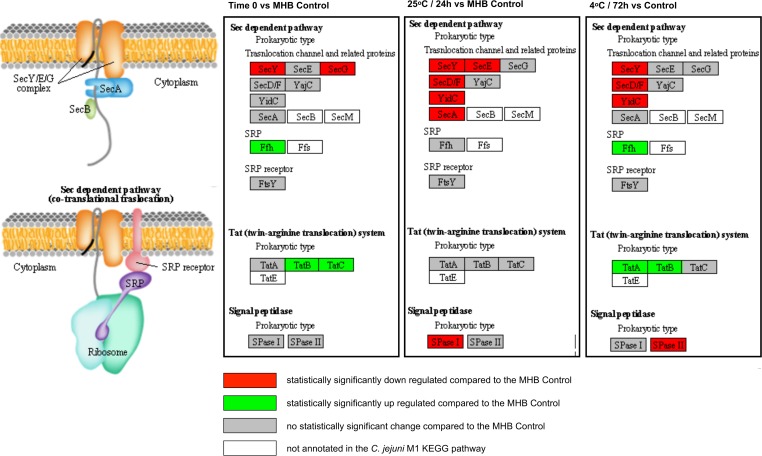
One of the overarching factors important in stress survival is the ability to transport proteins across membranes. The twin-arginine translocation (TAT) system in particular is vital for stress survival [79]. Our data show that components of the TAT system were upregulated at both Time 0 and at 4°C (72 h), compared to the MHB control (Fig 4), suggesting an increased need for translocation of proteins across the cell membrane at these time points and/or temperatures. In contrast, components of the Sec pathway were downregulated in all three conditions (Fig 4), confirming the crucial role of TAT during stressful conditions.
Fig 4. Significant expression changes determined through BitSeq analysis of Sec-dependent and twin-arginine (TAT) protein export pathway components mapped in KEGG.
Results are shown for Time 0, 25°C (24 h) and 4°C (72 h) in sterile distilled water.
The TAT system is an inner membrane translocase that transports proteins folded in the cytoplasm across the inner membrane. In contrast to Sec, which cannot accept tightly folded pre-proteins for translocation, the TAT system can translocate folded enzymes. Several substrates for the C. jejuni TAT system have been identified, including PhoX [80–83], the only alkaline phosphatase identified in Campylobacter species. Upon transport into the periplasm PhoX becomes active, providing Campylobacter with that vital energy source, inorganic phosphate (Pi). The gene encoding PhoX (CJM1_0145) was significantly upregulated at Time 0 compared to the MHB control.
Motility and chemotaxis
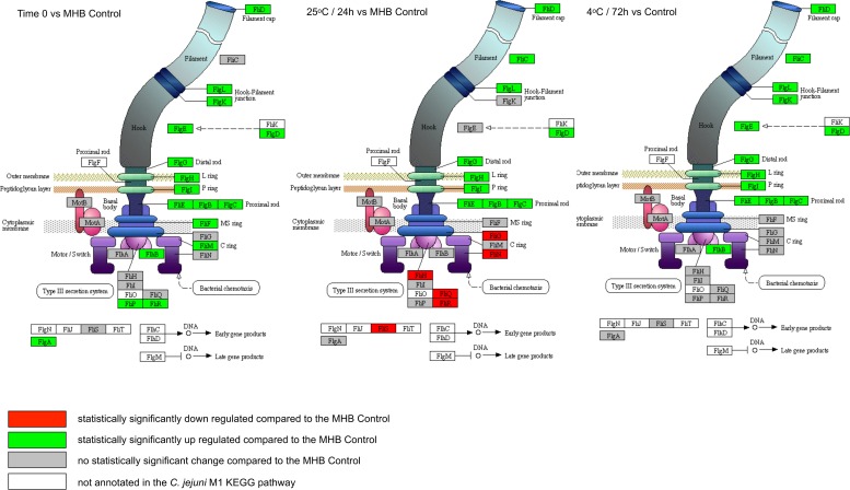
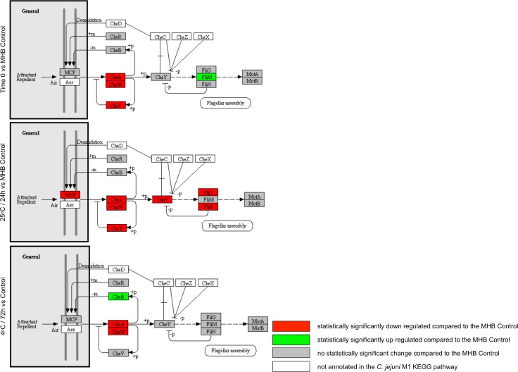
We observed the upregulation of many flagellar assembly genes across all experimental conditions (Fig 5); however, expression of both the flagellar motor genes and components of the chemotaxis pathway (Fig 6) was either downregulated or unchanged, indicating an alternative role to motility, as has been suggested previously [84]. The structural components of flagella are important for the secretion of virulence factors such as the Campylobacter invasion antigen (CiaB) [85]. However, under the conditions tested here, ciaB expression was not significantly upregulated (S3 Table). It has been suggested that C. jejuni could also play an important role in adhesion in the early stages of biofilm formation [86]. Further work is needed to determine whether the gene expression changes that we have seen in water are indicative of the bacteria aggregating as a precursor to forming biofilms.
Fig 5. Significant expression changes determined through BitSeq analysis of C. jejuni M1 flagellar assembly components mapped in KEGG.
Results are shown for Time 0, 25°C (24 h) and 4°C (72 h) in sterile distilled water.
Fig 6. Significant expression changes determined through BitSeq analysis of C. jejuni M1 chemotaxis components mapped in KEGG.
Results are shown for Time 0, 25°C (24 h) and 4°C (72 h) in sterile distilled water.
Studies in H. pylori have shown that the AI-2 encoded by luxS, which is highly upregulated across conditions in this study, targets expression of flagellar genes, and it has been suggested that H. pylori can regulate the composition of its flagella in response to environmental clues [87, 88]. Hence, there may be a link between the LuxS and flagellar component expression changes that we observed.
Electron transport pathways and metabolism
The respiratory chain in C. jejuni is highly branched, with a number of potential electron acceptors, including fumarate, nitrate, nitrite, trimethylamine-N-oxide (TMAO) and dimethylsulphoxide (DMSO), involved in growth under severely oxygen-limited conditions [89]. In our study, three genes (napG, napH, napB) encoding enzymes that play an important role in major electron transport pathways were down-regulated in response to the water environment (Fig 3; S2 Table). nrfA was also down-regulated in all three conditions, whilst nrfH was significantly down-regulated in two of the three conditions compared to the MHB control. These genes encode nitrate (Nap) and nitrite (Nrf) reductases involved in the use of nitrate and nitrite as electron acceptors. The Nap nitrate reductase is a two subunit enzyme comprising NapA and NapB, requiring NapD for proofreading. It is thought that the iron-sulphur proteins NapH and NapG assume the role of electron door to the NapAB complex [90]. The nitrite reductase NrfA is the terminal enzyme in the reduction of nitrite to ammonia, and is thought to play a role in defence against nitrosative stress [90]. It is thought that NrfH is the sole electron donor to NrfA [90]. Hence, our data suggest that nitrate or nitrite electron acceptors are not being utilised during survival in water.
The expression of many of the genes involved in central carbon metabolism was down-regulated in water. These included the genes encoding succinate dehydrogenase (SdhABC), malate dehydrogenase (Mdh) and formate dehydrogenase (FdhABC). Interestingly, the genes encoding PutP (proline transport) and PutA (proline dehydrogenase) were up-regulated, suggesting that proline metabolism was active. PutA catalyses the oxidation of proline to glutamate. Glutamine synthase (GlnA), which can catalyse conversion of glutamate to glutamine, and gamma-glutamyl transpeptidase (GGT), which can catalyse the hydrolysis of glutamine to form glutamate and ammonia, were also up-regulated. In H. pylori, it has been shown that the likely role of GGT is to supply the bacteria with glutamate for catabolism by the hydrolysis of extracellular glutathione or glutamine, with the substrates being hydrolysed in the periplasm before glutamate is transported into the cell [91]. Hence, there is some evidence for metabolism involving proline, glutamine and glutamate, but the full pathway is not clear.
Pathogenicity and virulence factors
Unlike other enteropathogenic bacteria, C. jejuni does not possess many conventional pathogenicity factors. The cells in VBNC state may play an important role in the pathogenicity of C. jejuni; for example, the expression of the virulence gene cadF, encoding an outer-membrane protein of C. jejuni involved in adhesion to intestinal fibronectin has still been detected at high levels up to the third week of entering a VBNC state [92]. We observed consistently high levels of expression of cadF across all conditions tested (S1 Table). However, we did not detect any statistically significant changes in cadF expression (S3 Table). In contrast, the genes encoding cytolethal distending toxin (cdtABC) were down-regulated in response to water (S3 Table).
Do genomic Regions of Difference (RODs) in the C. jejuni M1 genome play a role in its enhanced survival in the experimental conditions tested?
To further investigate the water survival response of C. jejuni M1, we identified RODs within the C. jejuni M1 genome compared to the other C. jejuni strains tested in this experiment, C. jejuni 414 and 1336 (Fig 1A and 1B), as well as the well characterized reference strain NCTC11168 [30, 38, 45]. We identified a total of 45 RODs in the C. jejuni M1 genome; 13 of these RODs are variable regions in all four genomes tested. The results have been summarized in S5 Table where shared RODs have been highlighted.
The currently published C. jejuni M1 genome (CP001900) contains 234 CDS annotated as “hypothetical protein” or “putative uncharacterized protein”. Albeit many of the functions and products of these genes have been described since, for ease of discussion we re-annotated the genome using a combination of PROKKA [93], and searches in stringDB and BLAST (BLASTP and BLASTX). Putative functions and expression profiles are summarised in S5 Table. An overall summary of the M1 genome, including RODs, hypothetical genes, and BitSeq expression data is shown in S2 Fig.
There were a number of examples of genes, or clusters of genes, within RODs that were either up- or down-regulated (S5 Table; S6 Table). It has been reported previously that the genomes of strains 1336 and 414 lack some genes that are widely distributed in C. jejuni [30]. Hence, some RODs include genes that have known putative functions, such as the cdtABC genes, which are down-regulated in water. Since M1 behaves differently to strains 1336 and 414 in water, it is possible that genes present in only strain M1 might contribute to these differences. For example, ROD-6 (comprising four putative genes) is absent from strains 414 and 1336, and is upregulated in water. However, the gene functions are not known. Further work would be needed to determine which, if any, RODs play a significant role in the strain M1 loss of culturabilityphenotype.
Conclusions
Our data suggest that C. jejuni M1 adapts rapidly to introduction into a water environment, instigating gene expression changes that allow it to adapt to the stressful conditions, whilst maintaining viability. A recent complementary study by de Vries et al (2017) used Transposon Directed Insertion Sequencing (TRADIS) to identify genes essential for survival in vitro and in vivo [94]. Although comparing knockout studies to whole-transcriptome studies is difficult because gene knockouts will likely produce different transcriptional profiles due to deregulation or disruption of genetic pathways, in both studies a gene involved in the oxidative stress response: trxC, was identified as a key candidate gene for survival in model water systems. trxC was significantly upregulated in our study and was identified as ‘essential’ in de Vries et al (2017). Thus, it will be important to follow up these studies, particularly focusing on oxidative stress, using targeted techniques such as real-time PCR, and comparing environmental water samples of different kinds.
In addition to the up-regulation of stress responses, a preference for secretion via the Tat pathway and the down-regulation of many (but not all) metabolic genes, the bacteria adapt in some more surprising ways. We observed down-regulation of chemotaxis genes coupled to up-regulation of flagellar genes, suggesting a role for flagella that is not linked to motility. In addition to the secretion of virulence factors, flagella have been implicated in autoagglutination and microcolony formation as a precursor to biofilm formation [95]. Hence, it is possible that the observed gene expression changes are indicative of the bacteria starting to instigate a lifestyle change from sessile to biofilm as a survival strategy. We also found evidence that the putative quorum sensing system protein LuxS plays a role during the adaptation to water.
Genes in the accessory genomes, including genes encoding hypothetical proteins of no known function, may play a role in the variable survival phenotypes that are observed between strains. Hence, despite the loss of culturability, strain M1 remains viable and adapts to suspension in water via multiple specific changes in gene expression. Further work is needed to ascertain which responses are shared by all C. jejuni strains, and which are specific to a sub-set of strains sharing the characteristics of M1.
Supporting information
(XLSX)
Cpm displayed are normalised by the Trimmed Mean of M-values (TMM) method, implemented in the edgR Bioconductor package. In brackets the log-fold-change cpm relative to the MHB is shown. Cells are shaded according to log-fold-change (yellow = negative, green = positive).
(DOCX)
Results are shown in Probability of Positive Log Ratio (PPLR), PPLR ≥0.95 shows a high probability that the transcript is upregulated (green shading) compared to the first condition; PPLR ≤0.05 shows a very low probability that the transcript is upregulated compared to the first condition and therefore the probability of the transcript being downregulated (red shading) is very high.
(XLSX)
Fold change compared to the MHB Control, as determined by edgeR, the Paired student’s T-test and PPLR determined through BitSeq analysis are shown in brackets. Stressor abbreviations: S, starvation; O, osmotic; OX, oxidative; A, aerobic; HS, heat-shock; F/T; freeze-thaw; I, iron stress; CD, cell density.
(DOCX)
RODs shaded in teal are divergent from M1 in the other three strains (414, 1336 and NCTC11168).
(XLSB)
Green shading indicates PPLR ≥0.95, red shading indicates ≤0.05, grey shading indicates no statistically significant difference.
(XLSB)
Example samples are shown for a sample from 25°C (24 h) (D1 3), Time 0 (D0 2), 4°C (72 h) (D3 1) and the Mueller Hinton Broth Control (C1 107). There was no evidence for RNA degradation in the 24 h or 72 h water samples compared to the time zero or control samples.
(TIF)
From the outside in: Track 1 C. jejuni M1 genome; Track 2 CDS forward strand; Track 3 CDS reverse strand; Track 4 hypothetical/ putative uncharacterized genes (CDS); Track 5 and 6 Regions of Difference (RODs) in the C. jejuni M1 genome compared to C. jejuni 414 (turquoise), M1 compared to NCTC11168 (magenta) and M1 compared to 1336 (blue); Track 7 operons in M1 as predicted by Rockhopper; Track 8 genes that are statistically significantly upregulated (green) or downregulated (red) at Time 0 only, compared to the Control; Track 9 genes that are statistically significantly upregulated (green) or downregulated (red) in 25°C (24 h) only compared to the Control; Track 10 genes that are statistically significantly upregulated (green) or downregulated (red) in 4°C (72 h) only compared to the Control; Track 11 genes that are statistically significantly upregulated (green) or downregulated (red) in 25°C (24 h) only compared to Time 0; Track 12 genes that are statistically significantly upregulated (green) or downregulated (red) in 4°C (72 h) only compared to Time 0; Track 13 genes that are statistically significantly upregulated (green) or downregulated (red) in 4°C (72 h) only compared to 25°C (24 h). Colouring of Tracks 2 and 3: replication related = bright red; efflux pumps = bright green; chemotaxis = bright blue; hydrogenases = pale pink; iron or heme-related = light brown; hydrogenases = rose; chaperone = orange; lipoproteins = yellow; membrane or periplasmic proteins = turquoise; ATP-/ ABC transporters = light green; ribosomal/RNA/ribonuclease = light blue; flagellar-related = magenta; hypotheticals = salmon.
(TIFF)
Acknowledgments
We would like to thank John Kenny at the CGR for help and advice.
We thank members of the ENIGMA Consortium for helpful discussion. The ENIGMA Consortium investigators comprise Sarah O’Brien (PI), Rob Christley, Christiane Hertz-Fowler, Paul Wigley, Nicola Williams and Craig Winstanley (University of Liverpool), Peter Diggle (Lancaster University), Iain Lake, Kevin Hiscock and Paul Hunter (University of East Anglia), Ken Forbes and Norval Strachan (University of Aberdeen), Rachel Griffith and Dan Rigby (University of Manchester), Paul Cross (Bangor University), Stephen Rushton (Newcastle University), Tom Humphrey (Swansea University), Malcolm Bennett (University of Nottingham), David Howard (Centre for Ecology and Hydrology), and Brendan Wren (London School of Hygiene and Tropical Medicine).
We acknowledge the Medical Research Council, Natural Environment Research Council, Economic and Social Research Council, Biotechnology and Biosciences Research Council and Food Standards Agency for the funding received for this project through the Environmental & Social Ecology of Human Infectious Diseases Initiative (Enigma; Grant Reference: G1100799/1).
Data Availability
RNA Sequencing data are available at the European Nucleotide Archive (http://www.ebi.ac.uk/ena) under Primary Accession PRJEB17925. Other data supporting the results are available as Supporting Information.
Funding Statement
Medical Research Council, Natural Environment Research Council, Economic and Social Research Council, Biotechnology and Biosciences Research Council and Food Standards Agency for the funding received for this project through the Environmental & Social Ecology of Human Infectious Diseases Initiative (Enigma; Grant Reference: G1100799/1)
References
- 1.Tam CC, O'Brien SJ, Tompkins DS, Bolton FJ, Berry L, Dodds J, et al. Changes in causes of acute gastroenteritis in the United Kingdom over 15 years: microbiologic findings from 2 prospective, population-based studies of infectious intestinal disease. Clinical infectious diseases: an official publication of the Infectious Diseases Society of America. 2012;54(9):1275–86. doi: 10.1093/cid/cis028 . [DOI] [PubMed] [Google Scholar]
- 2.Tam CC, Rodrigues LC, Viviani L, Dodds JP, Evans MR, Hunter PR, et al. Longitudinal study of infectious intestinal disease in the UK (IID2 study): incidence in the community and presenting to general practice. Gut. 2012;61(1):69–77. doi: 10.1136/gut.2011.238386 ; PubMed Central PMCID: PMC3230829. [DOI] [PMC free article] [PubMed] [Google Scholar]
- 3.Crim SM, Iwamoto M, Huang JY, Griffin PM, Gilliss D, Cronquist AB, et al. Incidence and trends of infection with pathogens transmitted commonly through food—Foodborne Diseases Active Surveillance Network, 10 U.S. sites, 2006–2013. MMWR Morbidity and mortality weekly report. 2014;63(15):328–32. . [PMC free article] [PubMed] [Google Scholar]
- 4.Wingstrand A, Neimann J, Engberg J, Nielsen EM, Gerner-Smidt P, Wegener HC, et al. Fresh chicken as main risk factor for campylobacteriosis, Denmark. Emerging infectious diseases. 2006;12(2):280–5. doi: 10.3201/eid1202.050936 ; PubMed Central PMCID: PMC3373097. [DOI] [PMC free article] [PubMed] [Google Scholar]
- 5.Taylor EV, Herman KM, Ailes EC, Fitzgerald C, Yoder JS, Mahon BE, et al. Common source outbreaks of Campylobacter infection in the USA, 1997–2008. Epidemiol Infect. 2013;141(5):987–96. doi: 10.1017/S0950268812001744 . [DOI] [PMC free article] [PubMed] [Google Scholar]
- 6.Bronowski C, James CE, Winstanley C. Role of environmental survival in transmission of Campylobacter jejuni. FEMS microbiology letters. 2014;356(1):8–19. doi: 10.1111/1574-6968.12488 . [DOI] [PubMed] [Google Scholar]
- 7.Pearson AD, Greenwood M, Healing TD, Rollins D, Shahamat M, Donaldson J, et al. Colonization of broiler chickens by waterborne Campylobacter jejuni. Applied and environmental microbiology. 1993;59(4):987–96. [DOI] [PMC free article] [PubMed] [Google Scholar]
- 8.Van de Giessen AW, Bloemberg BPM, Ritmeester WS, Tilburg J. Epidemiological study on risk factors and risk reducing measures for Campylobacter infections in Dutch broiler flocks. Epidemiol Infect. 1996;117(02):245–50. [DOI] [PMC free article] [PubMed] [Google Scholar]
- 9.Miettinen I, Zacheus O, Von Bonsdorff C, Vartiainen T. Waterborne epidemics in Finland in 1998–1999. Water Science & Technology. 2001;43(12):67–71. [PubMed] [Google Scholar]
- 10.Martin S, Penttinen P, Ljungstrom M, Allestam G, Andersson Y, Giesecke J, et al. A case-cohort study to investigate concomitant waterborne outbreaks of Campylobacter and gastroenteritis in Suderhamn, Sweden, 2002–3. J Water Health. 2006;4:417–24. [PubMed] [Google Scholar]
- 11.Hnninen M, Krenlampi R. Campylobacter in waterborne epidemics in Finland. Water Supply. 2004;4(2):39–45. [Google Scholar]
- 12.Said B, Wright F, Nichols GL, Reacher M, Rutter M. Outbreaks of infectious disease associated with private drinking water supplies in England and Wales 1970–2000. Epidemiol Infect. 2003;130(03):469–79. [PMC free article] [PubMed] [Google Scholar]
- 13.Smith A, Reacher M, Smerdon W, Adak GK, Nichols G, Chalmers RM. Outbreaks of waterborne infectious intestinal disease in England and Wales, 1992–2003. Epidemiol Infect. 2006;134(06):1141–9. [DOI] [PMC free article] [PubMed] [Google Scholar]
- 14.Jones IG, Roworth M. An outbreak of< i> Escherichia coli 0157 and campylobacteriosisassociated with contamination of a drinking water supply. Public health. 1996;110(5):277–82. [DOI] [PubMed] [Google Scholar]
- 15.Taylor EV, Herman KM, Ailes EC, Fitzgerald C, Yoder JS, Mahon BE, et al. Common source outbreaks of Campylobacter infection in the USA, 1997–2008. Epidemiol Infect. 2013;141(05):987–96. [DOI] [PMC free article] [PubMed] [Google Scholar]
- 16.Kassem II, Rajashekara G. An ancient molecule in a recalcitrant pathogen: the contributions of poly-P to the pathogenesis and stress responses of Campylobacter jejuni. Future Microbiol. 2011;6(10):1117–20. doi: 10.2217/fmb.11.94 [DOI] [PubMed] [Google Scholar]
- 17.Murphy C, Carroll C, Jordan KN. The effect of different media on the survival and induction of stress responses by Campylobacter jejuni. J Microbiol Methods. 2005;62(2):161–6. doi: 10.1016/j.mimet.2005.02.005 . [DOI] [PubMed] [Google Scholar]
- 18.Jones K. Campylobacters in water, sewage and the environment. Symp Ser Soc Appl Microbiol. 2001;(30):68S–79S. . [DOI] [PubMed] [Google Scholar]
- 19.Guillou S, Leguérinel I, Garrec N, Renard MA, Cappelier JM, Federighi M. Survival of< i> Campylobacter jejuni in mineral bottled water according to difference in mineral content: Application of the Weibull model. Water Res. 2008;42(8):2213–9. [DOI] [PubMed] [Google Scholar]
- 20.Rollins DM, Colwell RR. Viable but nonculturable stage of Campylobacter jejuni and its role in survival in the natural aquatic environment. Applied and environmental microbiology. 1986;52(3):531–8. . [DOI] [PMC free article] [PubMed] [Google Scholar]
- 21.Chaisowwong W, Kusumoto A, Hashimoto M, Harada T, Maklon K, Kawamoto K. Physiological characterization of Campylobacter jejuni under cold stresses conditions: its potential for public threat. The Journal of veterinary medical science/the Japanese Society of Veterinary Science. 2012;74(1):43–50. [DOI] [PubMed] [Google Scholar]
- 22.Cools I, Uyttendaele M, Caro C, D'Haese E, Nelis HJ, Debevere J. Survival of Campylobacter jejuni strains of different origin in drinking water. Journal of applied microbiology. 2003;94(5):886–92. . [DOI] [PubMed] [Google Scholar]
- 23.Lazaro B, Carcamo J, Audicana A, Perales I, Fernandez-Astorga A. Viability and DNA maintenance in nonculturable spiral Campylobacter jejuni cells after long-term exposure to low temperatures. Appl Environ Microbiol. 1999;65(10):4677–81. ; PubMed Central PMCID: PMC91624. [DOI] [PMC free article] [PubMed] [Google Scholar]
- 24.Tholozan JL, Cappelier JM, Tissier JP, Delattre G, Federighi M. Physiological characterization of viable-but-nonculturable Campylobacter jejuni cells. Appl Environ Microbiol. 1999;65(3):1110–6. ; PubMed Central PMCID: PMC91151. [DOI] [PMC free article] [PubMed] [Google Scholar]
- 25.Thomas C, Hill D, Mabey M. Culturability, injury and morphological dynamics of thermophilic Campylobacter spp. within a laboratory‐based aquatic model system. Journal of Applied Microbiology. 2002;92(3):433–42. [DOI] [PubMed] [Google Scholar]
- 26.Buswell CM, Herlihy YM, Lawrence LM, McGuiggan JTM, Marsh PD, Keevil CW, et al. Extended Survival and Persistence of Campylobacterspp. in Water and Aquatic Biofilms and Their Detection by Immunofluorescent-Antibody and-rRNA Staining. Applied and environmental microbiology. 1998;64(2):733–41. [DOI] [PMC free article] [PubMed] [Google Scholar]
- 27.Stahl M, Butcher J, Stintzi A. Nutrient acquisition and metabolism by Campylobacter jejuni. Frontiers in cellular and infection microbiology. 2012;2:5 doi: 10.3389/fcimb.2012.00005 ; PubMed Central PMCID: PMC3417520. [DOI] [PMC free article] [PubMed] [Google Scholar]
- 28.Colles FM, Jones K, Harding RM, Maiden MC. Genetic diversity of Campylobacter jejuni isolates from farm animals and the farm environment. Appl Environ Microbiol. 2003;69(12):7409–13. Epub 2003/12/09. doi: 10.1128/AEM.69.12.7409-7413.2003 ; PubMed Central PMCID: PMC309911. [DOI] [PMC free article] [PubMed] [Google Scholar]
- 29.French N, Barrigas M, Brown P, Ribiero P, Williams N, Leatherbarrow H, et al. Spatial epidemiology and natural population structure of Campylobacter jejuni colonizing a farmland ecosystem. Environ Microbiol. 2005;7(8):1116–26. doi: 10.1111/j.1462-2920.2005.00782.x . [DOI] [PubMed] [Google Scholar]
- 30.Hepworth PJ, Ashelford KE, Hinds J, Gould KA, Witney AA, Williams NJ, et al. Genomic variations define divergence of water/wildlife-associated Campylobacter jejuni niche specialists from common clonal complexes. Environ Microbiol. 2011;13(6):1549–60. Epub 2011/03/23. doi: 10.1111/j.1462-2920.2011.02461.x ; PubMed Central PMCID: PMCPMC3569610. [DOI] [PMC free article] [PubMed] [Google Scholar]
- 31.Hofreuter D, Tsai J, Watson RO, Novik V, Altman B, Benitez M, et al. Unique features of a highly pathogenic Campylobacter jejuni strain. Infection and immunity. 2006;74(8):4694–707. doi: 10.1128/IAI.00210-06 ; PubMed Central PMCID: PMC1539605. [DOI] [PMC free article] [PubMed] [Google Scholar]
- 32.Fouts DE, Mongodin EF, Mandrell RE, Miller WG, Rasko DA, Ravel J, et al. Major structural differences and novel potential virulence mechanisms from the genomes of multiple campylobacter species. PLoS biology. 2005;3(1):e15 doi: 10.1371/journal.pbio.0030015 ; PubMed Central PMCID: PMC539331. [DOI] [PMC free article] [PubMed] [Google Scholar]
- 33.Ahmed IH, Manning G, Wassenaar TM, Cawthraw S, Newell DG. Identification of genetic differences between two Campylobacter jejuni strains with different colonization potentials. Microbiology. 2002;148(Pt 4):1203–12. doi: 10.1099/00221287-148-4-1203 [DOI] [PubMed] [Google Scholar]
- 34.Hofreuter D. Defining the metabolic requirements for the growth and colonization capacity of Campylobacter jejuni. Frontiers in cellular and infection microbiology. 2014;4:137 doi: 10.3389/fcimb.2014.00137 ; PubMed Central PMCID: PMC4178425. [DOI] [PMC free article] [PubMed] [Google Scholar]
- 35.Hofreuter D, Novik V, Galan JE. Metabolic diversity in Campylobacter jejuni enhances specific tissue colonization. Cell host & microbe. 2008;4(5):425–33. doi: 10.1016/j.chom.2008.10.002 . [DOI] [PubMed] [Google Scholar]
- 36.Wilson DL, Rathinam VA, Qi W, Wick LM, Landgraf J, Bell JA, et al. Genetic diversity in Campylobacter jejuni is associated with differential colonization of broiler chickens and C57BL/6J IL10-deficient mice. Microbiology. 2010;156(Pt 7):2046–57. doi: 10.1099/mic.0.035717-0 ; PubMed Central PMCID: PMC3068676. [DOI] [PMC free article] [PubMed] [Google Scholar]
- 37.Van Dyke MI, Morton VK, McLellan NL, Huck PM. The occurrence of Campylobacter in river water and waterfowl within a watershed in southern Ontario, Canada. Journal of Applied Microbiology. 2010;109(3):1053–66. doi: 10.1111/j.1365-2672.2010.04730.x [DOI] [PubMed] [Google Scholar]
- 38.Friis C, Wassenaar TM, Javed MA, Snipen L, Lagesen K, Hallin PF, et al. Genomic characterization of Campylobacter jejuni strain M1. PLoS One. 2010;5(8):e12253 Epub 2010/09/25. doi: 10.1371/journal.pone.0012253 ; PubMed Central PMCID: PMCPMC2928727. [DOI] [PMC free article] [PubMed] [Google Scholar]
- 39.Martin M. Cutadapt removes adapter sequences from high-throughput sequencing reads. EMBnetjournal. 2011;17(1):2 http://dx.doi.org/10.14806/ej.17.1.200. [Google Scholar]
- 40.Anders S, Pyl PT, Huber W. HTSeq—a Python framework to work with high-throughput sequencing data. Bioinformatics. 2015;31(2):166–9. doi: 10.1093/bioinformatics/btu638 ; PubMed Central PMCID: PMC4287950. [DOI] [PMC free article] [PubMed] [Google Scholar]
- 41.Langmead B, Salzberg SL. Fast gapped-read alignment with Bowtie 2. Nat Methods. 2012;9(4):357–9. doi: 10.1038/nmeth.1923 ; PubMed Central PMCID: PMCPMC3322381. [DOI] [PMC free article] [PubMed] [Google Scholar]
- 42.Glaus P, Honkela A, Rattray M. Identifying differentially expressed transcripts from RNA-seq data with biological variation. Bioinformatics. 2012;28(13):1721–8. doi: 10.1093/bioinformatics/bts260 ; PubMed Central PMCID: PMC3381971. [DOI] [PMC free article] [PubMed] [Google Scholar]
- 43.Carver T, Berriman M, Tivey A, Patel C, Bohme U, Barrell BG, et al. Artemis and ACT: viewing, annotating and comparing sequences stored in a relational database. Bioinformatics. 2008;24(23):2672–6. Epub 2008/10/11. doi: 10.1093/bioinformatics/btn529 ; PubMed Central PMCID: PMC2606163. [DOI] [PMC free article] [PubMed] [Google Scholar]
- 44.Gundogdu O, Bentley SD, Holden MT, Parkhill J, Dorrell N, Wren BW. Re-annotation and re-analysis of the Campylobacter jejuni NCTC11168 genome sequence. BMC genomics. 2007;8:162 doi: 10.1186/1471-2164-8-162 ; PubMed Central PMCID: PMC1899501. [DOI] [PMC free article] [PubMed] [Google Scholar]
- 45.Parkhill J, Wren BW, Mungall K, Ketley JM, Churcher C, Basham D, et al. The genome sequence of the food-borne pathogen Campylobacter jejuni reveals hypervariable sequences. Nature. 2000;403(6770):665–8. doi: 10.1038/35001088 [DOI] [PubMed] [Google Scholar]
- 46.Carver TJ, Rutherford KM, Berriman M, Rajandream MA, Barrell BG, Parkhill J. ACT: the Artemis Comparison Tool. Bioinformatics. 2005;21(16):3422–3. Epub 2005/06/25. doi: 10.1093/bioinformatics/bti553 . [DOI] [PubMed] [Google Scholar]
- 47.Williams NJ, Jones TR, Leatherbarrow HJ, Birtles RJ, Lahuerta-Marin A, Bennett M, et al. Isolation of a novel Campylobacter jejuni clone associated with the bank vole, Myodes glareolus. Appl Environ Microbiol. 2010;76(21):7318–21. doi: 10.1128/AEM.00511-10 ; PubMed Central PMCID: PMC2976251. [DOI] [PMC free article] [PubMed] [Google Scholar]
- 48.Goon S, Kelly JF, Logan SM, Ewing CP, Guerry P. Pseudaminic acid, the major modification on Campylobacter flagellin, is synthesized via the Cj1293 gene. Mol Microbiol. 2003;50(2):659–71. . [DOI] [PubMed] [Google Scholar]
- 49.Young KT, Davis LM, Dirita VJ. Campylobacter jejuni: molecular biology and pathogenesis. Nat Rev Microbiol. 2007;5(9):665–79. doi: 10.1038/nrmicro1718 . [DOI] [PubMed] [Google Scholar]
- 50.Newell DG, Koopmans M, Verhoef L, Duizer E, Aidara-Kane A, Sprong H, et al. Food-borne diseases—the challenges of 20 years ago still persist while new ones continue to emerge. Int J Food Microbiol. 2010;139 Suppl 1:S3–15. doi: 10.1016/j.ijfoodmicro.2010.01.021 . [DOI] [PMC free article] [PubMed] [Google Scholar]
- 51.Chan KF, Le Tran H, Kanenaka RY, Kathariou S. Survival of clinical and poultry-derived isolates of Campylobacter jejuni at a low temperature (4 degrees C). Appl Environ Microbiol. 2001;67(9):4186–91. doi: 10.1128/AEM.67.9.4186-4191.2001 ; PubMed Central PMCID: PMCPMC93146. [DOI] [PMC free article] [PubMed] [Google Scholar]
- 52.Blaser MJ, Hardesty HL, Powers B, Wang WL. Survival of Campylobacter fetus subsp. jejuni in biological milieus. Journal of clinical microbiology. 1980;11(4):309–13. ; PubMed Central PMCID: PMC273394. [DOI] [PMC free article] [PubMed] [Google Scholar]
- 53.Hazeleger WC, Wouters JA, Rombouts FM, Abee T. Physiological activity of Campylobacter jejuni far below the minimal growth temperature. Appl Environ Microbiol. 1998;64(10):3917–22. ; PubMed Central PMCID: PMC106578. [DOI] [PMC free article] [PubMed] [Google Scholar]
- 54.Flint A, Sun Y-Q, Stintzi A. Cj1386 is an ankyrin-containing protein involved in heme trafficking to catalase in Campylobacter jejuni. J Bacteriol. 2012;194(2):334–45. doi: 10.1128/JB.05740-11 [DOI] [PMC free article] [PubMed] [Google Scholar]
- 55.Garénaux A, Jugiau F, Rama F, De Jonge R, Denis M, Federighi M, et al. Survival of Campylobacter jejuni strains from different origins under oxidative stress conditions: effect of temperature. Curr Microbiol. 2008;56(4):293–7. doi: 10.1007/s00284-007-9082-8 [DOI] [PubMed] [Google Scholar]
- 56.Purdy D, Cawthraw S, Dickinson JH, Newell DG, Park SF. Generation of a superoxide dismutase (SOD)-deficient mutant of Campylobacter coli: evidence for the significance of SOD in Campylobacter survival and colonization. Applied and environmental microbiology. 1999;65(6):2540–6. . [DOI] [PMC free article] [PubMed] [Google Scholar]
- 57.Bakshi CS, Malik M, Regan K, Melendez JA, Metzger DW, Pavlov VM, et al. Superoxide dismutase B gene (sodB)-deficient mutants of Francisella tularensis demonstrate hypersensitivity to oxidative stress and attenuated virulence. J Bacteriol. 2006;188(17):6443–8. doi: 10.1128/JB.00266-06 [DOI] [PMC free article] [PubMed] [Google Scholar]
- 58.Pesci EC, Cottle DL, Pickett CL. Genetic, enzymatic, and pathogenic studies of the iron superoxide dismutase of Campylobacter jejuni. Infection and immunity. 1994;62(7):2687–94. [DOI] [PMC free article] [PubMed] [Google Scholar]
- 59.Day WA, Sajecki JL, Pitts TM, Joens LA. Role of catalase in Campylobacter jejuniintracellular survival. Infection and immunity. 2000;68(11):6337–45. [DOI] [PMC free article] [PubMed] [Google Scholar]
- 60.Li S-R, Dorrell N, Everest PH, Dougan G, Wren BW. Construction and characterization of a Yersinia enterocolitica O: 8 high-temperature requirement (htrA) isogenic mutant. Infection and immunity. 1996;64(6):2088–94. [DOI] [PMC free article] [PubMed] [Google Scholar]
- 61.Laskowska E, Kuczyńska‐Wiśnik D, Skórko‐Glonek J, Taylor A. Degradation by proteases Lon, Clp and HtrA, of Escherichia coli proteins aggregated in vivo by heat shock; HtrA protease action in vivo and in vitro. Mol Microbiol. 1996;22(3):555–71. doi: 10.1046/j.1365-2958.1996.1231493.x [DOI] [PubMed] [Google Scholar]
- 62.Brøndsted L, Andersen MT, Parker M, Jørgensen K, Ingmer H. The HtrA protease of Campylobacter jejuni is required for heat and oxygen tolerance and for optimal interaction with human epithelial cells. Applied and environmental microbiology. 2005;71(6):3205–12. doi: 10.1128/AEM.71.6.3205-3212.2005 [DOI] [PMC free article] [PubMed] [Google Scholar]
- 63.Bui XT, Qvortrup K, Wolff A, Bang DD, Creuzenet C. Effect of environmental stress factors on the uptake and survival of Campylobacter jejuni in Acanthamoeba castellanii. BMC Microbiol. 2012;12(1):232. [DOI] [PMC free article] [PubMed] [Google Scholar]
- 64.Baek KT, Vegge CS, Skorko-Glonek J, Brondsted L. Different contributions of HtrA protease and chaperone activities to Campylobacter jejuni stress tolerance and physiology. Appl Environ Microbiol. 2011;77(1):57–66. Epub 2010/11/16. doi: 10.1128/AEM.01603-10 ; PubMed Central PMCID: PMC3019702. [DOI] [PMC free article] [PubMed] [Google Scholar]
- 65.Park SF. The physiology of Campylobacter species and its relevance to their role as foodborne pathogens. Int J Food Microbiol. 2002;74(3):177–88. . [DOI] [PubMed] [Google Scholar]
- 66.Birk T, Ingmer H, Andersen MT, Jorgensen K, Brondsted L. Chicken juice, a food-based model system suitable to study survival of Campylobacter jejuni. Lett Appl Microbiol. 2004;38(1):66–71. . [DOI] [PubMed] [Google Scholar]
- 67.Alter T, Scherer K. Stress response of Campylobacter spp. and its role in food processing. J Vet Med B Infect Dis Vet Public Health. 2006;53(8):351–7. doi: 10.1111/j.1439-0450.2006.00983.x . [DOI] [PubMed] [Google Scholar]
- 68.Berrier C, Besnard M, Ajouz B, Coulombe A, Ghazi A. Multiple mechanosensitive ion channels from Escherichia coli, activated at different thresholds of applied pressure. J Membr Biol. 1996;151(2):175–87. . [DOI] [PubMed] [Google Scholar]
- 69.Kloda A, Martinac B. Structural and functional differences between two homologous mechanosensitive channels of Methanococcus jannaschii. EMBO J. 2001;20(8):1888–96. doi: 10.1093/emboj/20.8.1888 ; PubMed Central PMCID: PMCPMC125423. [DOI] [PMC free article] [PubMed] [Google Scholar]
- 70.Booth IR, Louis P. Managing hypoosmotic stress: aquaporins and mechanosensitive channels in Escherichia coli. Curr Opin Microbiol. 1999;2(2):166–9. . [DOI] [PubMed] [Google Scholar]
- 71.Kakuda T, Koide Y, Sakamoto A, Takai S. Characterization of two putative mechanosensitive channel proteins of Campylobacter jejuni involved in protection against osmotic downshock. Vet Microbiol. 2012;160(1–2):53–60. doi: 10.1016/j.vetmic.2012.04.044 . [DOI] [PubMed] [Google Scholar]
- 72.Ridley KA, Rock JD, Li Y, Ketley JM. Heme utilization in Campylobacter jejuni. J Bacteriol. 2006;188(22):7862–75. doi: 10.1128/JB.00994-06 ; PubMed Central PMCID: PMC1636299. [DOI] [PMC free article] [PubMed] [Google Scholar]
- 73.Kornberg A. Inorganic polyphosphate: a molecule of many functions. Inorganic polyphosphates: Springer; 1999. p. 1–18. [DOI] [PubMed] [Google Scholar]
- 74.Candon HL, Allan BJ, Fraley CD, Gaynor EC. Polyphosphate kinase 1 is a pathogenesis determinant in Campylobacter jejuni. J Bacteriol. 2007;189(22):8099–108. doi: 10.1128/JB.01037-07 ; PubMed Central PMCID: PMCPMC2168705. [DOI] [PMC free article] [PubMed] [Google Scholar]
- 75.Gangaiah D, Kassem II, Liu Z, Rajashekara G. Importance of polyphosphate kinase 1 for Campylobacter jejuni viable-but-nonculturable cell formation, natural transformation, and antimicrobial resistance. Appl Environ Microbiol. 2009;75(24):7838–49. doi: 10.1128/AEM.01603-09 ; PubMed Central PMCID: PMCPMC2794102. [DOI] [PMC free article] [PubMed] [Google Scholar]
- 76.Vendeville A, Winzer K, Heurlier K, Tang CM, Hardie KR. Making 'sense' of metabolism: autoinducer-2, LuxS and pathogenic bacteria. Nat Rev Microbiol. 2005;3(5):383–96. doi: 10.1038/nrmicro1146 . [DOI] [PubMed] [Google Scholar]
- 77.Xavier KB, Bassler BL. LuxS quorum sensing: more than just a numbers game. Curr Opin Microbiol. 2003;6(2):191–7. . [DOI] [PubMed] [Google Scholar]
- 78.Hwang S, Kim M, Ryu S, Jeon B. Regulation of oxidative stress response by CosR, an essential response regulator in Campylobacter jejuni. PLoS One. 2011;6(7):e22300 doi: 10.1371/journal.pone.0022300 ; PubMed Central PMCID: PMCPMC3139631. [DOI] [PMC free article] [PubMed] [Google Scholar]
- 79.Rajashekara G, Drozd M, Gangaiah D, Jeon B, Liu Z, Zhang Q. Functional characterization of the twin-arginine translocation system in Campylobacter jejuni. Foodborne pathogens and disease. 2009;6(8):935–45. doi: 10.1089/fpd.2009.0298 . [DOI] [PubMed] [Google Scholar]
- 80.Hitchcock A, Hall SJ, Myers JD, Mulholland F, Jones MA, Kelly DJ. Roles of the twin-arginine translocase and associated chaperones in the biogenesis of the electron transport chains of the human pathogen Campylobacter jejuni. Microbiology. 2010;156(Pt 10):2994–3010. doi: 10.1099/mic.0.042788-0 . [DOI] [PubMed] [Google Scholar]
- 81.Drozd M, Gangaiah D, Liu Z, Rajashekara G. Contribution of TAT system translocated PhoX to Campylobacter jejuni phosphate metabolism and resilience to environmental stresses. PLoS One. 2011;6(10):e26336 doi: 10.1371/journal.pone.0026336 ; PubMed Central PMCID: PMCPMC3197622. [DOI] [PMC free article] [PubMed] [Google Scholar]
- 82.Hall SJ, Hitchcock A, Butler CS, Kelly DJ. A Multicopper oxidase (Cj1516) and a CopA homologue (Cj1161) are major components of the copper homeostasis system of Campylobacter jejuni. J Bacteriol. 2008;190(24):8075–85. doi: 10.1128/JB.00821-08 ; PubMed Central PMCID: PMCPMC2593206. [DOI] [PMC free article] [PubMed] [Google Scholar]
- 83.Wosten MM, Parker CT, van Mourik A, Guilhabert MR, van Dijk L, van Putten JP. The Campylobacter jejuni PhosS/PhosR operon represents a non-classical phosphate-sensitive two-component system. Mol Microbiol. 2006;62(1):278–91. doi: 10.1111/j.1365-2958.2006.05372.x . [DOI] [PubMed] [Google Scholar]
- 84.Guerry P. Campylobacter flagella: not just for motility. Trends Microbiol. 2007;15(10):456–61. doi: 10.1016/j.tim.2007.09.006 . [DOI] [PubMed] [Google Scholar]
- 85.Konkel ME, Klena JD, Rivera-Amill V, Monteville MR, Biswas D, Raphael B, et al. Secretion of virulence proteins from Campylobacter jejuni is dependent on a functional flagellar export apparatus. J Bacteriol. 2004;186(11):3296–303. doi: 10.1128/JB.186.11.3296-3303.2004 ; PubMed Central PMCID: PMCPMC415756. [DOI] [PMC free article] [PubMed] [Google Scholar]
- 86.Svensson SL, Pryjma M, Gaynor EC. Flagella-mediated adhesion and extracellular DNA release contribute to biofilm formation and stress tolerance of Campylobacter jejuni. PLoS One. 2014;9(8):e106063 doi: 10.1371/journal.pone.0106063 ; PubMed Central PMCID: PMCPMC4148357. [DOI] [PMC free article] [PubMed] [Google Scholar]
- 87.Rader BA, Campagna SR, Semmelhack MF, Bassler BL, Guillemin K. The quorum-sensing molecule autoinducer 2 regulates motility and flagellar morphogenesis in Helicobacter pylori. J Bacteriol. 2007;189(17):6109–17. doi: 10.1128/JB.00246-07 ; PubMed Central PMCID: PMCPMC1951907. [DOI] [PMC free article] [PubMed] [Google Scholar]
- 88.Suerbaum S. The complex flagella of gastric Helicobacter species. Trends Microbiol. 1995;3(5):168–70; discussion 70–1. . [DOI] [PubMed] [Google Scholar]
- 89.Sellars MJ, Hall SJ, Kelly DJ. Growth of Campylobacter jejuni supported by respiration of fumarate, nitrate, nitrite, trimethylamine-N-oxide, or dimethyl sulfoxide requires oxygen. JBacteriol. 2002;184(15):4187–96. [DOI] [PMC free article] [PubMed] [Google Scholar]
- 90.Pittman MS, Elvers KT, Lee L, Jones MA, Poole RK, Park SF, et al. Growth of Campylobacter jejuni on nitrate and nitrite: electron transport to NapA and NrfA via NrfH and distinct roles for NrfA and the globin Cgb in protection against nitrosative stress. Mol Microbiol. 2007;63(2):575–90. doi: 10.1111/j.1365-2958.2006.05532.x . [DOI] [PubMed] [Google Scholar]
- 91.Shibayama K, Wachino J, Arakawa Y, Saidijam M, Rutherford NG, Henderson PJ. Metabolism of glutamine and glutathione via gamma-glutamyltranspeptidase and glutamate transport in Helicobacter pylori: possible significance in the pathophysiology of the organism. Mol Microbiol. 2007;64(2):396–406. doi: 10.1111/j.1365-2958.2007.05661.x . [DOI] [PubMed] [Google Scholar]
- 92.Patrone V, Campana R, Vallorani L, Dominici S, Federici S, Casadei L, et al. CadF expression in Campylobacter jejuni strains incubated under low-temperature water microcosm conditions which induce the viable but non-culturable (VBNC) state. Antonie Van Leeuwenhoek. 2013;103(5):979–88. doi: 10.1007/s10482-013-9877-5 [DOI] [PubMed] [Google Scholar]
- 93.Seemann T. Prokka: rapid prokaryotic genome annotation. Bioinformatics. 2014;30(14):2068–9. doi: 10.1093/bioinformatics/btu153 . [DOI] [PubMed] [Google Scholar]
- 94.de Vries SP, Gupta S, Baig A, Wright E, Wedley A, Jensen AN, et al. Genome-wide fitness analyses of the foodborne pathogen Campylobacter jejuni in in vitro and in vivo models. Sci Rep. 2017;7(1):1251 Epub 2017/04/30. doi: 10.1038/s41598-017-01133-4 ; PubMed Central PMCID: PMCPMC5430854. [DOI] [PMC free article] [PubMed] [Google Scholar]
- 95.Haddock G, Mullin M, MacCallum A, Sherry A, Tetley L, Watson E, et al. Campylobacter jejuni 81–176 forms distinct microcolonies on in vitro-infected human small intestinal tissue prior to biofilm formation. Microbiology. 2010;156(Pt 10):3079–84. doi: 10.1099/mic.0.039867-0 . [DOI] [PubMed] [Google Scholar]
Associated Data
This section collects any data citations, data availability statements, or supplementary materials included in this article.
Supplementary Materials
(XLSX)
Cpm displayed are normalised by the Trimmed Mean of M-values (TMM) method, implemented in the edgR Bioconductor package. In brackets the log-fold-change cpm relative to the MHB is shown. Cells are shaded according to log-fold-change (yellow = negative, green = positive).
(DOCX)
Results are shown in Probability of Positive Log Ratio (PPLR), PPLR ≥0.95 shows a high probability that the transcript is upregulated (green shading) compared to the first condition; PPLR ≤0.05 shows a very low probability that the transcript is upregulated compared to the first condition and therefore the probability of the transcript being downregulated (red shading) is very high.
(XLSX)
Fold change compared to the MHB Control, as determined by edgeR, the Paired student’s T-test and PPLR determined through BitSeq analysis are shown in brackets. Stressor abbreviations: S, starvation; O, osmotic; OX, oxidative; A, aerobic; HS, heat-shock; F/T; freeze-thaw; I, iron stress; CD, cell density.
(DOCX)
RODs shaded in teal are divergent from M1 in the other three strains (414, 1336 and NCTC11168).
(XLSB)
Green shading indicates PPLR ≥0.95, red shading indicates ≤0.05, grey shading indicates no statistically significant difference.
(XLSB)
Example samples are shown for a sample from 25°C (24 h) (D1 3), Time 0 (D0 2), 4°C (72 h) (D3 1) and the Mueller Hinton Broth Control (C1 107). There was no evidence for RNA degradation in the 24 h or 72 h water samples compared to the time zero or control samples.
(TIF)
From the outside in: Track 1 C. jejuni M1 genome; Track 2 CDS forward strand; Track 3 CDS reverse strand; Track 4 hypothetical/ putative uncharacterized genes (CDS); Track 5 and 6 Regions of Difference (RODs) in the C. jejuni M1 genome compared to C. jejuni 414 (turquoise), M1 compared to NCTC11168 (magenta) and M1 compared to 1336 (blue); Track 7 operons in M1 as predicted by Rockhopper; Track 8 genes that are statistically significantly upregulated (green) or downregulated (red) at Time 0 only, compared to the Control; Track 9 genes that are statistically significantly upregulated (green) or downregulated (red) in 25°C (24 h) only compared to the Control; Track 10 genes that are statistically significantly upregulated (green) or downregulated (red) in 4°C (72 h) only compared to the Control; Track 11 genes that are statistically significantly upregulated (green) or downregulated (red) in 25°C (24 h) only compared to Time 0; Track 12 genes that are statistically significantly upregulated (green) or downregulated (red) in 4°C (72 h) only compared to Time 0; Track 13 genes that are statistically significantly upregulated (green) or downregulated (red) in 4°C (72 h) only compared to 25°C (24 h). Colouring of Tracks 2 and 3: replication related = bright red; efflux pumps = bright green; chemotaxis = bright blue; hydrogenases = pale pink; iron or heme-related = light brown; hydrogenases = rose; chaperone = orange; lipoproteins = yellow; membrane or periplasmic proteins = turquoise; ATP-/ ABC transporters = light green; ribosomal/RNA/ribonuclease = light blue; flagellar-related = magenta; hypotheticals = salmon.
(TIFF)
Data Availability Statement
RNA Sequencing data are available at the European Nucleotide Archive (http://www.ebi.ac.uk/ena) under Primary Accession PRJEB17925. Other data supporting the results are available as Supporting Information.


