Abstract
A 4-step enantioselective approach was developed to synthesize anti (1R, 2S)-1a and (1S, 2R)-1b containing a β-O-4 linkage in good yields. A significant difference was observed for the apparent binding affinities of four stereospecific lignin model compounds with TcDyP by surface plasmon resonance, which was not translated into a significant difference in enzyme activities. The discrepancy may be attributed to the conformational change involving a loop widely present in DyPs upon H2O2 binding.
Graphical abstract

Dye-decolorizing peroxidases (DyPs) are a new family of heme peroxidases that catalyze H2O2-dependent oxidation of anthraquinone-based dyes under acidic conditions.1–3 They have been reported to degrade phenolic lignin model compounds and wheat straw lignocellulose, though the efficiency is low.4–7 Our interest in DyPs stems from their potential applications in lignin degradation,8, 9 a critical step in converting lignocellulose biomass to biofuels.8, 10–13 Lignin biosynthesis produces a β-O-4 interunit linkage up to 50–65% with α and β carbons exhibiting (R,R), (R,S), (S,R), and (S,S) stereochemistry.11, 14 While the lignin polymer is a racemic mixture,15, 16 studies have suggested that the localized stereochemistry affects its enzymatic digestion.17–23 However, stereochemical relationships between β-O-4 dilignols and DyPs remained unknown due to limited access to these stereospecific compounds in sufficient amounts.
While many endeavors had been pursued to synthesize stereospecific lignin model compounds containing a β-O-4 linkage,21, 24–31 significant progress was not made until recently by Njiojob et al, who had successfully prepared the syn (1S, 2S)-dilignol using asymmetric epoxidation followed by kinetic resolution.32 However, attempts to scale up this 9-step reaction resulted in a low yield.33 Later, the same syn compounds were obtained by a 5-step synthesis similar to the method B (Scheme 1) described in this report.33 However, they had to employ a Mitsunobu reaction to produce anti (1R, 2S)-dilignol,33 which resulted in additional three steps and is disadvantageous for reaction scale-up. Thus, we decided to develop a short approach to synthesize anti dilignols containing a β-O-4 linkage. Here a 4-step enantioselective approach is reported to prepare anti (1R, 2S)-1a and (1S, 2R)-1b (Scheme 1), achieving the goal of obtaining all four stereoisomers in gram scales by controlling the conditions of enolate formation in the presence of Evans chiral auxiliaries. These model compounds were studied for their stereodiscrimination by the A-class TcDyP from Thermomonspora curvata using surface plasmon resonance (SPR) and enzyme activity assays.
Scheme 1.
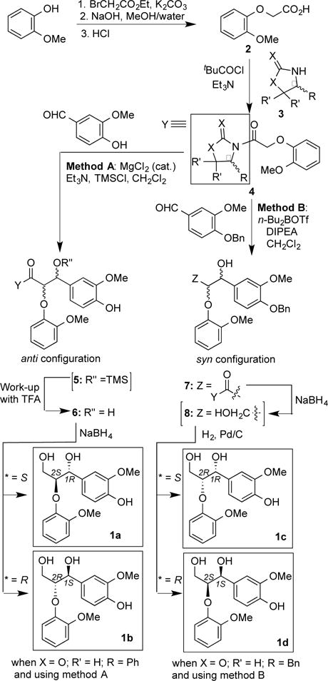
Enantioselective synthesis of β-O-4 dilignolsa
aCompounds 5 and 8 were used without purification.
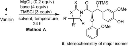
As shown in Scheme 1, guaiacol was converted to acid 2 and then reacted with oxazolidinone/thiazolidinethione 3 to yield adduct 4 (Figure 1),28, 34 which was screened for its efficiency in diastereoselectivity by HPLC. A representative HPLC profile is shown in Figure 2 and the results are summarized in Table 1. For method A involving trimethylsilyl chloride (TMSCl) and catalytic MgCl2,35, 36 4a (X=O, R=R-Ph, R´=H) was found to be the best for diastereoselectivity to yield an anti-aldol product. When 4a was reacted with vanillin, di-TMS-protected aldol product was formed initially (see NMR in SI). However, the phenolic TMS was not stable and could be easily cleaved to produce mono-TMS- protected 5a (X =O, R=R-Ph, R´= H). It has to be noted that isolation of the major diastereomer in 5 by silica-gel chromatography was difficult. Therefore, without purification, 5 was directly treated with trifluoroacetic acid (TFA) to afford compound 6, in which the major diastereomer with anti-configuration was isolated in 75% yields. To further optimize diastereoselectivity, effects of organic bases and solvents were also screened. The results are summarized in Table 1. It was concluded that Et3N and CH2Cl2 were the best base and solvent, respectively. It was also found that low temperature slightly improved diastereoselectivity (entries 1 and 2 in Table 1).
Figure 1.
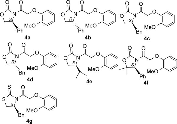
Structures of 4a–4g containing Evans auxiliaries
Figure 2.
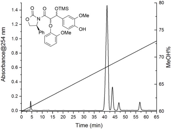
Representative HPLC profile of diastereoselectivity using method A. The straight line indicates MeOH gradient. The peak at 4.8 min represents unreacted aldehyde.
Table 1.
Optimization of aldol reactions using method Aa
| ||||
|---|---|---|---|---|
|
| ||||
| entry | compd | base | solvent | diastereoselectivityb |
| 1 | 4a | Et3N | CH2Cl2 | 78:7:6:9 (rt) |
| 2 | 4a | Et3N | CH2Cl2 | 80:12:4:4 |
| 3 | 4c | Et3N | CH2Cl2 | 78:4:16:2 (rt) |
| 4 | 4e | Et3N | CH2Cl2 | 72:9:10:9 (rt) |
| 5 | 4f | Et3N | CH2Cl2 | 57:27:16 (rt)c |
| 6 | 4g | Et3N | CH2Cl2 | 31:16:53 (rt)c |
| 7 | 4a | BuNH2 | CH2Cl2 | trace product |
| 8 | 4a | PhNH2 | CH2Cl2 | no product |
| 9 | 4a | i-Pr2NH | CH2Cl2 | 73:15:4:8 |
| 10 | 4a | DIPEAd | CH2Cl2 | no product |
| 11 | 4a | pyridine | CH2Cl2 | no product |
| 12 | 4a | DBUe | CH2Cl2 | no product |
| 13 | 4a | Et3N | ClCH2CH2Cl | 72:16:7:5 |
| 14 | 4a | Et3N | EtOAc | 76:7:8:9 |
| 15 | 4a | Et3N | THF | 71:2:7:20 |
| 16 | 4a | Et3N | Et2O | 64:6:14:16 |
| 17 | 4a | Et3N | toluene | 57:6:6:31 |
| 18 | 4a | Et3N | CH3CN | no product |
Unless indicated, all reactions were carried out at 0 °C;
Diastereoselectivity was determined by HPLC. See SI for details;
Diastereomers were inseparable under this HPLC condition;
Diisopropylethylamine;
1,8-Diazabicyclo[5.4.0]undec-7-ene.
Reductive cleavage of the auxiliary in compound 6 with NaBH4 gave the desired stereoisomer, (1R, 2S)-1a or (1S, 2R)-1b, depending on the configuration of phenyl group. The enantiomeric excess (ee) of the final compounds was determined to be > 99% using chiral HPLC. Assignment of the configurations in each stereoisomer was based on the reported optical rotations, which was also consistent with theoretical predictions.35, 37 These results are summarized in supplemental Figure S1 and Table S1. Thus, the stereospecific anti-dilignols were obtained in just four steps with an overall 42% yield, which was a significant improvement over the other asymmetric methods reported so far.32, 33
Method B employing n-Bu2BOTf and DIPEA to form enolates has been recently reported to synthesize syn-aldol 7e (X = O, R = R-iPr, R´ = H) using chiral auxiliary 4e.33 However, our screen on auxiliaries found that 4c had a slightly better diastereoselectivity than 4e (see Figure S2 and Table S2 in SI). Thus, 4c was selected as the chiral auxiliary for subsequent preparations. After the chiral auxiliary in 7 was cleaved by NaBH4 to give 8, a subsequent hydrogenesis yielded (1R, 2R)-1c or (1S, 2S)-1d depending on the configuration of the benzyl (Bn) group in the auxiliary.
Surface plasmon resonance (SPR) is a highly sensitive technique to study non-covalent interactions of biomolecules, especially protein-protein and protein-small molecule interactions in real time.38 Purified TcDyP (Figure S3 in SI) was immobilized on a CMD500M sensor chip (Xantec Bioanalytics GmbH) via amine coupling chemistry.39 The synthesized stereoisomers 1a-1d at a series of concentrations were then injected and sensorgrams are shown in supplemental Figure S4. Steady-state affinity analysis based on a 1:1 binding model (Figure S5 in SI) was used to determine the apparent binding affinity (KD). The results and parameters evaluating global fittings of the data are summarized in Table 2. It was revealed that all stereoisomers had weak interactions with TcDyP. For stereoisomer 1c, its affinity was too weak to be determined. Among the other three stereoisomers, 1a appeared to bind the strongest with the enzyme at submillimolar concentrations. The KD values of 1b and 1d are at least 10-fold higher than that of 1a, suggesting that stereochemistry at Cα and Cβ does affect the substrate binding in the absence of H2O2. Such observations may shed light on catalysis by DyPs.
Table 2.
SPR analysis and enzyme assays with TcDyP
| parameters | 1a (1R, 2S) |
1b (1S, 2R) |
1c (1R, 2R) |
1d (1S, 2S) |
|
|---|---|---|---|---|---|
| SPR | KD (μM) | 0.166 | 2.37 | NDc | 2.06 |
| RUmaxa | 35.6 | 163 | 68.8 | ||
| Chi2(RU2)b | 1.51 | 0.280 | 0.575 | ||
| SA (mU/mg) | 232 | 161 | 142 | 215 | |
RUmax corresponds to the unconstrained value for the fitted data at saturation, for which the theoretical values are 105 for TcDyP;
The fitting of the observed data to theoretical model is evaluated by Chi2;
ND: not determined.
We have previously reported that racemic mixtures of β-O-4 dilignol 1 can be degraded by TcDyP in the presence of H2O2.6 Based on the MS results, Cα−Cβ was proposed to be cleaved during degradation.6 To investigate the stereochemical effects of TcDyP, the four stereoisomers were individually incubated with the enzyme in the presence of H2O2. Reaction rates were determined based on disappearance of the peak at 13.2 min corresponding to the substrate (Figure 3). Specific activities (SAs) of TcDyP toward the four stereoisomers are summarized in Table 2, which are in the order of 1a > 1d > 1b > 1c, consistent with the order of apparent binding affinities (KD) obtained by SPR. However, one important difference was found between SPR and enzyme activity assays. It was observed that 1a bound TcDyP by 14- and 12-fold better than 1b and 1d, respectively. Stereoisomer 1c displayed very weak binding with TcDyP. Therefore, the significant difference observed in SPR experiments was not reflected in enzyme activities toward these stereoisomers, in which only 1.6-fold difference was observed for the ones with the highest (1a) and lowest (1c) activities.
Figure 3.
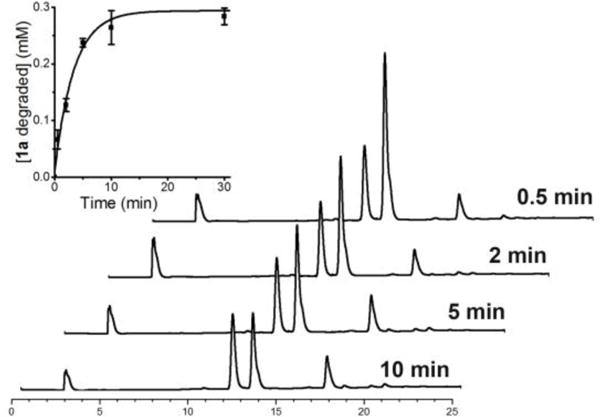
HPLC profiles of TcDyP incubating with 1a and H2O2. Peaks at 3.1, 12.0, 13.2, and 17.4-20.5 min correspond to impurity from citrate buffer, internal standard, substrate 1a, and degradation products, respectively. The inset shows the rate of 1a degradation.
We recently determined the crystal structure of TcDyP,40 which revealed that a large loop consisting of residues 277-310 is present next to an access channel leading to the propionate group on pyrrole ring C in the heme active site (Figure 4). The entrance of this channel in D-class DyP from Bjerkandera adusta has been shown to be a substrate binding site.41 The same propionate group has also been demonstrated to provide a direct electron transfer from the substrate to porphyrin radical in heme-containing manganese and ascorbate peroxidases.42, 43 Additionally, this 34-residue loop is close to W263, which has been characterized as a surface-exposed substrate oxidation site in TcDyP.40 The closest distance between the loop (L279) and W263 is 6.1 Å. Moreover, this loop is widely present in DyPs although the size varies between classes. Deletion of the loop in TcDyP has resulted in the loss of enzyme activity (data not shown). Thus, it is highly possible that DyP enzymes undergo a conformational change involving this large loop upon H2O2 binding. Such H2O2-induced conformational changes have been reported for a number of other heme peroxidases including cytochrome c peroxidase and horseradish peroxidase.44–48 Since our SPR experiments were carried out in the absence of H2O2, the significant difference in KD with the four stereoisomers may not be translated into a significant difference in enzyme activities toward them, though the overall order of stereochemical preference is retained in both SPR and activity assay experiments.
Figure 4.
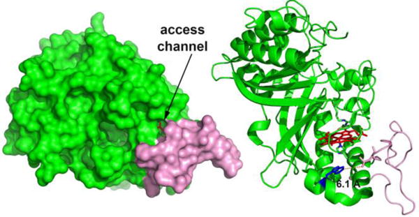
Crystal structure of TcDyP (PDB 5JXU) in surface (left) and cartoon (right) representations. The loop consisting of residues 277-310 is colored in pink. The heme b, catalytic residues (D220, H312 and R327), W263, and L279 are shown in red, green, blue, and pink sticks.
In summary, a 4-step enantioselective method has been developed to prepare anti (1R, 2S)-1a and (1S, 2R)-1b in the presence of TMSCl and catalytic MgCl2, achieving the goal of synthesizing all four stereospecific β-O-4 dilignols in gram scales by controlling conditions of enolate formation. The TcDyP displayed different stereodiscrimination against these stereoisomers in the absence and presence of H2O2, which may be attributed to the conformational change involving a loop widely present in DyP enzymes upon H2O2 binding. The overall weak stereodiscrimination by DyPs may appear advantageous in their applications as a biocatalyst for lignin degradation, as they will not discriminate the source of biomass that may have different localized stereochemistry.
Supplementary Material
Acknowledgments
This work was supported by awards to P.L from the pilot project of NIH P30GM110761 and Johnson Cancer Research Center. G.H. is supported by an NIH K-INBRE fellowship under grant number P20GM103418.
Footnotes
Supporting Information
The Supporting Information is available free of charge on the ACS Publications website at DOI:
Detailed experimental procedures and spectroscopic data (PDF)
Notes
The authors declare no competing financial interest.
References
- 1.Strittmatter E, Plattner DA, Piontek K. Encyclopedia of Inorganic and Bioinorganic Chemistry. John Wiley and Sons, Ltd; New York: 2011. Dye-Decolorizing Peroxidase (DyP) [Google Scholar]
- 2.Sugano Y. Cell Mol Life Sci. 2009;66:1387–1403. doi: 10.1007/s00018-008-8651-8. [DOI] [PMC free article] [PubMed] [Google Scholar]
- 3.Colpa DI, Fraaije MW, van Bloois E. J Ind Microbiol Biot. 2014;41:1–7. doi: 10.1007/s10295-013-1371-6. [DOI] [PubMed] [Google Scholar]
- 4.Ahmad M, Roberts JN, Hardiman EM, Singh R, Eltis LD, Bugg TDH. Biochemistry. 2011;50:5096–5107. doi: 10.1021/bi101892z. [DOI] [PubMed] [Google Scholar]
- 5.Brown ME, Barros T, Chang MCY. ACS Chem Biol. 2012;7:2074–2081. doi: 10.1021/cb300383y. [DOI] [PubMed] [Google Scholar]
- 6.Chen C, Shrestha R, Jia K, Gao PF, Geisbrecht BV, Bossmann SH, Shi JS, Li P. J Biol Chem. 2015;290:23447–23463. doi: 10.1074/jbc.M115.658807. [DOI] [PMC free article] [PubMed] [Google Scholar]
- 7.Min K, Gong G, Woo HM, Kim Y, Um Y. Sci Rep. 2015;5:8245. doi: 10.1038/srep08245. [DOI] [PMC free article] [PubMed] [Google Scholar]
- 8.Brown ME, Chang MCY. Curr Opin Chem Biol. 2014;19:1–7. doi: 10.1016/j.cbpa.2013.11.015. [DOI] [PubMed] [Google Scholar]
- 9.de Gonzalo G, Colpa DI, Habib MHM, Fraaije MW. J Biotechnol. 2016;236:110–119. doi: 10.1016/j.jbiotec.2016.08.011. [DOI] [PubMed] [Google Scholar]
- 10.Paliwal R, Rawat AP, Rawat M, Rai JPN. Appl Biochem Biotech. 2012;167:1865–1889. doi: 10.1007/s12010-012-9735-3. [DOI] [PubMed] [Google Scholar]
- 11.Wong DWS. Appl Biochem Biotech. 2009;157:174–209. doi: 10.1007/s12010-008-8279-z. [DOI] [PubMed] [Google Scholar]
- 12.ten Have R, Teunissen PJ. Chem Rev. 2001;101:3397–413. doi: 10.1021/cr000115l. [DOI] [PubMed] [Google Scholar]
- 13.Bugg TD, Ahmad M, Hardiman EM, Singh R. Curr Opin Biotech. 2011;22:394–400. doi: 10.1016/j.copbio.2010.10.009. [DOI] [PubMed] [Google Scholar]
- 14.Boerjan W, Ralph J, Baucher M. Annu Rev Plant Biol. 2003;54:519–546. doi: 10.1146/annurev.arplant.54.031902.134938. [DOI] [PubMed] [Google Scholar]
- 15.Akiyama T, Magara K, Matsumoto Y, Meshitsuka G, Ishizu A, Lundquist K. J Wood Sci. 2000;46:414–415. [Google Scholar]
- 16.Akiyama T, Sugimoto T, Matsumoto Y, Meshitsuka G. J Wood Sci. 2002;48:210–215. [Google Scholar]
- 17.Gall DL, Kim H, Lu FC, Donohue TJ, Noguera DR, Ralph J. J Biol Chem. 2014;289:8656–8667. doi: 10.1074/jbc.M113.536250. [DOI] [PMC free article] [PubMed] [Google Scholar]
- 18.Gall DL, Ralph J, Donohue TJ, Noguera DR. Environ Sci Technol. 2014;48:12454–12463. doi: 10.1021/es503886d. [DOI] [PMC free article] [PubMed] [Google Scholar]
- 19.Masai E, Ichimura A, Sato Y, Miyauchi K, Katayama Y, Fukuda M. J Bacteriol. 2003;185:1768–1775. doi: 10.1128/JB.185.6.1768-1775.2003. [DOI] [PMC free article] [PubMed] [Google Scholar]
- 20.Katayama T, Tsutsui J, Tsueda K, Miki T, Yamada Y, Sogo M. J Wood Sci. 2000;46:458–465. [Google Scholar]
- 21.Hishiyama S, Otsuka Y, Nakamura M, Ohara S, Kajita S, Masai E, Katayama Y. Tetrahedron Lett. 2012;53:842–845. [Google Scholar]
- 22.Sato Y, Moriuchi H, Hishiyama S, Otsuka Y, Oshima K, Kasai D, Nakamura M, Ohara S, Katayama Y, Fukuda M, Masai E. Appl Environ Microbiol. 2009;75:5195–5201. doi: 10.1128/AEM.00880-09. [DOI] [PMC free article] [PubMed] [Google Scholar]
- 23.Jonsson L, Karlsson O, Lundquist K, Nyman PO. FEBS Lett. 1990;276:45–8. doi: 10.1016/0014-5793(90)80503-b. [DOI] [PubMed] [Google Scholar]
- 24.Yue FX, Lu FC, Sun RC, Ralph J. Chem Eur J. 2012;18:16402–16410. doi: 10.1002/chem.201201506. [DOI] [PubMed] [Google Scholar]
- 25.Kawai S, Jensen KA, Bao W, Hammel KE. Appl Environ Microbiol. 1995;61:3407–3414. doi: 10.1128/aem.61.9.3407-3414.1995. [DOI] [PMC free article] [PubMed] [Google Scholar]
- 26.Forsythe WG, Garrett MD, Hardacre C, Nieuwenhuyzen M, Sheldrake GN. Green Chem. 2013;15:3031–3038. [Google Scholar]
- 27.Kishimoto T, Uraki Y, Ubukata M. Org Biomol Chem. 2005;3:1067–1073. doi: 10.1039/b416699j. [DOI] [PubMed] [Google Scholar]
- 28.Buendia J, Mottweiler J, Bolm C. Chem Eur J. 2011;17:13877–13882. doi: 10.1002/chem.201101579. [DOI] [PubMed] [Google Scholar]
- 29.Li KC, Helm RF. Holzforschung. 2000;54:597–603. [Google Scholar]
- 30.Li KC, Helm RF. J Chem Soc Perkin Trans. 1996;1:2425–2426. [Google Scholar]
- 31.Nakatsubo F, Sato K, Higuchi T. Holzforschung. 1975;29:165–168. [Google Scholar]
- 32.Njiojob CN, Rhinehart JL, Bozell JJ, Long BK. J Org Chem. 2015;80:1771–1780. doi: 10.1021/jo502685k. [DOI] [PubMed] [Google Scholar]
- 33.Njiojob CN, Bozell JJ, Long BK, Elder T, Key RE, Hartwig WT. Chem Eur J. 2016;22:12506–12517. doi: 10.1002/chem.201601592. [DOI] [PubMed] [Google Scholar]
- 34.Prashad M, Kim HY, Har D, Repic O, Blacklock TJ. Tetrahedron Lett. 1998;39:9369–9372. [Google Scholar]
- 35.Evans DA, Tedrow JS, Shaw JT, Downey CW. J Am Chem Soc. 2002;124:392–393. doi: 10.1021/ja0119548. [DOI] [PubMed] [Google Scholar]
- 36.Evans DA, Downey CW, Shaw JT, Tedrow JS. Org Lett. 2002;4:1127–1130. doi: 10.1021/ol025553o. [DOI] [PubMed] [Google Scholar]
- 37.Evans DA, Rieger DL, Bilodeau MT, Urpi F. J Am Chem Soc. 1991;113:1047–1049. [Google Scholar]
- 38.Nguyen HH, Park J, Kang S, Kim M. Sensors-BASEL. 2015;15:10481–10510. doi: 10.3390/s150510481. [DOI] [PMC free article] [PubMed] [Google Scholar]
- 39.Johnsson B, Lofas S, Lindquist G. Anal Biochem. 1991;198:268–277. doi: 10.1016/0003-2697(91)90424-r. [DOI] [PubMed] [Google Scholar]
- 40.Shrestha R, Chen XJ, Ramyar KX, Hayati Z, Carlson EA, Bossmann SH, Song LK, Geisbrecht BV, Li P. ACS Catal. 2016;6:8036–8047. doi: 10.1021/acscatal.6b01952. [DOI] [PMC free article] [PubMed] [Google Scholar]
- 41.Yoshida T, Tsuge H, Hisabori T, Sugano Y. FEBS Lett. 2012;586:4351–4356. doi: 10.1016/j.febslet.2012.10.049. [DOI] [PubMed] [Google Scholar]
- 42.Sharp KH, Mewies M, Moody PCE, Raven EL. Nat Struct Biol. 2003;10:303–307. doi: 10.1038/nsb913. [DOI] [PubMed] [Google Scholar]
- 43.Sundaramoorthy M, Kishi K, Gold MH, Poulos TL. J Biol Chem. 1994;269:32759–32767. [PubMed] [Google Scholar]
- 44.Miyazaki C, Takahashi H. FEBS Lett. 2001;509:111–114. doi: 10.1016/s0014-5793(01)03127-1. [DOI] [PubMed] [Google Scholar]
- 45.Wang X, Wang LK, Wang X, Sun F, Wang CC. Biochem J. 2012;441:113–118. doi: 10.1042/BJ20110380. [DOI] [PubMed] [Google Scholar]
- 46.Hassler K, Rigler P, Blom H, Rigler R, Widengren J, Lasser T. Opt Express. 2007;15:5366–5375. doi: 10.1364/oe.15.005366. [DOI] [PubMed] [Google Scholar]
- 47.Wen F, Dong YH, Feng L, Wang S, Zhang SC, Zhang XR. Anal Chem. 2011;83:1193–1196. doi: 10.1021/ac1031447. [DOI] [PubMed] [Google Scholar]
- 48.Bonagura CA, Bhaskar B, Shimizu H, Li HY, Sundaramoorthy M, McRee DE, Goodin DB, Poulos TL. Biochemistry. 2003;42:5600–5608. doi: 10.1021/bi034058c. [DOI] [PubMed] [Google Scholar]
Associated Data
This section collects any data citations, data availability statements, or supplementary materials included in this article.


