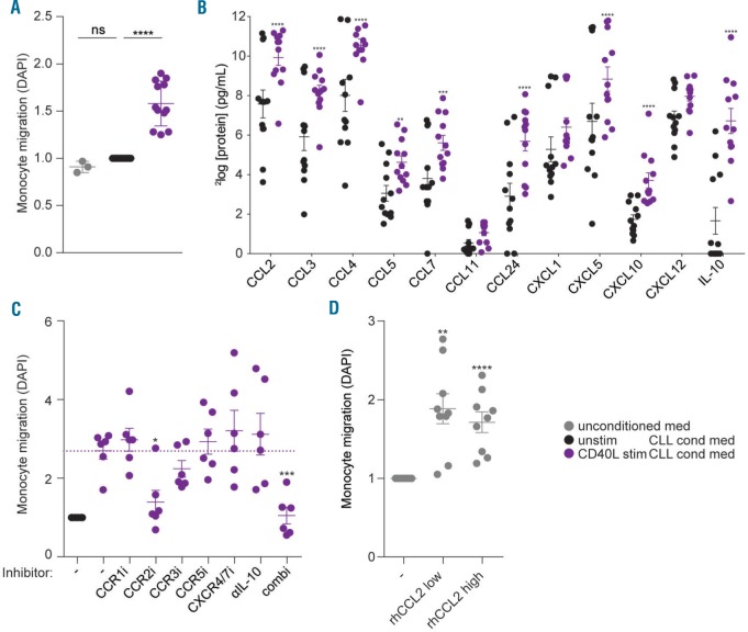
Figure 3.
CD40L-stimulated chronic lymphocytic leukemia (CLL) cells attract monocytes as a result of CCR2 axis signaling. (A) Freshly isolated healthy donor (HD) monocytes were seeded in the upper chambers of a trans-well migration plate to migrate towards conditioned media (cond med) obtained from PBMC samples from CLL patients (for patients’ characteristics see Online Supplementary Table S1) that were cultured for 16 hours (h) on CD40L-overexpressing (CD40L stim) or parental NIH-3T3 cells (unstim). Next, the amount of migrated monocytes was quantified using DAPI staining. Each dot represents the relative [compared to the unstimulated (unstim) CLL condition] DAPI signals of 12 different CLL conditioned media or 3 control media in 3 independent experiments using monocytes from 3 different donors and mean±Standard Error of Mean (SEM) are shown. All measurements were performed in triplicate. ****P<0.0001 in t-tests. (B) Protein levels of chemokines involved in monocyte migration23–26 were determined in the conditioned media that were used to perform the migration assays in (A) by using Luminex. Dots represent protein levels and mean±SEM are shown for 12 CLL conditioned media; **P<0.01, ***P<0.001, ****P<0.0001 in a two-way ANOVA test with Bonferroni post hoc analysis. (C) Freshly isolated monocytes and conditioned media were pre-incubated for 30 minutes (min) with individual small-molecule inhibitors directed against indicated chemokine receptors, with an IL-10 neutralizing antibody, or a combination of all inhibitors (combi), before performing migration assays as in (A). Each dot represents the relative (compared to the unstimulated CLL condition) DAPI signals obtained in 4 independent experiments using monocytes from 3 different donors and different CLL supernatants; mean±SEM are shown. All measurements were performed in triplicate; *P<0.05, ***P<0.001, in a one-way ANOVA test with Bonferroni post hoc analysis. (D) Monocytes were seeded in the upper chambers of a trans-well migration plate to migrate towards migration medium without or with 10 ng/mL recombinant human CCL2 (rhCCL2 low) or 100 ng/mL rhCCL2 (rhCCL2 high). Next, the amount of migrated monocytes was quantified using DAPI staining. Each dot represents the relative (compared to condition without rhCCL2) DAPI signals of 9 separate read-outs in 3 independent experiments using monocytes from 3 different donors and mean±SEM are shown; **P<0.01, ****P<0.0001 in t-tests.

