The diagnosis of autoimmune hemolytic anemia (AIHA) is based on the evidence of anemia, hemolysis and the detection of red blood cell (RBCs) autoantibodies.1–2 The absence of detectable RBC autoantibodies does not always exclude autoimmune hemolysis. Conversely, the presence of RBC autoantibodies is not always associated with hemolytic anemia. Silent RBC autoantibodies have been detected in healthy blood donors,3,4 in pregnant females5 and in patients with autoimmune disorders.6 While the occurrence of clinically non-significant RBC autoantibodies is a well-known phenomenon in immunohematology laboratories, there is little information about the clinical course of subjects with silent RBC autoantibodies and whether disorders known to be associated with AHIA could also be present in these subjects. To address these issues, we retrospectively analyzed the characteristics and the long-term outcome of 60 subjects with silent RBC autoantibodies who were referred to our Hematology Center between January 1995 and December 2012 and followed on a regular basis over time.
Our results showed that the development of AHIA in subjects with silent RBC autoantibodies was a rare event and that disorders known to be associated with AHIA were present in a relevant proportion of cases with silent agglutinins.
Subjects with silent RBC autoantibodies showed normal Hb values, no signs of hemolysis, acrocyanosis and/or Raynaud phenomena, and no previous history of drugs known to be associated with AHIA, vaccinations, infections, lymphoproliferative diseases or autoimmune disorders.
The initial assessment included complete blood count, immunohematologic evaluation, reticulocyte count, haptoglobin, bilirubin, LDH, serum electrophoresis and clinical examination. Data on blood counts, bilirubin levels and immunohematologic tests performed during follow up were also recorded. More details about the clinical workout performed at baseline and during follow up are reported in the Online Supplementary data.
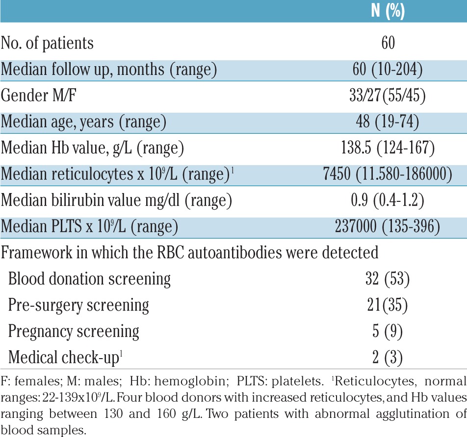
Silent RBC autoantibodies were detected in 5 (8%) pregnant females, 34 (57%) healthy individuals (blood donors, 32; subjects with an abnormal agglutination of blood samples, 2) and 21 (35%) subjects screened prior to surgery (benign disorder, 16; malignancy, 5).
The median follow up was 60 months (range, 10–204 months), the median age 48 years (range, 19–74 years), and the median Hb value 138.5 g/L (range, 124–167 g/L) with a median reticulocyte count of 74.5 × 10⁹/L (range:11.580-186000). Four blood donors showed an increased reticulocyte count with Hb values ranging between 130 and 160 g/L. Haptoglobin, bilirubin, and LDH levels were normal in all cases (Table 1).
Table 1.
Clinical characteristics of subjects at the time of the first detection of the silent red blood cell autoantibodies.

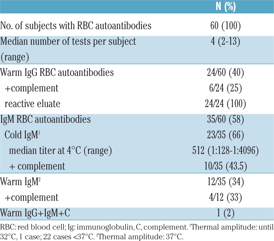
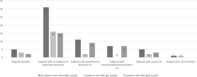
IgG autoantibodies were detected in 24 (40%) cases, IgM in 35 (58%) and IgM+IgG in 1 (2%). Cold agglutinins with a median titer of 1:512 and a thermal amplitude around 30°C were recorded in 23/35 (66%) cases, and warm agglutinins in 12 (34%). The median number of immunohematologic tests per patient was 4 (range 2–13) (Table 2). Clinical and serologic characteristics of subjects with silent RBC autoantibodies according to the associated condition or disorder are described in Figure 1.
Table 2.
Serological characteristics of the silent red blood cell autoantibodies.

Figure 1.
Serologic characteristics of subjects with silent red blood cell autoantibodies according to the associated condition or disorder. (1) Autoimmune disorders: Hashimoto thyroiditis, 4 cases; anti-phospholipid syndrome, 5; systemic lupus erythematosus, 1; autoimmune glomerulonephritis, 1. (2) Lymphoproliferative disorders: bone marrow involvement with MBL, 6 cases, marginal cell lymphoma, 1. (3) Cancers: breast cancer, 2; thyroid cancers, 3.
Asymptomatic RBC autoantibodies were detected in 5 (8%) pregnant females at the time of the first trimester screening (IgG 3 cases, IgM 1, IgG+IgM 1) and persisted during and after pregnancy. No signs of hemolytic anemia were observed in either the mothers or the newborns.
In 31 of the remaining 55 (56%) cases, no evidence of an underlying disorder was observed, while it was present in 24 (44%) cases; in 5/21 (24%) with IgG autoantibodies and in 19/34 (56%) with IgM autoantibodies (P<0.05).
Five (5/55, 9%) cancer-bearing subjects (breast cancer 2; thyroid cancer, 3) revealed silent RBC autoantibodies (IgG, 2; IgM, 3) at the time of the pre-surgery screening for cancer removal. RBC autoantibodies disappeared in 2 cases (IgG 1; IgM 1) after removal of thyroid cancer, while they persisted in 3 with active disease.
The significant positivity of other autoantibodies directed further serologic and clinical evaluations allowing the identification of an autoimmune disorder in 11 (11/55, 20%) subjects (Hashimoto thyroiditis, 4; anti-phospholipid syndrome, 5; systemic lupus erythematosus, 1; autoimmune glomerulonephritis, 1). IgM autoantibodies were detected in the majority of cases (9/11 cases, 82%).
Before surgery to remove colonic polyps, IgG RBC autoantibodies associated with an active HCV hepatitis were detected in 1 (1/55, 1.8%) patient. Both the HCV virus and the RBC autoantibodies disappeared after treatment with peg alpha2-interferon.
Bone marrow (BM) biopsy or aspirate with a flow-cytometry analysis revealed the presence of a lymphoproliferative disorder in 7 (7/55, 13%) cases, all with RBC IgM autoantibodies (cold IgM, 4 cases; warm IgM, 3).
A marginal zone lymphoma was diagnosed in 1 patient who also showed enlarged abdominal lymph-nodes. In the other 6 cases, all with no enlarged lymph-nodes, a limited infiltration of small clonal B-lymphocytes was detected in the BM. In all cases, monoclonal B-cell lymphocytes (MBLs) were also detected in the peripheral blood by flow-cytometry. MBLs showed lymphoma-like features in 3 cases and chronic lymphocytic leukemia-like features in 3 (Online Supplementary Table S1). The median MBL count was low (0.135 ×10⁹/L; range, 0.56–0.215 ×10⁹/L) and remained stable during follow up in 5 cases, while it gradually increased over 10 years in 1. (Online Supplementary Figure S1). None of these subjects developed a clinically overt lymphoma/chronic lymphocytic leukemia.
The IgG autoantibodies disappeared in 10/24 (42%) cases; in 7 subjects without an associated disorder, in 2 after the removal of a thyroid cancer and in 1 cured of the HCV hepatitis. Agglutinins persisted in all cases after a median follow up of 93 months (range, 10–204). Only two patients, both with agglutinins (2/60 total cases 3%; 2/35 IgM cases, 6%) and no evidence of a lymphoproliferative disorder, experienced AHIA after 14 and 17 years, respectively, from the first detection of cold autoantibodies.
Survival of subjects with silent agglutinins was lower, though not significantly lower, than those with silent warm autoantibodies (at 15 years, 75% vs. 100%; P=0.2) (Online Supplementary Figure S2).
The retrospective nature of this study, and the fact that not all cases with silent RBC were referred to our institute, should be underlined. This is an understandable phenomenon as healthy subjects are not usually investigated for asymptomatic RBC autoantibodies. The lack of a denominator did not permit us to ascertain the incidence and prevalence of silent autoantibodies among different patient populations. In addition, the number of cases was too small to be able to define the relative distribution of different disease states among subjects with silent RBC autoantibodies.
The abnormal agglutination of blood samples in the presence of cold agglutinins was common. This phenomenon was influenced both by the elapse of time from obtaining samples and the temperature on storage.
The development of a clinically manifest AHIA was recorded in only 2 (6%) of the 35 subjects with asymptomatic agglutinins. Multiple factors are likely to have played a role in preventing hemolysis. As observed in some cases of CAD, the complement pathway probably could not be activated given the protective effect of the physiological complement inhibitors CD55 and CD59 on the RBC membrane.7 The lack of hemolysis could also be due to an ineffective complement activation by some IgG subclasses or binding by the FC-receptors of phagocytic cells.7 In some cases, cross-reacting antiphospholipid autoantibodies, adsorbing non-specifically onto the erythrocyte membrane, may be an incidental cause of false positive tests.8 In addition, an effective erythropoiesis could play a relevant compensatory effect in preventing anemia.
As in a previous study,5 RBC autoantibodies detected in 5 pregnant females had no effect on the course of pregnancy, on the fetus development, or on the health of the newborns.
An associated disorder was observed in 24 (44%) of the remaining 55 cases and was significantly more frequent in subjects with silent agglutinins.
Given the known relationship between AHIA and autoimmune diseases,6 cancers,9 HCV infection,10 and lymphoproliferative disorders,11,12 the detection of these conditions in subjects with silent RBC autoantibodies was not surprising.
A lymphoproliferative disorder was detected only in subjects with silent agglutinins. Subjects with cold agglutinin and a limited clonal B-cell disorder involving the BM were described by Randen et al.13 as having a primary cold agglutinin disease (CAD) or “cold agglutinin (CA)-associated lymphoproliferative disease”. The 6 cases with asymptomatic agglutinins, BM involvement and MBL showed similar characteristics of primary CAD, even in the absence of hemolytic anemia. Interestingly, none of these subjects developed clinical signs of hemolysis or clinical evidence of lymphoma during follow up. Low count MBL have also been detected in patients with AIHA, immune thrombocytopenic purpura,14 and in healthy blood donors.15 Although a low count MBL is associated with a negligible risk of progression to a lymphoproliferative disease, they may have a pathogenetic relationship in the development of RBC autoantibodies. IgG autoantibodies disappeared after the cure of hepatitis in 1 patient and the removal of a thyroid cancer in 2. This finding suggests that, in some cases, the removal of an immune stimulation could have led to the clearance of the autoantibodies.
Taken together, the results of this study showed that in subjects with silent RBC autoantibodies, the development of a clinically overt AHIA was a rare event, while disorders known to be associated with AHIA were present in a relevant proportion of cases and more frequently in subjects with silent agglutinins.
Supplementary Material
Footnotes
Information on authorship, contributions, and financial & other disclosures was provided by the authors and is available with the online version of this article at www.haematologica.org.
References
- 1.Packman CH. The clinical pictures of autoimmune hemolytic anemia. Transfus Med Hemother. 2015;42(5):317–324. [DOI] [PMC free article] [PubMed] [Google Scholar]
- 2.Go RS, Winters JL, Kay NE. How I treat autoimmune hemolytic anemia. Blood. 2017;129(22):2971–2979. [DOI] [PubMed] [Google Scholar]
- 3.Gorst DW, Rawlinson VI, Merry AH, Stratton F. Positive direct antiglobulin test in normal individuals. Vox Sang. 1980;38(2):99–105. [DOI] [PubMed] [Google Scholar]
- 4.Bareford D, Longster G, Gilks L, Tovey LA. Follow-up of normal individuals with a positive antiglobulin test. Scand J Haematol. 1985;35(3):348–353. [DOI] [PubMed] [Google Scholar]
- 5.Hoppe B, Stibbe W, Bielefeld A, Pruss A, Salama A. Increased RBC autoantibody production in pregnancy. Transfusion. 2001; 41(12):1559–1561. [DOI] [PubMed] [Google Scholar]
- 6.Comellas-Kirkerup L, Hernández-Molina G, Cabral AR. Antiphospholipid-associated thrombocytopenia or autoimmune hemolytic anemia in patients with or without definite primary antiphospholipid syndrome according to the Sapporo revised classification criteria: a 6-year follow-up study. Blood. 2010;116(16):3058–3063. [DOI] [PubMed] [Google Scholar]
- 7.Berentsen S. Complement, cold agglutinins, and therapy. Blood. 2014;123(26):4010–4012. [DOI] [PubMed] [Google Scholar]
- 8.Ünlü O, Zuily S, Erkan D. The clinical significance of antiphospholipid antibodies in systemic lupus erythematosus. Eur J Rheumatol. 2016;3(2):75–84. [DOI] [PMC free article] [PubMed] [Google Scholar]
- 9.Nenova IS, Valcheva MY, Beleva EA, et al. Autoimmune phenomena in patients with solid tumors. Folia Medica (Plovdiv). 2016;58(3):195–199. [DOI] [PubMed] [Google Scholar]
- 10.Basseri RJ, Schmidt MT, Basseri B. Autoimmune hemolytic anemia in treatment-naive chronic hepatitis C infection: a case report and review of literature. Clin J Gastroenterol. 2010:3(5):237–2421. [DOI] [PubMed] [Google Scholar]
- 11.Mauro FR, Foa R, Cerretti R, et al. Autoimmune hemolytic anemia in chronic lymphocytic leukemia: clinical, therapeutic, and prognostic features. Blood. 2000;95(9):2786–2792. [PubMed] [Google Scholar]
- 12.Barcellini W, Capalbo S, Agostinelli RM, et al. GIMEMA Chronic Lymphocytic Leukemia Group. Relationship between autoimmune phenomena and disease stage and therapy in B-cell chronic lymphocytic leukemia. Hematologica. 2006;91(12):689–692. [PubMed] [Google Scholar]
- 13.Randen U, Trøen G, Tierens A, et al. Primary cold agglutinin-associated lymphoproliferative disease: a B-cell lymphoma of the bone marrow distinct from lymphoplasmacytic lymphoma. Haematologica. 2014;99(3):497–504. [DOI] [PMC free article] [PubMed] [Google Scholar]
- 14.Mittal S, Blaylock MG, Culligan DJ, Barker RN, Vickers MA. A high rate of CLL phenotype lymphocytes in autoimmune hemolytic anemia and immune thrombocytopenic purpura. Hematologica. 2008; 93(1):151–152. [DOI] [PubMed] [Google Scholar]
- 15.Shim YK, Rachel JM, Ghia P, et al. Monoclonal B-cell lymphocytosis in healthy blood donors: an unexpectedly common finding. Blood. 2014;123(9):1319–1326. [DOI] [PMC free article] [PubMed] [Google Scholar]
Associated Data
This section collects any data citations, data availability statements, or supplementary materials included in this article.


