Abstract
Liver plays a crucial role in controlling energy homeostasis in mammals, although the exact mechanism by which it influences other peripheral tissues has yet to be addressed. Here we show that Creb regulates transcriptional co-activator (Crtc) 2 is a major regulator of whole-body energy metabolism. Crtc2 liver-specific knockout lowers blood glucose levels with improved glucose and insulin tolerance. Liver-specific knockout mice display increased energy expenditure with smaller lipid droplets in adipose depots. Both plasma and hepatic Fgf21 levels are increased in Crtc2 liver-specific knockout mice, as a result of the reduced miR-34a expression regulated by Creb/Crtc2 and the induction of Sirt1 and Pparα. Ectopic expression of miR-34a reverses the metabolic changes in knockout liver. We suggest that Creb/Crtc2 negatively regulates the Sirt1/Pparα/Fgf21 axis via the induction of miR-34a under diet-induced obesity and insulin-resistant conditions.
CREB-regulated transcription coactivator 2, CRTC2, has been associated with regulation of glucose and lipid homeostasis. Here Han et al. show that Creb/Crtc2 modulates lipid and glucose metabolism by inhibiting the expression of mi-R34 that, in turn, represses the expression of Sirt1 and PPARα and consequently Fgf21 levels.
Introduction
Under fasting conditions, liver plays a major role in maintaining energy homeostasis in part via activation of gluconeogenesis and ketogenesis to provide enough fuels for critical organs including brain and skeletal muscles1, 2. The activation of these processes is achieved in part via an increased transcription of genes encoding key rate-limiting enzymes in the pathway. For example, among various transcription factors, cAMP-mediated activation of cAMP response element binding protein (Creb) has been shown to be responsible for the increased hepatic gluconeogenesis, while free fatty acid-dependent induction of peroxisome proliferator-activated receptor (Ppar) α functions as a key transcription factor for the activation of fatty acid β-oxidation and ketogenesis in the liver.
Fibroblast growth factor (Fgf) 21 belongs to a subfamily of Fgf proteins that functions as an endocrine hormone3. It is mainly produced in the liver upon starvation or ketogenic diet via the transcriptional control by Pparα, Foxo1, and Atf44–7. Interestingly, carbohydrate ingestion also enhances expression of this factor in a carbohydrate response element binding protein (Chrebp)-dependent manner8. Besides in the liver, cold exposure or mitochondrial stress also induce expression of Fgf21 in adipocytes and skeletal muscles, mainly functioning in an autocrine or paracrine manner9–12. Fgf21 has been shown to improve energy homeostasis by increasing hepatic fatty acid oxidation and ketogenesis in the liver, thermogenesis of brown adipose tissue (BAT), and browning of white adipose tissue (WAT), thereby increasing whole-body energy expenditure in mammals.
Previously, Creb co-activator Crtc2 was shown to be essential in the regulation of chronic activation of gluconeogenesis in the liver, and it was suggested that prolonged activation of Crtc2 under insulin resistance might be crucial for hyperglycemia in that setting13–15. However, the physiological importance of Crtc2 in the liver was not indisputably verified due to the lack of studies that utilized liver-specific Crtc2 knockout mice16, 17. Given the recent report suggesting a role of Crtc2 in the regulation of lipid metabolism, which also utilized a systemic Crtc2 knockout model, it is necessary to delineate the role of hepatic Crtc2 in the control of various metabolic pathways in more appropriate mouse models.
In this study, by utilizing the liver-specific Crtc2 knockout mice, we uncovered a previously unidentified mechanism by which Crtc2 controls whole-body energy metabolism. We found that the depletion of hepatic Crtc2 is beneficial in relieving not only hyperglycemia but also improving whole-body energy metabolism in diet-induced obesity (DIO) mouse models by promoting an Fgf21-dependent pathway.
Results
Hepatic depletion of Crtc2 improves glucose homeostasis
We generated liver-specific Crtc2 knockout (Crtc2 LKO) mice to investigate the physiological role of this factor in energy metabolism. Albumin promoter-mediated expression of Cre recombinase effectively deleted Crtc2 expression in the liver, but not in other tissues (Supplementary Fig. 1a–c). As expected, mice with hepatic depletion of Crtc2 displayed reduced blood glucose levels and improved insulin tolerance and pyruvate tolerance compared with the control, with concomitant reduction in gluconeogenic genes such as phosphoenolpyruvate carboxykinase (Pepck) and glucose-6-phosphatase catalytic subunit (G6pase) in the liver of mice under normal chow diet (NCD) (Supplementary Fig. 1d–f). Interestingly, lipid droplets in the brown and WATs were smaller in Crtc2 LKO mice compared with the control (Crtc2 f/f) mice, while hepatic and plasma triglycerides (TG) were unchanged (Supplementary Fig. 2a, b). In spite of the improved insulin tolerance, we did not observe changes in the insulin signaling pathway as confirmed by the lack of changes in p-Akt levels between livers of Crtc2 LKO mice and those of Crtc2 f/f mice (Supplementary Fig. 2c).
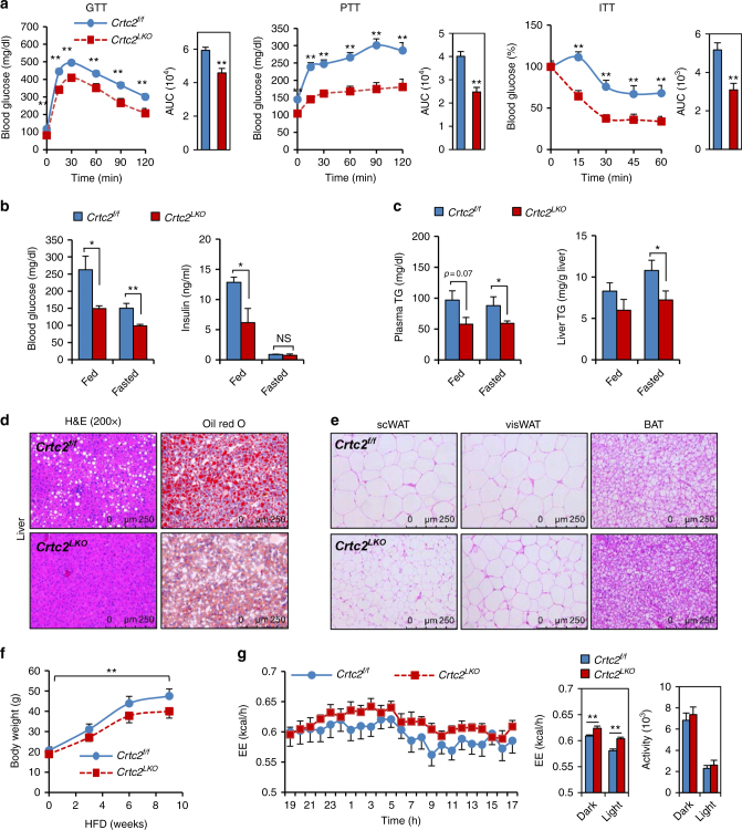
To elaborate the potential function of hepatic Crtc2 in the obese setting, we fed mice with a high-fat diet (HFD) for 8 weeks. We observed a significant impact on glucose metabolism by liver-specific depletion of Crtc2, as evidenced by the improved glucose, pyruvate, glucagon, and insulin tolerance in Crtc2 LKO mice compared with Crtc2 f/f mice in HFD conditions (Fig. 1a; Supplementary Fig. 3a). While Crtc2 LKO mice displayed reduced fasting hepatic glycogen levels compared with the control, hepatic glycogen levels were not different between the two genotypes during feeding (Supplementary Fig. 3b). In line with the improved insulin tolerance, the peripheral insulin signaling might be more active in Crtc2 LKO mice in comparison to Crtc2 f/f mice, as shown by the increased tyrosine phosphorylation of insulin receptor (IR) and the increased serine phosphorylation of Akt in response to insulin in the liver or visceral WAT (Supplementary Fig. 3c, d). Blood glucose and insulin levels were also lower in Crtc2 LKO mice, corroborating the suggested role of Crtc2 in glucose metabolism (Fig. 1b).
Fig. 1.
Hepatic Crtc2 controls systemic glucose and lipid metabolism in mammals. a Glucose tolerance test (GTT, left), pyruvate tolerance test (PTT, middle), and insulin tolerance test (ITT, right) showing effects of chronic depletion of hepatic Crtc2 in mice under HFD for 8 weeks on glucose metabolism and insulin signaling (n = 13 for Crtc2 f/f mice and n = 11 for Crtc2 LKO mice). Area under the curve (AUC) for each test was also shown. b Ad libitum (fed) or 16 h-fasting (fasted) blood glucose levels (left), and ad libitum (fed) or 16 h-fasting (fasted) insulin levels (right) from either Crtc2 f/f mice or Crtc2 LKO mice under HFD for 9 weeks (n = 13 for Crtc2 f/f mice and n = 11 for Crtc2 LKO mice). c Plasma triglycerides (TG) levels (left) and hepatic TG levels (right) from either Crtc2 f/f mice or Crtc2 LKO mice under ad libitum (fed) or 16 h-fasting (fasted) conditions under HFD for 9 weeks (n = 13 for Crtc2 f/f mice and n = 11 for Crtc2 LKO mice). d Paraffin-embedded sections of liver tissues from 8-week HFD-fed mice were stained with hematoxylin and eosin (H&E, left). Frozen liver sections were also stained with oil red O (right). Data represent three independent experiments (n = 3 mice per group). e Paraffin-embedded sections of subcutaneous white adipose tissues (scWAT, left), visceral white adipose tissues (visWAT, middle), and brown adipose tissues (BAT, right) from 16 h-fasted, 9-week HFD-fed Crtc2 f/f mice or Crtc2 LKO mice were stained with H&E. Data represent three independent experiments (n = 3 mice per group). f Body weight from either Crtc2 f/f mice or Crtc2 LKO mice under HFD (n = 13 for Crtc2 f/f mice and n = 11 for Crtc2 LKO mice). g Energy expenditure (top and bottom left for quantitation) and locomotor activity (bottom right) were measured from 7-week HFD-fed Crtc2 f/f mice or Crtc2 LKO mice by using metabolic cage (n = 10 for Crtc2 f/f mice and n = 9 for Crtc2 LKO mice). Note that the HFD was initiated at 4 weeks of age and maintained throughout the experimental period. Data in a, b, c, f and g represent mean ± s.e.m. (*P < 0.05, **P < 0.01, t-test a–c and g or Tukey–Kramer multiple comparisons f)
Hepatic Crtc2 deficiency enhances energy expenditure
Unexpectedly, we found that both plasma and hepatic triacylglycerol levels were considerably lower in Crtc2 LKO mice compared with Crtc2 f/f mice (Fig. 1c), arguing against a recent report showing an inhibitory role of Crtc2 on hepatic lipogenesis, a study that utilized systemic Crtc2 knockout mice18. In fact, we observed rather reduction in lipogenic gene expression in livers of Crtc2 LKO mice in comparison to those of Crtc2 f/f mice, suggesting that effects of liver-specific depletion of Crtc2 on hepatic lipogenesis are different from those of systemic Crtc2 knockout mice, perhaps due in part to the improved insulin sensitivity in Crtc2 LKO mice (Supplementary Fig. 3e). In line with the data with the improved lipid metabolism upon chronic depletion of Crtc2 in the liver, we observed reduced size in lipid droplets in the liver, BAT, subcutaneous white adipose tissue (scWAT), and visceral white adipose tissue (visWAT) (Fig. 1d, e). Furthermore, we observed that the fat body mass was also reduced in Crtc2 LKO mice compared with Crtc2 f/f mice (Supplementary Fig. 3f). Reduced lipid droplet size in the liver may suggest the increased degradation of lipid droplets by autophagy (or lipophagy) in the Crtc2 LKO liver compared with the Crtc2 f/f liver19. However, we did not observe consistent changes in expression of key components of autophagy between livers of Crtc2 LKO mice and those of Crtc2 f/f mice (i.e., reduced Atg7 expression and induced Atg12 expression, together with no changes in p62 levels or LC3-II levels), arguing against the potential involvement of lipophagy in this setting (Supplementary Fig. 4a, b). As the reduced lipid droplets in fat cells and reduced body weight gain upon HFD in Crtc2 LKO mice may indicate an increased energy expenditure (Fig. 1f), we performed an indirect calorimetric assay by using mice under HFD conditions. Indeed, we observed a small but significant increase in energy expenditure from Crtc2 LKO mice compared with Crtc2 f/f mice, which could lead to the reduced body weight for the mutant group, without thereby changes in food intake, water consumption, or locomotor activity (Fig. 1g; Supplementary Fig. 4c, d).
Depletion of hepatic Crtc2 promotes a Pparα-dependent pathway
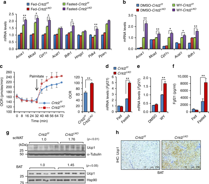
Since Crtc2 is a transcriptional co-activator, we reasoned that the impact of hepatic Crtc2 depletion on energy metabolism should be stemmed from changes in gene expression. Thus, we performed an extensive quantitative RT-PCR (Q-PCR) analysis to detect potential target genes of Creb/Crtc2 that could be causal to the current metabolic phenotype shown in this study. We noticed that genes that code for enzymes in the fatty acid oxidation and ketogenesis were generally increased in livers of Crtc2 LKO mice in comparison to those of Crtc2 f/f mice (Fig. 2a). In compliance with this data, we observed higher plasma ketone body levels in Crtc2 LKO mice, and the increased expression of fatty acid oxidation genes together with the increased oxygen consumption in response to palmitate in primary hepatocytes from Crtc2 LKO mice compared with Crtc2 f/f mice (Fig. 2b, c; Supplementary Fig. 4e). Specifically, the expression of Fgf21, a hepatokine that was shown to improve whole-body energy metabolism by accelerating lipid usage and non-shivering thermogenesis in adipocytes11, 20, was induced more than two-fold in livers of Crtc2 LKO mice in comparison to those of Crtc2 f/f mice during fasting conditions, which was confirmed in isolated primary hepatocytes (Fig. 2d, e). Plasma Fgf21 levels were also higher in Crtc2 LKO mice compared with Crtc2 f/f mice under fasting (Fig. 2f). Indeed, we observed increased Ucp1 protein levels in BAT and subcutaneous WAT, suggesting that higher plasma concentration of Fgf21 in Crtc2 LKO mice greatly affected lipid metabolism in adipocytes (Fig. 2g, h). Unexpectedly, expression of gluconeogenic genes was largely unaltered by chronic depletion of Crtc2 in the liver under HFD, although the primary hepatocytes from Crtc2 LKO mice showed a dampened transcriptional response to forskolin (Supplementary Fig. 5a, b). We noticed a potential compensatory increase in messenger RNA (mRNA) levels of C/ebpβ (Supplementary Fig. 5c), a transcription factor that was also shown to enhance transcription of gluconeogenic genes21. In addition, higher plasma cortisol levels in Crtc2 LKO mice may ensure the normal expression levels of gluconeogenic genes even in the absence of Crtc2 (Supplementary Fig. 5d), as suggested by the recent report showing that Fgf21 can induce hepatic gluconeogenesis by inducing secretion of cortisol from the adrenal gland in vivo22.
Fig. 2.
Chronic depletion of hepatic Crtc2 promotes a Pparα-dependent transcriptional program in the liver. a Effects of chronic depletion of hepatic Crtc2 in mice under HFD for 9 weeks on fatty acid beta oxidation genes (Q-PCR, n = 5–7 mice per group). b Effects of Crtc2 knockout on fatty acid beta oxidation genes in primary hepatocytes in the absence or in the presence of Pparα agonist WY14643 (10 μM for 16 h) (Q-PCR, n = 3 sets of cells per group). c Oxygen consumption rate was measured from primary hepatocytes of Crtc2 f/f mice or Crtc2 LKO mice as a surrogate for the rate of fatty acid oxidation (n = 5 sets of cells per group). Area under the curve was also shown at right. d Effects of chronic depletion of hepatic Crtc2 in mice under HFD for 9 weeks on Fgf21 expression (Q-PCR, n = 5–7 mice per group). e Effects of Crtc2 knockout on Fgf21 expression in primary hepatocytes in the absence or in the presence of Pparα agonist WY14643 (10 μM for 16 h) (Q-PCR, n = 3 sets of cells per group). f Effects of chronic depletion of hepatic Crtc2 on plasma Fgf21 levels in 16 h-fasted mice under HFD for 8 weeks (n = 7–11 mice per group). g Effects of hepatic Crtc2 knockout on Ucp1 protein levels in the subcutaneous WAT from 6 h-fasted mice under HFD for 8 weeks (top). Quantitation of Ucp1 levels of each group was shown above the Ucp1 bands (P < 0.01, t-test). Effects of hepatic Crtc2 knockout on Ucp1 protein levels in the BAT from 6 h-fasted mice under HFD for 8 weeks (bottom). Quantitation of Ucp1 levels of each group was shown above the Ucp1 bands (P < 0.05, t-test). h Immunohistochemistry analysis showing effects of chronic liver-specific depletion of Crtc2 in mice on Ucp1 expression in BAT. Data represent four independent experiments (n = 4 mice per group). Note that the HFD was initiated at 4 weeks of age and maintained throughout the experimental period. Data in a–e and g represent mean ± s.d. (*P < 0.05, **P < 0.01, t-test (c) or Tukey–Kramer multiple comparisons a, b, d and e), and data in f represent mean ± s.e.m. (**P < 0.01, Tukey–Kramer multiple comparisons)
Crtc2 regulates Sirt1–Pparα–Fgf21 axis by controlling miR-34a
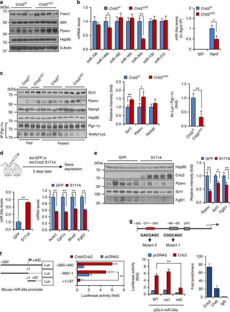
Various transcription factors such as Pparα, Atf4, and Foxo1 were shown to be directly involved in the regulation of Fgf21 expression4–7. While neither Atf4 nor Foxo1 levels were altered, Pparα protein levels were higher in the livers of Crtc2 LKO mice in comparison to those of Crtc2 f/f mice under fasting conditions (Fig. 3a). In addition, we did not observe any changes in hepatic Chrebp expression between the two groups of mice (data not shown). These data suggest that Crtc2 could directly control expression of Pparα at the protein levels, perhaps through a direct regulation of microRNAs. Thus, we performed small RNA sequencing analysis by using livers of either Crtc2 LKO mice or Crtc2 f/f mice under HFD. We noticed a significant change in the expression of several microRNAs in livers of Crtc2 LKO mice including miR-34a, miR-10b, and miR-146b (Supplementary Table 1). Among the potential microRNAs, only miR-34a level was confirmed to be reduced more than two-fold upon depletion of Crtc2 by Q-PCR (Fig. 3b). Primary miR-34a levels were also reduced in the livers of Crtc2 LKO mice in comparison to those of Crtc2 f/f mice under HFD-fed conditions, corroborating our hypothesis that the transcription of miR-34a requires the Creb/Crtc2 transcriptional machinery (Supplementary Fig. 5e). As shown before, miR-34a level was higher in the livers of HFD-fed mice compared with the control (Supplementary Fig. 5f)23–25. Interestingly, previous reports suggested that miR-34a reduces expression of various targets such as Sirt1, Pparα, and Nampt in the liver, which could directly affect activity of Pparα23, 24, 26. Indeed, we observed that protein levels of Sirt1, Pparα, and Nampt were higher in livers of Crtc2 LKO mice in comparison to those of Crtc2 f/f mice (Fig. 3c). Furthermore, we observed the decreased acetylation of peroxisome proliferator activating receptor gamma (Pgc)-1α, a known co-activator for Pparα, presumably due to the increased expression of Sirt1 and Nampt proteins. These data corroborate our hypothesis that reduced expression of miR-34a resulted in the activation of Pparα-transcriptional machinery, leading to the enhanced expression of Fgf21 in the Crtc2 LKO liver during fasting. Overexpression of miR-34a in the liver resulted in the decreased expression of Pparα and Fgf21 as well as Pparα target genes, confirming that miR-34a directly controls Fgf21 expression via a Pparα-dependent transcriptional pathway (Supplementary Fig. 6a–c).
Fig. 3.
Hepatic miR-34a is critical in reducing Sirt1-Pparα-mediated expression of Fgf21 in a Creb/Crtc2-dependent manner. a Effects of hepatic Crtc2 knockout on protein levels of Atf4, Foxo1, and Pparα in the livers of 16 h-fasted, 9-week HFD-fed mice. b Effects of hepatic Crtc2 knockout on mature microRNA expression in the livers of 16 h-fasted, 9-week HFD-fed mice (left) (Q-PCR, n = 4 mice per group). Levels of AGO2-associated miR-34a were also shown (right) (Q-PCR, n = 4 mice per group). c Effects of hepatic Crtc2 knockout on protein levels in the livers of 16 h fasted, 9-week HFD-fed mice. Quantitation of protein levels of each condition was shown at right. d, e Effects of expression of constitutively active form of Crtc2 (S171A) on hepatic genes. 8-week old C57BL/6 mice were injected with adenovirus for 5 days before being killed after 24 h fasting. Hepatic miR-34a levels (d, left), hepatic expression of Pparα target genes (d, right), hepatic protein levels (e, left), and quantitation of protein levels (e, right) from C57BL/6 mice that were infected with Ad-GFP or Ad-Crtc2 S171A adenovirus (n = 5 mice per group). f 5′- and 3′-deletion analysis was performed to map the putative Creb/Crtc2 response element on the promoter of mouse miR-34a (top). Luciferase reporter assay was performed in 293T cells to determine the effects of Creb/Crtc2 on miR-34a promoter activity (bottom). N = 3 independent experiments in triplicate. g Location of putative CREs on the mouse miR-34a promoter (top), the effects of CRE mutations on the mouse miR-34a promoter activity (left), and the chromatin immunoprecipitation assay showing the occupancy of Creb/Crtc2 over mouse miR-34a promoter (right) were shown. N = 3 independent experiments in triplicate. Note that the HFD was initiated at 4 weeks of age and maintained throughout the experimental period (a–c). Data in b–g represent mean ± s.d. (*P < 0.05, **P < 0.01, t-test)
We did not observe significant changes in miR-34a expression either by fasting or the liver-specific depletion of Crtc2 under NCD conditions (data not shown). We reasoned that Creb/Crtc2 transcriptional machinery per se might not be sufficient to enhance transcription of miR-34a but could be responsible for the increased expression of hepatic miR-34a in the DIO conditions, in which conditions a consistent activation of Crtc2 was observed14. To mimic the DIO conditions that results in the consistent activation of Crtc2, we utilized adenovirus expressing Crtc2 S171A, a constitutively active form of Crtc215. Overexpression of Crtc2 S171A resulted in the increased expression of miR-34a in the liver, with concomitant decrease in protein levels of Pparα, Sirt1, and Fgf21, together with Pparα target genes (Fig. 3d, e; Supplementary Fig. 6d). Luciferase reporter assay also showed that promoter activity of miR-34a was induced by co-expression of Creb and Crtc2 in cultured cells (Fig. 3f; Supplementary Fig. 7a). p53 has been listed as a major transcriptional regulator for miR-34a transcription27, 28. It is of note that Crtc2 did not further activate p53-dependent increase in miR-34a promoter activity, showing that these two pathways do not converge on the transcriptional regulation of miR-34a (Supplementary Fig. 7b). Promoter sequences for miR-34a were previously delineated, and we identified several potential cAMP response elements (CRE). The mutagenesis study revealed that among the potential CREs, the element at −132 for the human promoter and the one at −271 for the mouse promoter were critical in Creb/Crtc2 transcriptional response (Fig. 3g; Supplementary Fig. 7c). The result was also confirmed by chromatin immunoprecipitation assay both in HepG2 cells and mouse primary hepatocytes. These data collectively suggest that Creb/Crtc2 is critical in the regulation of Pparα-dependent lipid metabolism not only in the liver but also at the whole-body level by controlling hepatic expression of Fgf21 in the DIO conditions.
Depletion of Fgf21 reverts the effect of Crtc2 knockout in DIO mice
We then wanted to confirm whether the effect of liver-specific depletion of Crtc2 on the whole-body energy metabolism is due to the elevation in Fgf21 levels. To this end, we generated Fgf21 liver-specific mice (Fgf21 LKO mice) together with Crtc2/Fgf21 double liver-specific knockout mice (Crtc2/Fgf21 LKO mice) by using albumin-Cre transgene. While single knockout of Fgf21 in mice almost completely diminished the plasma Fgf21 levels compared with WT mice, we failed to observe any significant changes in either blood glucose levels or body weight between the two genotypes. Similarly, we did not observe any differences in body weight or lipid droplet size in the liver, scWAT, visWAT, and BAT between Crtc2 f/f mice and Fgf21 LKO mice, suggesting that the simple depletion of hepatic Fgf21 per se might not provoke metabolic phenotypes under DIO conditions (Supplementary Fig. 8a–e). On the other hand, concomitant depletion of Crtc2 and Fgf21 in the liver, which also almost completely diminished the plasma Fgf21 levels, reversed the beneficial effect of liver-specific depletion of Crtc2 in blood glucose levels, body weight, and whole-body lipid metabolism and adiposity, as we observed the increased lipid droplet size in liver, WAT, and BAT of Crtc2/Fgf21 LKO mice compared with the Crtc2 LKO mice (Supplementary Fig. 8a–e). In addition, Ucp1 protein levels (BAT) were increased by single knockout of Crtc2 in the liver, which were reduced by double knockout of Crtc2 and Fgf21 in the liver of mice (Supplementary Fig. 8f). Again, a single depletion of hepatic Fgf21 only did not affect BAT Ucp1 levels in DIO conditions. These data suggest that the effect of liver-specific depletion of Crtc2 on the lipid homeostasis and the energy metabolism might be mainly functioning via the increased plasma Fgf21. Knockout of Fgf21 in the liver effectively removed the beneficial effect of the increased Fgf21 in this setting, reversed the effect of Crtc2 depletion on hepatic lipid contents and overall adiposity under DIO conditions. These data suggest that increased plasma Fgf21 under fasting could promote improved lipid homeostasis while the depletion of hepatic Fgf21 (and thus reducing plasma Fgf21 levels) per se might not instigate metabolic disorders.
Effect of miR-34a on adiposity is blocked by co-expression of Fgf21
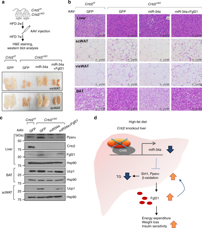
To test whether increased Pparα/Fgf21 program under Crtc2 depletion is indeed through miR-34a-dependent pathway, we expressed miR-34a or miR-34a plus Fgf21 in the liver of Crtc2 LKO mice via adeno-associated virus-mediated gene delivery. As shown before, plasma Fgf21 levels were increased in Crtc2 LKO mice in comparison to the control, which were reduced significantly by re-expression of miR-34a. As expected, co-expression of Fgf21 together with miR-34a restored higher plasma Fgf21 levels (Supplementary Fig. 9a). Body weight, as well as size of WATs, was reduced in Crtc2 LKO mice compared with the control. Re-expression of miR-34a reversed this phenotype, and co-expression of Fgf21 inhibited the effect of miR-34a (Fig. 4a; Supplementary Fig. 9b–d). The changes in lipid homeostasis were indeed prominent as shown by hematoxylin and eosin (H&E) staining data of liver and adipose tissues (Fig. 4b). MiR-34a effectively blocked the changes in lipid droplet size of liver, WAT, and BAT in hepatic Crtc2 depleted mice, and co-expression of Fgf21 also interfered with the effect of miR-34a on adiposity in mice. Mechanistically, the levels of Pparα protein and its target Fgf21 protein were induced in livers of Crtc2 LKO mice in comparison to those of Crtc2 f/f mice. Re-expression of miR-34a in Crtc2 LKO led to the reduction of both proteins, while co-expression of miR-34a and Fgf21 restored hepatic Fgf21 protein levels but not Pparα protein levels. As well, increased Ucp1 protein levels in BAT and scWAT of Crtc2 LKO mice were reduced by miR-34a expression, and they were restored by co-expression of miR-34a and Fgf21, confirming our hypothesis that Crtc2-miR-34a axis interferes with Pparα-Fgf21 pathway under HFD-induced obesity and insulin-resistant conditions (Fig. 4c).
Fig. 4.
Effect of miR-34a on adiposity is reversed by co-expression of Fgf21 in Crtc2 liver-specific knockout mice. a–d Effects of miR-34a and/or Fgf21 on Crtc2 LKO mice under 9-week HFD (n = 5 mice per group). Schematics of AAV injection (a, top) and the effect of miR-34a and Fgf21 on fat size (a, bottom) were shown. H&E staining of the liver, scWAT, visWAT, BAT of each group of mice were shown (b). c Western blot analysis showing hepatic protein levels, as well as Ucp1 protein levels in scWAT and BAT of each genotype. Data represent four independent experiments (n = 4 mice per group). Note that the HFD was initiated at 4 weeks of age and maintained throughout the experimental period. d Schematic diagram showing the effects of Creb/Crtc2-driven expression of miR-34a on hepatic fatty acid metabolism and whole-body energy homeostasis under diet-induced obesity (DIO) and insulin-resistant conditions. Depletion of Crtc2 promotes the activation of Sirt1/Pparα pathway under DIO conditions, leading to the increased hepatic fatty acid beta oxidation and the amelioration of fatty liver symptoms. In addition, enhanced hepatic Fgf21 expression is also essential in improving lipid metabolism in the peripheral tissues including the liver and adipose tissues, resulting in the increased energy expenditure and the improved insulin sensitivity
Discussion
In this study, we identified a novel mechanism by which Crtc2 controls whole-body energy homeostasis via controlling expression of hepatokine Fgf21. Fgf21 has been shown to improve energy homeostasis in mammals by acting directly on the central nervous systems as well as peripheral tissues including adipose tissues20. These studies were corroborated by use of tissue-specific βKlotho knockout mouse models, a co-receptor for Fgf19 and 21 that is crucial in mediating intracellular hormonal signals29, 30. Interestingly, a recent study disputed the role of βKlotho in mediating Fgf21 response in WATs, suggesting that further study is necessary to clarify the molecular characteristics of plasma membrane receptors of Fgf21 in tissue-specific manners31. Although it was shown that Fgf21 improves hepatic lipid metabolism by enhancing fatty acid oxidation and ketogenesis and reducing lipogenesis, the effects of Fgf21 on the liver may be indirect via the action of sympathetic nerve activity30, 32. On the other hand, a recent report showed a role of direct action of Fgf21 on hepatic metabolism, suggesting that the issue is yet to be clearly determined33.
It is noteworthy to mention that the effect of Crtc2 depletion on Fgf21 levels was only prominent under fasting conditions (Fig. 2f). Being regarded as an endocrine hormone, this type of regulation of Fgf21 levels is not unprecedented. Indeed, most endocrine hormones are regulated in response to the nutrient status. Examples include insulin (during feeding), glucagon (during fasting), and many incretins such as GLP-1 and GIP-1 (during feeding), which mainly function in the specific nutrient status and countered by counter-regulatory hormones upon changes in nutrient state. While the secretion of such hormones are regulated at the level of exocytosis, there are examples of endocrine hormones such as GLP-1 that are also regulated at the level of transcription. In addition, counter-regulatory hormones exist to limit the action of certain hormones for a prolonged period of time. Crtc2 liver-specific knockout mice showed increased insulin sensitivity, thus we suspected that insulin could counteract to reduce Fgf21 levels during feeding by reducing transcriptional activation of hepatic Fgf21 via the inhibition of Pparα activity.
Based on our data, we would like to propose that the tight regulation of hepatic Creb/Crtc2 function is critical in the maintenance of energy homeostasis in mammals. Under hepatic insulin resistant state, prolonged activation of Crtc2 is not only responsible for the increased glucose production from the liver14, but also critical in promoting the accumulation of lipids in the liver and peripheral tissues in part by activating miR-34a-mediated repression of Sirt1/Pparα activity and Fgf21 expression (Fig. 4d). As noted before, Fgf21 can function to promote hepatic fatty acid oxidation and induce adaptive thermogenesis in adipocytes, leading to the decreased peripheral fat mass, increased energy expenditure, and improved insulin sensitivity11, 33. Thus, the inhibition of miR-34a by reducing Crtc2 activity in the liver could be beneficial in combating obesity and insulin resistance by promoting whole-body energy metabolism in an Fgf21-dependent manner.
Methods
Animal experiments
Crtc2 conditional knockout mice were generated by Macrogen (Seoul, Korea) by using Crtc2 mutant ES cell clone (#EPD0197_3_A08) from EUCOMM (European conditional mouse consortium). The knockout first line was first mated with CAG-Flp transgenic mice (Jackson lab) to discard the FRT sites, resulting in the generation of Crtc2 flox/flox mice that contain the loxP sites surrounding the critical exon 4 of Crtc2 gene. Crtc2 flox/flox mice were then crossed with albumin-Cre transgenic mice (Jackson lab) to generate liver-specific Crtc2 knockout mice. Fgf21 flox/flox mice were purchased from the Jackson lab. Fgf21 flox/flox mice were crossed with Crtc2 LKO mice to produce Crtc2/Fgf21 LKO mice. All mouse lines were backcrossed with C57BL/6 for at least five times before being used for the experiment.
For induction of obesity and insulin resistance, male 4-week-old mice were fed a HFD (60% fat diet, D12492; Research Diets) for 3–12 weeks (specific conditions are shown in the figure legends). For the glucose tolerance test (GTT) or pyruvate tolerance test (PTT), 16 h-fasted mice were intraperitoneally injected with a bolus of glucose or pyruvate (2 g/kg of body weight for NCD and 1.5 g/kg for HFD). For the insulin tolerance test (ITT) or the glucagon tolerance test, 6 h-fasted mice were intraperitoneally injected with a bolus of insulin (0.75 units of insulin/kg body weight) or glucagon (15 μg of glucagon/kg body weight). Blood glucose was measured from tail vein blood with an automatic glucose monitor (One Touch, LifeScan). For insulin signaling, either PBS or insulin (0.1 units per mouse) was injected intraperitoneally for 10 min, and the liver and the WAT were collected for the further analyses. Neither randomization nor blinding was used for the animal experiments in the study. Sample size was determined using previous experimental studies for metabolic assessment. All procedures were performed in a specific pathogen-free facility at the Gyerim Experimental Animal Resource Center, Korea University (12:12 h light–dark cycle, maintained at 22 °C), based on the protocols that were approved by the Korea University Institutional Animal Care and Use Committee.
Culture of primary hepatocytes
Mouse primary hepatocytes from 8- to 10-week-old C57BL/6N mice were isolated by collagenase perfusion methods34. After the isolation, the perfused liver was minced and was subject to the percoll gradient to selectively purify hepatocytes. Cells were then maintained in medium 199 (M199; Sigma-Aldrich, St. Louis, MO) with 10% fetal bovine serum (FBS; HyClone, Logan, UT), 100 U/ml penicillin, 100 μg/ml streptomycin, and 10 nM of dexamethasone. After the attachment, cells were then treated with various reagents as well as adenoviruses as shown in the figure legends.
Western blot analysis and immunoprecipitation
Western blot analyses were performed by using 10–60 μg of protein extracts as described previously35. Antibodies against Crtc2 and phosphor-Tyr IR were purchased from Calbiochem, an antibody against Crtc3 was from Bethyl lab, antibodies against Akt, phosphor Akt (T308, S473), Foxo1, acetyl lysine, autophagy proteins were from Cell Signaling, antibodies against β-actin and α-tubulin were from Sigma, an antibody against Sirt1 was from Millipore, antibodies against Hsp90, Atf4, and IRβ were from Santa Cruz, an antibody against Nampt was from Enzo life sciences, antibodies against Pparα and Ucp1 were from Abcam, an antibody against Fgf21 was from R&D systems. Acetylation of Pgc-1α was determined by the immunoprecipitation with Pgc-1α antibody36, followed by the detection of acetylated lysine residues with an antibody against acetyl lysine. Band density was quantified by using the Image J software (NIH). Specific conditions for antibodies used in the western blot analysis are summarized in Supplementary Table 2.
Histological analysis
Liver and fat tissues were isolated from mice and were fixed with 10% formalin (Sigma). Histological changes were examined by H&E stain. Neutral lipid accumulation in liver was analyzed by Oil red O (ORO) staining (Sigma) on the frozen liver tissues. Mayer’s hematoxylin was used as counterstaining, respectively. Slides were observed with a light microscope (Leica DMi 8).
Plasmids
Human miR-34a promoter was excised from the pGL3-PMT-miR-34a plasmid (a gift from Judy Lieberman (Addgene plasmid #25799)37, and was subcloned into the pGL4 vector to generate the human full-length miR-34a luciferase plasmid. For the mouse miR-34a promoter analysis, genomic sequences containing the miR-34a promoter were amplified by PCR from the mouse genomic DNA (Promega G309a), and were cloned into the pGL4 vector. Mutagenesis on the CREB binding sites within the promoter was performed by using the QuikChange Site-Directed Mutagenesis Kit according to the manufacturer’s protocol (Stratagene). All constructs were confirmed by sequencing.
Luciferase reporter assays
293T cells were purchased directly from ATCC, and were routinely checked for the mycoplasma contamination. Cells were plated onto 24-well plates and then maintained in DMEM (Hyclone) supplemented with 10% FBS, penicillin (100 U/ml), and streptomycin (100 μg/ml). Each transfection was performed with 100 ng of pGL4-h/m miR-34a promoter, 50 ng of β-galactosidase, 50 ng of expression vector for Creb or Crtc2 by using TransIT-LT1 reagent (Mirus) for 48 h. Experiments were performed in triplicates and repeated at least three times. Luciferase activity was determined with the Promega Luciferase Assay Kit according to the manufacturer’s protocol and normalized by β-galactosidase activity.
RNA extraction and quantitative PCR
Total RNA was isolated by using Trisure (Bioline) and RNeasy Kit (Qiagen) according to the manufacturer’s protocols. Complimentary DNA was synthesized by using Goscript Reverse transcription systems (Promega) as described in the manufacturer’s protocol. Quantitative real-time PCR (Q-PCR) analysis was performed in triplicate by using a SensiFAST SYBR green mix (Bioline) and CFX connect real-time system (Bio-Rad). The mRNA levels were normalized to ribosomal L32 or Gapdh. For microRNA (miRNA) quantification, total RNA was reverse transcribed by using the miScript II RT Kit (Qiagen). Primers specific for mouse miR-34a, miR-10b, miR-146b, miR-212, miR-132, and miR-582 were purchased from Qiagen. The values were normalized to RNU6B (Qiagen). Sequences for gene-specific primers are shown in Supplementary Table 3.
Chromatin immunoprecipitation assay
Mouse primary hepatocytes or HepG2 were plated in 100 mm dish. After treatment with 100 nM glucagon or 10 μM Forskolin for 2 h, cells were cross-linked with 1% formaldehyde for 10 min at 37 °C and stopped by the addition of glycine to a final concentration of 0.125 M. The ChIP assay was performed by using the ChIP Assay Kit (Millipore) according to the manufacturer’s protocol as described previously. For human miR-34a promoter, primers −140(F): 5′-GATCCCGGGCTGGAGAGA-3′, and +120(R): 5′-CTGAGAAACACAAGCGTTTACCT-3′ were used. For mouse miR-34a promoter, primers −300(F): 5′-CTCCCTATTCCCCGCCTG-3′, and-1(R): 5′-CCCCCA ATCTGTGCAGTTAC-3′ were used. Total input was used as a normalization control.
Serum FGF21, corticosterone, insulin level
For detecting plasma levels of specific hormones, Corticosterone ELISA Kit (ab108821), Fgf21 Kit (R&D systems MF2100), and Insulin Kit (Alpco) were used according to the manufacturer’s protocols.
Triglycerides level
Total liver lipids were extracted according to the Folch method with a slight modification38. Briefly, mouse liver was homogenized with chloroform/methanol solution (2:1, v/v), and was centrifuged at room temperature. The supernatant was then washed with 1/5 volumes of 0.9% NaCl and was again centrifuged. After discarding the upper phase, the remaining solution was evaporated under vacuum. Hepatic and plasma TG contents were measured using an Enzymatic Colorimetric Assay Kit (Wako Chemicals).
Ketone body level
Plasma β-hydroxybutyrate concentrations were measured by using a β-Hydroxybutyrate Assay Kit (Abcam, ab83390).
Fatty acid oxidation
Primary hepatocytes isolated from Crtc2 f/f mice or Crtc2 LKO mice were seeded at 2 × 10⁴ cells per well on XF-24 cell culture plates (Seahorse Bioscience) in M199 medium (Sigma). After overnight incubation, cells were equilibrated with KHB buffer (111 mM NaCl, 4.7 mM KCl, 2 mM MgSO4, 1.2 mM Na2HPO4, 2.5 mM glucose, 0.5 mM carnitine, pH 7.4) and incubated at 37 °C for 1 h without CO2. For induction of fatty acid oxidation, palmitate–BSA complex was injected at a final concentration of 200 μM into the XF-24 cartridge (Seahorse Bioscience). Fatty acid oxidation capacity was represented as increased oxygen consumption rate in response to the palmitate–BSA complex.
Adenovirus, AAV virus
Adenovirus expressing constitutive active form of Crtc2 (Ad-Crtc2 S171A) and adenovirus expressing miR-34a (Ad-miR-34a) have been described previously15, 23. Recombinant adenovirus (0.5 × 10⁹ pfu) was delivered by tail vein injection to the mice. For AAV-miR34a, miR-34a was first subcloned into the pAAV-IRES, and was co-transfected with pAAV-DJ vector and the pAAV-Helper vector into HEK 293T cells to generate recombinant adeno-associated virus expressing miR-34a as described in the manufacturer’s protocol (Cell Biolabs). About 2 × 10¹⁰ vg (per mouse) of purified viruses was injected into the mice via tail vein injection. After 7 days, mice were fed a HFD for 7 weeks before being killed.
Ribonucleoprotein immunoprecipitation
AGO2 immunoprecipitation experiments after Ad-miR-34a infection were conducted by using the liver tissues as described previously39. Briefly, the liver tissue was triturated in 10 ml of HBSS and transferred to a 10 cm dish. Tissue suspensions were irradiated three times at 400 mJ/cm2 in the UV cross-linker. Cross-linked lysate was resuspended in 1×PXL (1× PBS, 0.1% SDS, 0.5% deoxycholate, 0.5% NP-40, w/o Mg2+, Ca2+, complete protease inhibitor cocktail, RNasin (Promega)). The lysate was incubated with premade AGO2 antibody (2E12, Abnova, H00027161-M01)-Dynabead Protein A complex for 4 h at 4 °C. The antibody corresponding protein–RNA complex was pull down with a magnetic bar (Invitrogen).The total RNA in the complex is purified by miRNeasy Kit (Qiagen) and reverse transcribed using the miScript II RT Kit (Qiagen) to analyze the miRNA expression.
Metabolic cage analysis
Indirect calorimetry was assessed by using indirect calorimetric chamber (OxyletPro System, Harvard Apparatus; Panlab) according to the manufacturer’s protocol40. Briefly, male 16-week-old, 8-week-HFD-fed Crtc2 f/f mice, and Crtc2 LKO mice were acclimated in the calorimetric chamber for 24 h prior to the measurement. VO2 and VCO2 were measured every 9 min for 4 h by an O2 and CO2 analyzer at a controlled flow rate of 600 ml/min. At each point of analysis, the installed software automatically calculated the RQ and EE. In addition, food intake and physical activity were also measured in the system. Data represent the single 24 h measurement.
Body composition analysis
Body composition was measured by using the NMR analyzer Bruker Minispec LF50 (Bruker Optics Inc.) at the Korea Mouse Phenotyping Center (KMPC), Seoul National University, Seoul, Korea.
Small RNA sequencing
For small RNA sequencing, RNAs were extracted from liver tissues of HFD-fed Crtc2 f/f mice and Crtc2 LKO mice by using miRNeasy Mini Kit (Qiagen). Total RNA quality and quantity were verified spectrophotometrically and electrophoretically (Bioanalyzer 2100). Sequencing library construction with the Illumina TruSeq small RNA Sample Prep Kit was performed as described in the manufacturer’s protocol. Libraries were sequenced on the Illumina MiSeq instrument. Sequence alignment and detection of known and novel microRNAs were performed by using miRDeep2 software algorithm. Prior to performing sequence alignment, the Mus musculus reference genome release mm10 was retrieved from UCSC genome browser and indexed using Bowtie (1.1.1), a bowtie for aligning sequencing reads to reference sequences. The pre-processed and clustered reads were aligned to Mus musculus reference genome. Those reads were then aligned to the Mus musculus matured and precursor miRNAs obtained from the miRBase v21. The miRDeep2 algorithm is based on the miRNA biogenesis model; it aligns reads to potential hairpin structures in a manner consistent with Dicer processing, and assigns scores that represent the probability that hairpins are true miRNA precursors. In addition to detecting known and novel miRNAs, miRDeep2 estimates their abundance. The experiment was performed by Macrogen, Seoul, Korea.
Statistics
Results are represented as either mean ± s.e.m.(for metabolites) or mean ± s.d. (for Q-PCR and luciferase assay). Comparison of different groups was carried out by using two-tailed unpaired Student’s t test for comparing two groups, or Tukey–Kramer multiple comparisons for comparing values among multiple groups as indicated in the figure legends.
Data availability
All the data and materials supporting this work are available upon reasonable request to the corresponding author. The data for the small RNA sequencing were deposited in the Sequence Read Archive (SRA), National Cancer Center for Biotechnology Information (accession number: SAMN07661813).
Electronic supplementary material
Acknowledgements
We thank Professor Sung-Gil Chi (Korea University, Seoul, Korea) for pCMV-flag-p53 construct and Professor Jongsook Kim Kemper (University of Illinois, Champaign, IL, USA) for miR-34a adenovirus construct. We thank Professor Je Kyung Seong (Korea Mouse Phenotyping Center, Seoul National University, Seoul, Korea) for the help to utilize NMR analyzer Bruker Minispec LF50, and Professor Sung-Wook Chi (Korea University, Seoul, Korea) for the help to analyze AGO2-associated miR-34a. We also thank Sun Myung Park (Korea University, Seoul, Korea) for technical assistance and members of our laboratory for the critical reading of the manuscript. This work was supported by the National Research Foundation of Korea (grant nos.: NRF-2012M3A9B6055345, NRF-2015R1A5A1009024, NRF- 2015R1A2A1A01006687, and NRF-2017M3A9D5A01052447), funded by the Ministry of Science and ICT, Republic of Korea, and a grant from Korea University, Seoul, Republic of Korea.
Author contributions
The project was conceived by S.-H.K. Experiments were designed and performed by H.-S.H., B.H.C., J.S.K., G.K. and S.-H.K., and were analyzed by H.-S.H. and S.-H.K. H.-S.H. and S.-H.K. wrote the paper.
Competing interests
The authors declare no competing financial interests.
Footnotes
Electronic supplementary material
Supplementary Information accompanies this paper at doi:10.1038/s41467-017-01878-6.
Publisher's note: Springer Nature remains neutral with regard to jurisdictional claims in published maps and institutional affiliations.
References
- 1.Cahill GF., Jr. Fuel metabolism in starvation. Annu. Rev. Nutr. 2006;26:1–22. doi: 10.1146/annurev.nutr.26.061505.111258. [DOI] [PubMed] [Google Scholar]
- 2.Marliss EB, Aoki TT, Unger RH, Soeldner JS, Cahill GF., Jr. Glucagon levels and metabolic effects in fasting man. J. Clin. Invest. 1970;49:2256–2270. doi: 10.1172/JCI106445. [DOI] [PMC free article] [PubMed] [Google Scholar]
- 3.Goetz R, et al. Molecular insights into the klotho-dependent, endocrine mode of action of fibroblast growth factor 19 subfamily members. Mol. Cell. Biol. 2007;27:3417–3428. doi: 10.1128/MCB.02249-06. [DOI] [PMC free article] [PubMed] [Google Scholar]
- 4.Badman MK, et al. Hepatic fibroblast growth factor 21 is regulated by PPARalpha and is a key mediator of hepatic lipid metabolism in ketotic states. Cell Metab. 2007;5:426–437. doi: 10.1016/j.cmet.2007.05.002. [DOI] [PubMed] [Google Scholar]
- 5.Inagaki T, et al. Endocrine regulation of the fasting response by PPARalpha-mediated induction of fibroblast growth factor 21. Cell Metab. 2007;5:415–425. doi: 10.1016/j.cmet.2007.05.003. [DOI] [PubMed] [Google Scholar]
- 6.De Sousa-Coelho AL, Marrero PF, Haro D. Activating transcription factor 4-dependent induction of FGF21 during amino acid deprivation. Biochem. J. 2012;443:165–171. doi: 10.1042/BJ20111748. [DOI] [PubMed] [Google Scholar]
- 7.Haeusler RA, Han S, Accili D. Hepatic FoxO1 ablation exacerbates lipid abnormalities during hyperglycemia. J. Biol. Chem. 2010;285:26861–26868. doi: 10.1074/jbc.M110.134023. [DOI] [PMC free article] [PubMed] [Google Scholar]
- 8.von Holstein-Rathlou S, et al. FGF21 mediates endocrine control of simple sugar intake and sweet taste preference by the liver. Cell Metab. 2016;23:335–343. doi: 10.1016/j.cmet.2015.12.003. [DOI] [PMC free article] [PubMed] [Google Scholar]
- 9.Chartoumpekis DV, et al. Brown adipose tissue responds to cold and adrenergic stimulation by induction of FGF21. Mol. Med. 2011;17:736–740. doi: 10.2119/molmed.2011.00075. [DOI] [PMC free article] [PubMed] [Google Scholar]
- 10.Hondares E, et al. Thermogenic activation induces FGF21 expression and release in brown adipose tissue. J. Biol. Chem. 2011;286:12983–12990. doi: 10.1074/jbc.M110.215889. [DOI] [PMC free article] [PubMed] [Google Scholar]
- 11.Fisher FM, et al. FGF21 regulates PGC-1 alpha and browning of white adipose tissues in adaptive thermogenesis. Genes Dev. 2012;26:271–281. doi: 10.1101/gad.177857.111. [DOI] [PMC free article] [PubMed] [Google Scholar]
- 12.Kim KH, et al. Autophagy deficiency leads to protection from obesity and insulin resistance by inducing Fgf21 as a mitokine. Nat. Med. 2013;19:83–92. doi: 10.1038/nm.3014. [DOI] [PubMed] [Google Scholar]
- 13.Dentin R, Hedrick S, Xie J, Yates J, 3rd, Montminy M. Hepatic glucose sensing via the CREB coactivator CRTC2. Science. 2008;319:1402–1405. doi: 10.1126/science.1151363. [DOI] [PubMed] [Google Scholar]
- 14.Dentin R, et al. Insulin modulates gluconeogenesis by inhibition of the coactivator TORC2. Nature. 2007;449:366–369. doi: 10.1038/nature06128. [DOI] [PubMed] [Google Scholar]
- 15.Koo SH, et al. The CREB coactivator TORC2 is a key regulator of fasting glucose metabolism. Nature. 2005;437:1109–1111. doi: 10.1038/nature03967. [DOI] [PubMed] [Google Scholar]
- 16.Le Lay J, et al. CRTC2 (TORC2) contributes to the transcriptional response to fasting in the liver but is not required for the maintenance of glucose homeostasis. Cell Metab. 2009;10:55–62. doi: 10.1016/j.cmet.2009.06.006. [DOI] [PMC free article] [PubMed] [Google Scholar]
- 17.Wang Y, et al. Targeted disruption of the CREB coactivator Crtc2 increases insulin sensitivity. Proc. Natl Acad. Sci. USA. 2010;107:3087–3092. doi: 10.1073/pnas.0914897107. [DOI] [PMC free article] [PubMed] [Google Scholar]
- 18.Han J, et al. The CREB coactivator CRTC2 controls hepatic lipid metabolism by regulating SREBP1. Nature. 2015;524:243–246. doi: 10.1038/nature14557. [DOI] [PubMed] [Google Scholar]
- 19.Singh R, et al. Autophagy regulates lipid metabolism. Nature. 2009;458:1131–1135. doi: 10.1038/nature07976. [DOI] [PMC free article] [PubMed] [Google Scholar]
- 20.Owen BM, Mangelsdorf DJ, Kliewer SA. Tissue-specific actions of the metabolic hormones FGF15/19 and FGF21. Trends Endocrinol. Metab. 2015;26:22–29. doi: 10.1016/j.tem.2014.10.002. [DOI] [PMC free article] [PubMed] [Google Scholar]
- 21.Arizmendi C, Liu S, Croniger C, Poli V, Friedman JE. The transcription factor CCAAT/enhancer-binding protein beta regulates gluconeogenesis and phosphoenolpyruvate carboxykinase (GTP) gene transcription during diabetes. J. Biol. Chem. 1999;274:13033–13040. doi: 10.1074/jbc.274.19.13033. [DOI] [PubMed] [Google Scholar]
- 22.Liang Q, et al. FGF21 maintains glucose homeostasis by mediating the cross talk between liver and brain during prolonged fasting. Diabetes. 2014;63:4064–4075. doi: 10.2337/db14-0541. [DOI] [PubMed] [Google Scholar]
- 23.Lee J, et al. A pathway involving farnesoid X receptor and small heterodimer partner positively regulates hepatic sirtuin 1 levels via microRNA-34a inhibition. J. Biol. Chem. 2010;285:12604–12611. doi: 10.1074/jbc.M109.094524. [DOI] [PMC free article] [PubMed] [Google Scholar]
- 24.Choi SE, et al. Elevated microRNA-34a in obesity reduces NAD+ levels and SIRT1 activity by directly targeting NAMPT. Aging Cell. 2013;12:1062–1072. doi: 10.1111/acel.12135. [DOI] [PMC free article] [PubMed] [Google Scholar]
- 25.Fu T, et al. Aberrantly elevated microRNA-34a in obesity attenuates hepatic responses to FGF19 by targeting a membrane coreceptor beta-Klotho. Proc. Natl Acad. Sci. USA. 2012;109:16137–16142. doi: 10.1073/pnas.1205951109. [DOI] [PMC free article] [PubMed] [Google Scholar]
- 26.Ding J, et al. Effect of miR-34a in regulating steatosis by targeting PPARalpha expression in nonalcoholic fatty liver disease. Sci. Rep. 2015;5:13729. doi: 10.1038/srep13729. [DOI] [PMC free article] [PubMed] [Google Scholar]
- 27.Chang TC, et al. Transactivation of miR-34a by p53 broadly influences gene expression and promotes apoptosis. Mol. Cell. 2007;26:745–752. doi: 10.1016/j.molcel.2007.05.010. [DOI] [PMC free article] [PubMed] [Google Scholar]
- 28.Raver-Shapira N, et al. Transcriptional activation of miR-34a contributes to p53-mediated apoptosis. Mol. Cell. 2007;26:731–743. doi: 10.1016/j.molcel.2007.05.017. [DOI] [PubMed] [Google Scholar]
- 29.Ding X, et al. betaKlotho is required for fibroblast growth factor 21 effects on growth and metabolism. Cell Metab. 2012;16:387–393. doi: 10.1016/j.cmet.2012.08.002. [DOI] [PMC free article] [PubMed] [Google Scholar]
- 30.Owen BM, et al. FGF21 acts centrally to induce sympathetic nerve activity, energy expenditure, and weight loss. Cell Metab. 2014;20:670–677. doi: 10.1016/j.cmet.2014.07.012. [DOI] [PMC free article] [PubMed] [Google Scholar]
- 31.Markan KR, et al. FGF21 resistance is not mediated by downregulation of beta-klotho expression in white adipose tissue. Mol. Metab. 2017;6:602–610. doi: 10.1016/j.molmet.2017.03.009. [DOI] [PMC free article] [PubMed] [Google Scholar]
- 32.Kurosu H, et al. Tissue-specific expression of betaKlotho and fibroblast growth factor (FGF) receptor isoforms determines metabolic activity of FGF19 and FGF21. J. Biol. Chem. 2007;282:26687–26695. doi: 10.1074/jbc.M704165200. [DOI] [PMC free article] [PubMed] [Google Scholar]
- 33.Fisher FM, et al. Integrated regulation of hepatic metabolism by fibroblast growth factor 21 (FGF21) in vivo. Endocrinology. 2011;152:2996–3004. doi: 10.1210/en.2011-0281. [DOI] [PMC free article] [PubMed] [Google Scholar]
- 34.Yoon YS, et al. Suppressor of MEK null (SMEK)/protein phosphatase 4 catalytic subunit (PP4C) is a key regulator of hepatic gluconeogenesis. Proc. Natl Acad. Sci. USA. 2010;107:17704–17709. doi: 10.1073/pnas.1012665107. [DOI] [PMC free article] [PubMed] [Google Scholar]
- 35.Choi D, et al. Protein arginine methyltransferase 1 regulates hepatic glucose production in a FoxO1-dependent manner. Hepatology. 2012;56:1546–1556. doi: 10.1002/hep.25809. [DOI] [PubMed] [Google Scholar]
- 36.Lee MW, et al. Regulation of hepatic gluconeogenesis by an ER-bound transcription factor, CREBH. Cell Metab. 2010;11:331–339. doi: 10.1016/j.cmet.2010.02.016. [DOI] [PubMed] [Google Scholar]
- 37.Navarro F, et al. miR-34a contributes to megakaryocytic differentiation of K562 cells independently of p53. Blood. 2009;114:2181–2192. doi: 10.1182/blood-2009-02-205062. [DOI] [PubMed] [Google Scholar]
- 38.Folch J, Lees M, Sloane Stanley GH. A simple method for the isolation and purification of total lipids from animal tissues. J. Biol. Chem. 1957;226:497–509. [PubMed] [Google Scholar]
- 39.Chi SW, Zang JB, Mele A, Darnell RB. Argonaute HITS-CLIP decodes microRNA-mRNA interaction maps. Nature. 2009;460:479–486. doi: 10.1038/nature08170. [DOI] [PMC free article] [PubMed] [Google Scholar]
- 40.Park J, et al. SIK2 is critical in the regulation of lipid homeostasis and adipogenesis in vivo. Diabetes. 2014;63:3659–3673. doi: 10.2337/db13-1423. [DOI] [PubMed] [Google Scholar]
Associated Data
This section collects any data citations, data availability statements, or supplementary materials included in this article.
Supplementary Materials
Data Availability Statement
All the data and materials supporting this work are available upon reasonable request to the corresponding author. The data for the small RNA sequencing were deposited in the Sequence Read Archive (SRA), National Cancer Center for Biotechnology Information (accession number: SAMN07661813).


