Dear Editor,
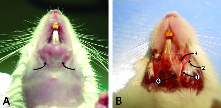
We would like to draw attention to the correct nomenclature for blood collection from superficial veins on the head of mice. In a September 2016 article,4 Regan and colleagues named a submental procedure for collecting blood from the ventral aspect of the chin. The correct terminology for the chin is the mental region, but the site the authors mark may be instead the mandibular region.1 The authors identify the vessel as the convergence of the facial and submental veins; however, they do not provide a photo of the dissected head revealing the anatomical site. The submental vein (v. submentalis) runs obliquely over the digastricus muscle, heading towards the chin. The caliber of this vein is too small for collecting blood in a suitable volume or drip rate. In a dissection performed by one of this letter’s authors,1 the site of blood collection (Figure 1) appears to be the inferior labial vein (v. labialis inferior) or its convergence with the facial vein (v. facialis). The inferior labial vein runs along the dorsal border of the body of the mandible, parallel to the ventral edge of the buccinator muscle (m. buccinator). The facial vein courses along the ventral aspect of the masseter muscle (m. masseter) and crosses the body of the mandible along the rostral edge of this muscle. Both veins have sufficient caliber for this blood collection procedure.
Figure 1.
(A) Target areas, 1-2 mm from midline, for vascular access (arrows) in a mouse. (B). Dissected lower jaw showing (arrows): 1. and 2. facial v.; 3. inferior labial v.; 4. submental v.
Photos kindly provided by Erika French, MS, LAT, printed with permission.
In the same article, Regan and colleagues refer to a submandibular [venous] plexus and submandibular region of the head. For information about the vasculature, we recall Jerald Silverman’s Letter to the Editor 5 in which he stated “the correct term is the facial vein (v. facialis) or linguofacial vein (v. linguofacialis), depending on the exact site of blood collection. It is also possible to obtain blood from the nearby maxillary vein (v. maxillaris). Using Nomina Anatomica Veterinaria as a guide, there is no submandibular vein in the mouse”.2,5
For illustrations of the anatomy of these veins and head regions mentioned here, the reader is referred to Popesko3and Constantinescu.1
Given the frequent confusion over the proper names of these two procedures, the authors suggest that simple and appropriate names be used to identify each technique, such as the “cheek bleed” and the “chin bleed.” The venipuncture of “cheek bleed” is targeting the facial vein, which curves ventrally and rostrally around the margins of the masseter muscle. The “chin bleed” is performed by the venipuncture of the inferior labial vein, a branch of the facial vein running rostrally alongside the dorsal border of the mandible. Both the masseter muscle and the mandible are easy palpable.
Letters to the Editor
Letters discuss material published in JAALAS in the previous 3 issues. They can be submitted through email (journals@aalas.org) or by regular mail (9190 Crestwyn Hills Dr, Memphis, TN 38125). Letters are not necessarily acknowledged upon receipt nor are the authors necessarily consulted before publication. Whether published in full or part, letters are subject to editing for clarity and space. The authors of the cited article will generally be given an opportunity to respond in the same issue in which the letter is published.
Contributor Information
Gheorghe M. Constantinescu, Professor Emeritus of Veterinary Anatomy Professional Member of the Association of Medical Illustrators Honorary Member of the Romanian Academy of Agricultural and Forestry Sciences; Department of Biomedical Sciences College of Veterinary Medicine University of Missouri-Columbia.
Nicole E. Duffee, Director, Education & Scientific Affairs AALAS.
References
- Constantinescu G. 2011. Comparative anatomy of the mouse and rat: a color atlas and text. Memphis (TN): American Association for Laboratory Animal Science. [Google Scholar]
- International Committee on Veterinary Gross Anatomical Nomenclature. [Internet].2012. Nomina anatomica veterinaria, 5th ed (revised version) [Cited 14 July 2017]. Available at: http://www.wava-amav.org/downloads/nav_2012.pdf.
- Popesko P, Rajtova V, Horak J. 1992. A colour atlas of the anatomy of small laboratory animals, vol. 2. London (United Kingdom): Wolfe Publishing. [Google Scholar]
- Regan RD, Fenyk-Melody JE, Tran SM, Chen G, Stocking KL. 2016. Comparison of submental blood collection with the retroorbital and submandibular methods in mice (Mus musculus). J Am Assoc Lab Anim Sci 55:570–576.PubMed [PMC free article] [PubMed] [Google Scholar]
- Silverman J. 2010. Letter to the editor: clinical biochemistry parameters in C57BL/6J mice after blood collection from the submandibular vein and retroorbital plexus. J Am Assoc Lab Anim Sci 49:400. [PMC free article] [PubMed] [Google Scholar]


