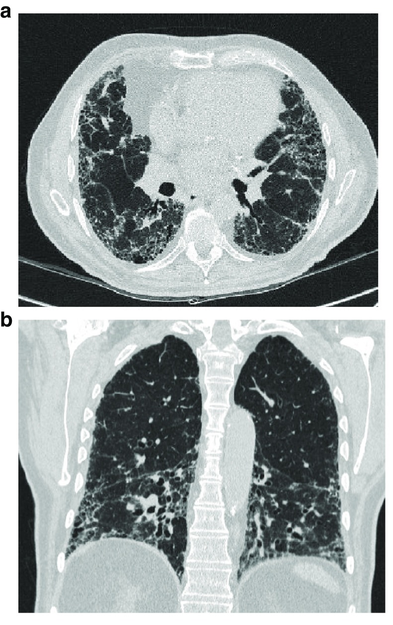
Figure 2. Typical high-resolution computed tomography pattern of usual interstitial pneumonia.
a) patchy, predominantly peripheral, sub-pleural and bibasal reticular abnormalities, traction bronchiectasis and bronchiolectasis, and irregular septal thickening associated with honey combing, b) coronal CT section showing bronchiectasis and bronchiolectasis in the bibasal zones.

