Key Points
Question
Does the aqueous humor of retinoblastoma eyes contain sufficient tumor-derived DNA to perform genetic analysis?
Findings
This study included 6 aqueous humor samples from 3 retinoblastoma eyes, 2 after primary enucleation and 4 before intravitreous injection of melphalan in an eye treated for vitreous seeding. Evaluation of aqueous humor demonstrated tumor-derived, cell-free DNA and chromosomal copy number variations (regions of chromosomal gains and losses), representing changes in the tumor.
Meaning
These findings suggest that aqueous humor can serve as a surrogate tumor biopsy for analyses of tumor-derived DNA in retinoblastoma eyes, which may be useful for eyes undergoing salvage therapy in which tumor tissue is unavailable.
Abstract
Importance
Retinoblastoma (Rb) is one of the first tumors to have a known genetic etiology. However, because biopsy of this tumor is contraindicated, it has not been possible to define the effects of secondary genetic changes on the disease course.
Objective
To investigate whether the aqueous humor (AH) of Rb eyes has sufficient tumor-derived DNA to perform genetic analysis of the tumor, including DNA copy number alterations.
Design, Setting, and Participants
This investigation was a case series study at a tertiary care hospital (Children’s Hospital Los Angeles) with a large Rb treatment center. Cell-free DNA (cfDNA) was isolated from 6 AH samples from 3 children with Rb, including 2 after primary enucleation and 1 undergoing multiple intravitreous injections of melphalan for vitreous seeding. Samples were taken between December 2014 and September 2015.
Main Outcomes and Measures
Measurable levels of nucleic acids in the AH and identification of tumor-derived DNA copy number variation in the AH. The AH was analyzed for DNA, RNA, and micro-RNA using Qubit high-sensitivity kits. Cell-free DNA was isolated from the AH, and sequencing library protocols were optimized. Shallow whole-genome sequencing was performed on an Illumina platform, followed by genome-wide chromosomal copy number variation profiling to assess the presence of tumor DNA fractions in the AH cfDNA of the 3 patients. One child’s cfDNA from the AH and tumor DNA were subjected to Sanger sequencing to isolate the RB1 mutation.
Results
Six AH samples were obtained from 3 Rb eyes in 3 children (2 male and 1 female; diagnosed at ages 7, 20, and 28 months). A corroborative pattern between the chromosomal copy number variation profiles of the AH cfDNA and tumor-derived DNA from the enucleated samples was identified. In addition, a nonsense RB1 mutation (Lys→STOP) from 1 child was also identified from the AH samples obtained during intravitreous injection of melphalan, which matched the tumor sample postsecondary enucleation. Sanger sequencing of the AH cfDNA and tumor DNA with polymerase chain reaction primers targeting RB1 gene c.1075A demonstrated this same RB1 mutation.
Conclusions and Relevance
In this study evaluating nucleic acids in the AH from Rb eyes undergoing salvage therapy with intravitreous injection of melphalan, the results suggest that the AH can serve as a surrogate tumor biopsy when Rb tumor tissue is not available. This novel method will allow for analyses of tumor-derived DNA in Rb eyes undergoing salvage therapy that have not been enucleated.
This case series study investigates whether the aqueous humor of retinoblastoma eyes has sufficient tumor-derived DNA to perform genetic analysis of the tumor, including DNA copy number alterations.
Introduction
Retinoblastoma (Rb) is a primary pediatric ocular cancer and one of the first tumors to have a known genetic etiology. Most germline and sporadic forms of Rb develop in response to biallelic mutations in RB1 (phenotype OMIM 180200 and gene/locus OMIM 614041), the first molecularly defined tumor suppressor gene. The elucidation of this gene and study of Rb provided a firm foundation for the field of cancer genetics. In addition to having RB1 mutations, Rb tumors analyzed after enucleation demonstrate recurrent chromosomal copy number variation (eg, regions of chromosomal gains and losses). These chromosomal changes underlie many human cancers and are frequent secondary genomic events during Rb tumorigenesis and contribute to disease progression. Yet, it has not been possible to define the effects of these chromosomal changes on treatment response and the disease course in Rb due to lack of access to tumor DNA. This lack of access is because, unlike many cancers, biopsy of Rb is contraindicated because of risk of tumor seeding outside the eye, possibly leading to orbital relapse.
Historically, any attempt to biopsy or obtain fluid from Rb eyes has been discouraged for concern of tumor dissemination. However, updates to intravitreous injection protocols for vitreous seeding include a paracentesis to increase safety of the injection. In 1995, intravitreous injection for Rb was initially described, but it was not widely accepted until 2012 when Munier et al published their safety-enhanced protocol, which recommended an initial paracentesis to withdraw aqueous humor (AH) to lower the intraocular pressure and thus prevent reflux of seeds to the pars plana injection site. Intravitreous injection of melphalan has revolutionized treatment of vitreous seeding and is now the standard of care for patients with Rb in the United States and internationally. The procedure is considered safe and effective in eradicating seeds. A systematic review found no reports of tumor spread after the safety-enhanced technique with an initial paracentesis. Therefore, the procedure not only provided an effective treatment for vitreous seeding but also facilitated safe access into the intraocular space and, specifically, the AH in Rb eyes. The procedure changed the dogma that the globe in Rb eyes was inviolable.
Like tumor, the AH is a rich source of information for intraocular disease. Previous studies demonstrate that the AH harbors markers of intraocular disease, such as cell-free nucleic acids and proteins. Micro-RNA (miRNA) has been detected from the AH in eyes with cataract and glaucoma. In enucleated eyes with Rb, greater AH lactate dehydrogenase is a marker for Rb activity. Increased epinephrine and norepinephrine and increased catabolic by-products secondary to the tumor have also been found in the AH. Higher protein levels are present in the AH of Rb eyes compared with eyes with cataracts. Furthermore, detection of the protein survivin, which suppresses apoptosis, has been detected in the AH of Rb eyes. Therefore, because nucleic acids have been successfully isolated from the AH, we hypothesized that the AH may harbor tumor-derived DNA that could allow the AH to serve as a surrogate tumor biopsy to detect genomic changes when tumor tissue is not available (eg, when the eye is not enucleated).
With increased use of intravitreous injection to treat vitreous seeding and thus greater access to the AH, we examined whether the AH can be used as a liquid biopsy of cell-free DNA (cfDNA) derived from Rb tumors. Specifically, after obtaining AH from Rb eyes after primary enucleation or before intravitreous injection of melphalan, we (1) found measurable levels of nucleic acids, including cfDNA, RNA, and miRNA; (2) defined genome-wide, chromosomal copy number variation profiles of tumor-derived cfDNA; and (3) detected the specific RB1 mutation in the AH of 1 patient. The application of this technique may allow us to better diagnose and monitor Rb as well as to develop novel insights into tumor progression at the molecular level.
Methods
This investigation was a case series study at a tertiary care hospital (Children’s Hospital Los Angeles [CHLA]) with a large Rb treatment center. Samples were taken between December 2014 and September 2015. Children’s Hospital Los Angeles Institutional Review Board approval was obtained for this study. Written informed consent was obtained from the parents of all participants, and that included permission for publication.
Surgical Procedure
A paracentesis with extraction of 0.1 mL of AH with a 32-gauge needle via the clear cornea is performed routinely as part of the procedure for the intravitreous injection of melphalan for treatment of vitreous seeding (or 0.1 mL from the anterior chamber after enucleation). There is no contact between the needles and the retinal tumor or the vitreous cavity.
Analysis of Cellular Content in the AH
Ten 0.1-mL samples of the AH were evaluated by CHLA clinical cytopathology laboratory staff immediately after extraction. Hematoxylin-eosin staining was performed. Bright-field microscopy was used to evaluate for cells, and none were found. These samples were used clinically, and further analyses were not performed. Subsequent AH samples were stored at −80°C in the original tuberculin syringes. After enucleation, tumor tissue was suspended in phosphate-buffered saline and then embedded in optimum cutting temperature compound (Sakura Finetek). Blocks were stored at −80°C. DNA was isolated using the QIAamp DNA Micro Kit (Qiagen).
Analysis of Nucleic Acid Content in the AH
DNA, RNA, and miRNA concentrations were assayed using the Qubit HS (High Sensitivity) Assay Kit (Thermo Fisher), which measures concentration of the assayed nucleic acid with the Qubit Fluorometer. The lower limit of detection is 10 pg/µL.
cfDNA Isolation and Purification
The AH was separated from the thawed sample by centrifugation for 10 minutes at 2000g. Collected AH was centrifuged again for 10 minutes at 14 000g to remove any cell debris and frozen at −80°C for further analysis. After centrifugation, AH supernatant was used for cfDNA extraction, while 5 µL from the bottom of the tube was suspended, plated on slides, and examined for intact cells with an IX81 Microscope (Olympus). No intact cells were found. The extracted AH volume was replaced with 1 × phosphate-buffered saline. Extraction of cfDNA was done with the QIAamp Circulating Nucleic Acid Kit (Qiagen) per the manufacturer’s instructions. This kit purifies DNA with a silica-based membrane. Concentration of cfDNA was measured using the Qubit Fluorometer System (Thermo Fisher). One AH sample provided enough cfDNA for size distribution evaluation with the Bioanalyzer 2100 High Sensitivity DNA Assay and Kit (Agilent Technologies).
Next-Generation Sequencing
DNA libraries for Illumina sequencing were constructed with the QIAseq Ultralow Input Library Kit (Qiagen) due to the small amount of extracted cfDNA from the AH samples. Each library was constructed with a sample bar code to permit pooling of multiple samples on a single Illumina HiSeq lane. DNA libraries were sequenced on the Illumina HiSeq platform for a single-end 50 base pair (bp) protocol.
Data Analysis
The bioinformatic procedures involved in the chromosomal copy number variation analysis have been previously published. Briefly, next-generation sequencing (NGS) reads from pooled DNA libraries were deconvoluted using bar codes. The reads were mapped to the human genome (hg19, Genome Reference Consortium GRCh37, University of California Santa Cruz Genome Browser database; https://genome.ucsc.edu), and polymerase chain reaction duplicates were removed. Normalization for guanine-cytosine content was performed, and DNA segment copy numbers were estimated by dividing the genome into 5000 variable-length bins and calculating the relative number of reads in each bin. Hierarchical clustering was performed using the heatmap.2 function in the R package gplots (https://cran.r-project.org/web/packages/gplots/index.html) on median-centered data using the method by Ward as the distance metric. Thresholds of 0.8 and 1.25 relative to the median were used to define deletions and amplifications, respectively.
Medical histories of the included patients were reviewed. Clinical details and treatment regimens were recorded.
Results
Patient and AH Sample Characteristics
Six AH samples from 3 Rb eyes in 3 children (2 after primary enucleation and 1 undergoing multiple intravitreous injections of melphalan for vitreous seeding) were obtained via a paracentesis. Two patients presented with unilateral disease. One of them had a constitutive 13q deletion, which predisposes to the development of Rb due to complete loss of the RB1 gene. Both patients underwent primary enucleation given the advanced nature of the tumor. The third patient presented with bilateral disease and underwent eye-salvaging systemic chemotherapy, followed by intravitreous melphalan treatment for tumor seeding. The AH was obtained from the eye with vitreous seeding only. This patient demonstrated 30% mosaicism for (c.1075A>T p.Lys359Ter /Lys→STOP) in the peripheral blood.
Evaluation of AH Nucleic Acid Content
Analysis of the 6 samples demonstrated measurable concentrations of DNA, RNA, and miRNA (eTable in the Supplement). The DNA concentration ranged from 0.084 to 56 ng/µL (median, 0.174 ng/µL). The AH samples from the patient treated with intravitreous injection had lower concentrations of all nucleic acids compared with the AH samples obtained after primary enucleation. There was no consistent decrease in AH nucleic acid concentration after subsequent intravitreous injection, although there was a decrease in active tumor seeding. The DNA size distribution peaked between 145 and 165 bp (median, 150 bp), consistent with cfDNA.
Tumor-Specific cfDNA in the AH After Primary Enucleation
Two eyes that were primarily enucleated were evaluated. Their case reports follow.
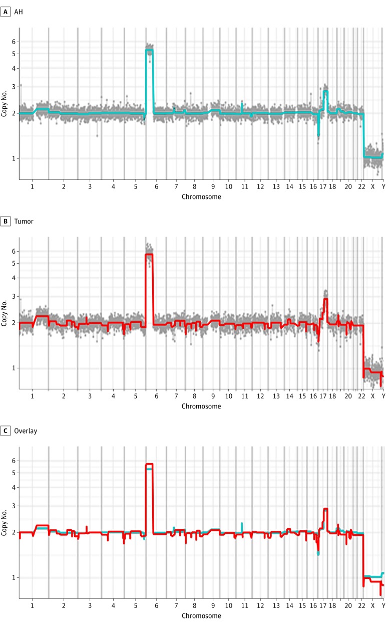
Case 1 is a boy diagnosed at age 7 months as having unilateral Group D Rb. The NGS of peripheral monocytes did not demonstrate a germline RB1 mutation. An AH paracentesis and tumor sample were taken immediately after enucleation and subsequently evaluated. Whole-genome copy number variation profiling was done, which showed the chromosomal changes (both gains and losses) that diverge from a diploid cell with a full complement of chromosomes. This analysis demonstrated that the chromosomal copy number gains and losses from the AH cfDNA (Figure 1A) correlated with the tumor DNA (Figure 1B). Profiles from both tissues showed a gain on chromosomes 1q and 6p, consistent with known chromosomal changes in Rb. There was also a gain on 17q22-17q25 and a loss of 17p13.3-p13.1 demonstrated in both the tumor and the AH (Table and Figure 1).
Figure 1. Case 1: Chromosomal Copy Number Variation Profile Demonstrating the Chromosomal Changes (Both Gains and Losses) That Vary From a Diploid Cell With a Full Complement of Chromosomes.
This profile is from a patient after primary enucleation showing overlapping gains and losses in the aqueous humor (AH) and tumor demonstrating that the cell-free DNA in the AH is derived from the tumor. A, Chromosomal copy number variation profile from cell-free DNA in the AH (gain of 1q, 6p, and 17q22-17q25 and loss of 17p13.3-p13.1). B, Chromosomal copy number variation profile from tumor (gain of 1q, 6p, and 17q22-17q25 and loss of 17p13.3-p13.1). C, Overlay of the chromosomal copy number variation profiles (AH is blue, and tumor is red).
Table. Chromosomal Copy Number Variations in Aqueous Humor (AH) and Tumor in the 3 Patients With Retinoblastoma.
| Variable | Case No./Sex/Age at Diagnosis, mo | Group Status | Blood Testing of RB1 Mutation | Treatment | No. of AH Samples | Copy No. | |
|---|---|---|---|---|---|---|---|
| Gains | Losses | ||||||
| AH | 1/M/7 | D | Negative | Primary enucleation | 1 | 1q, 6p, 17q22-17q25 | 17p13.3-p13.1 |
| Tumor | 1 | 1q, 6p, 17q22-17q25 | 17p13.3-p13.1 | ||||
| AH | 2/M/20 | E | 13q Deletion (13q13.3-q21.32) | Primary enucleation | 1 | 1q, 2p, 4q, 6p, 15q | 1p36.33-p32.3, 13q13.3-q21.32, 16q, 17p, 20p |
| Tumor | 1 | 1q, 6p, 7q31-q36 | 5q, 13q13.3-q21.32, 16q | ||||
| AH | 3/F/28 | D | 30% Mosaicism for c.1075A>T p.Lys359Ter point mutation in exon 11 of RB1 | Systemic plus intravitreous chemotherapy | 4 | 1q, 2p, 2q, 6p, 7q, 17q21.31-q25, 18q21.32-q23, Xq26-q28 | 11q14-q25, 16q, 19q |
| Tumor | 1 | 1q, 2p, 6p, 7q, 17q21.31-q25, 18q21.32-q23, Xq26-q28 | 11q14-q25, 16q, 19q | ||||
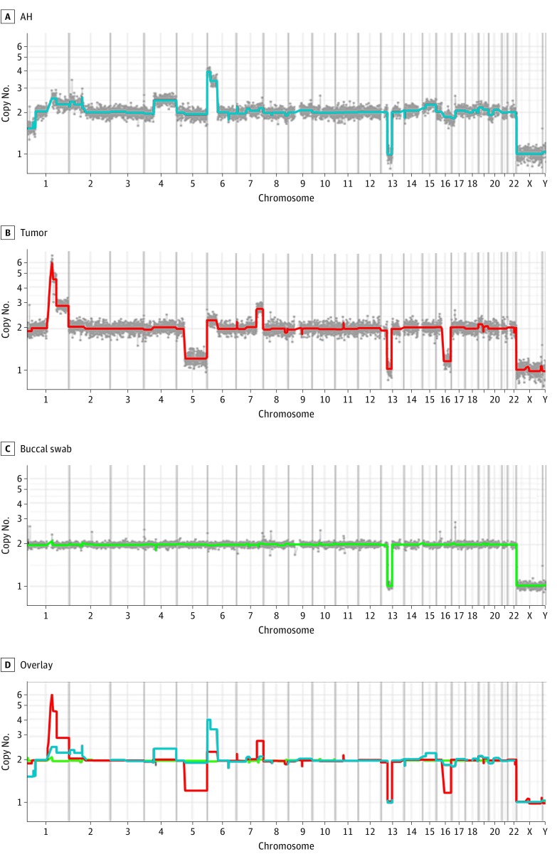
Case 2 is a boy diagnosed at age 20 months as having unilateral Group E Rb and treated with primary enucleation. The child was known to have a germline 13q deletion that predisposed to the development of Rb. Genomic DNA from a buccal swab (Figure 2C) confirmed the 13q deletion (13q13.3-q21.32), which was also seen in the AH (Figure 2A) and the tumor (Figure 2B). The AH and tumor samples were taken after enucleation and evaluated. Chromosomal analysis demonstrated a heterogeneous sample, with some differences in the copy number variation profile between the AH and the tumor. The chromosomal copy number variation in the AH sample showed a gain of 1q and 6p, which was also seen in the tumor (however, the amplitude of the 1q gain was higher in the tumor than in the AH; 6p was higher in the AH), but additional copy number amplification was seen at 2p, 4q, and 15q. The tumor showed a gain at 7q31-q36, which was not detected in the AH. In terms of chromosomal losses, aside from the 13q13.3-q21.32 loss seen in all tissues, the AH demonstrated additional loss of 1p36.33-p32.3, 16q, 17p, and 20p but did not demonstrate the 5q loss seen in the tumor (Table and Figure 2). Given the germline predisposition to Rb, the eye was filled with multiple large tumors, and we hypothesize that the chromosomal copy number variation in the AH reflected cfDNA from multiple tumor clones and not the single tumor biopsied after enucleation.
Figure 2. Case 2: Chromosomal Copy Number Variation Profile Taken After Primary Enucleation in a Patient With a 13q− Deletion Indicating Predisposition to the Development of Retinoblastoma.
A and B, Chromosomal analysis demonstrated a heterogeneous sample with clonal differences in the chromosomal copy number variations between the cell-free DNA in the aqueous humor (AH) (A) and tumor (B). C, Genomic DNA from a buccal swab demonstrates the same 13q deletion (13q13.3-q21.32). D, The overlay of the chromosomal copy number variation profiles is shown (AH is blue, tumor is red, and buccal swab is green).
Tumor-Specific cfDNA in the AH After Treatment With Intravitreous Melphalan for Vitreous Seeding
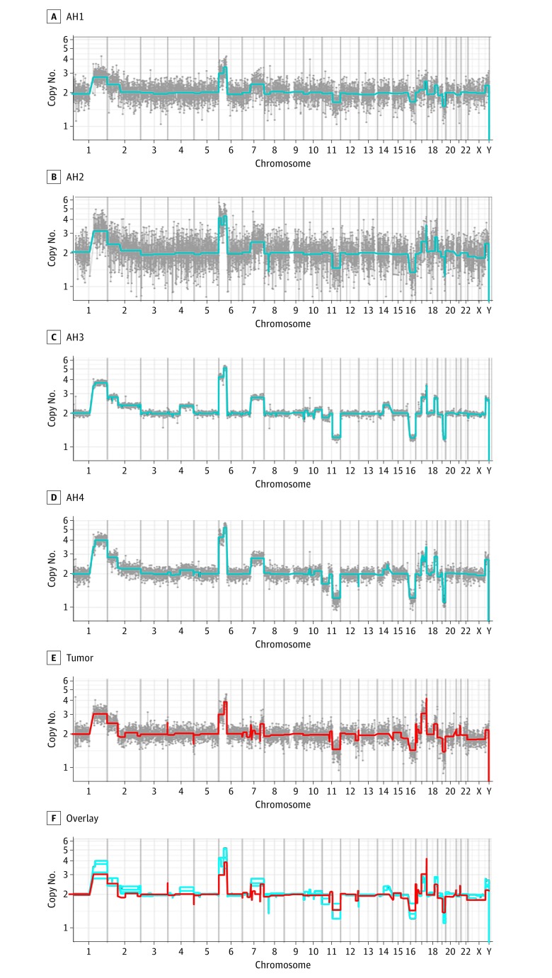
The AH samples were evaluated from Case 3 during serial intravitreous injections of melphalan for treatment of vitreous seeding. Case 3 is a girl diagnosed at age 28 months as having bilateral Group D Rb. The child was treated with 6 cycles of 3-drug chemotherapy per CHLA protocol, followed by multiple intravitreous injections of melphalan for vitreous seeding in the right eye. A secondary enucleation was subsequently required due to tumor relapse. Four of the AH samples taken during the course of serial intravitreous injections were analyzed for tumor-derived cfDNA. The first 3 samples were obtained 1 month apart, and the fourth sample was taken 2 weeks after the third sample. Evaluation of the AH cfDNA at all time points demonstrated a gain on chromosomes 1q, 2p, and 6p and a loss on chromosome 16q (Figure 3), consistent with known copy number variations in Rb. In addition, there were gains on 2q, 7q, 17q21.31-q25, 18q21.32-q23, and Xq26-q28 and losses on 11q14-q25 and 19q. This complex set of chromosomal gains and losses was also in the tumor sample (Figure 3E) taken after treatment at the secondary enucleation. The NGS clinical testing of the peripheral blood showed 30% mosaicism for an A-to-T point mutation in exon 11 (c.1075A>T p.Lys359Ter/Lys→STOP), causing an immediate termination codon that predisposed to the development of Rb. Sanger sequencing of the AH cfDNA and the tumor DNA with polymerase chain reaction primers targeting RB1 gene c.1075A demonstrated this same RB1 mutation (Table and eFigure in the Supplement).
Figure 3. Case 3: Chromosomal Copy Number Variation Profile Showing Similar Chromosomal Gains and Losses Over Time in a Patient Treated With Multiple Intravitreous Injections of Melphalan With Concurrent Aqueous Humor (AH) Extraction During Each Injection.
A-D, Chromosomal copy number variation profile from cell-free DNA in the AH from sequential samples (AH 1 through AH 4). E, Chromosomal copy number variation profile from tumor after subsequent (secondary) enucleation showing that the same chromosomal changes seen initially in the AH were also found in the tumor. F, Overlay of the AH and tumor chromosomal copy number variation profiles (AH is blue, and tumor is red).
Discussion
We evaluated the AH from 3 Rb eyes to assess whether the AH can serve as a surrogate Rb tumor biopsy. First, we demonstrated that there were measurable concentrations of nucleic acids in the small volume of AH sampled after primary enucleation and before intravitreous injection of melphalan. This finding is notable because the volume of active tumor in eyes undergoing intravitreous injection is significantly less than those with large, globe-filling tumors that undergo primary enucleation. While the volume of AH that can be extracted is small (0.1 mL), the fluid harbored measurable concentrations of DNA, RNA, and miRNA, raising the possibility that future studies might reveal diagnostic or prognostic roles for AH RNA and DNA. Based on this study evaluating nucleic acids purified from the AH in Rb eyes undergoing intravitreous injection of melphalan for vitreous seeding, we hypothesize that the source of cfDNA in the AH is from tumor cells in the posterior segment that have undergone necrosis and lysed. Subsequently, this necrosis and lysis allowed tumor-derived cfDNA to diffuse across the vitreous face into the AH. Another possibility is that tumor cells migrated to the anterior chamber and then lysed. However, no tumor cells were found in the 10 AH samples analyzed immediately after extraction. This result is consistent with published data on cytopathologic analysis of 122 AH samples extracted before intravitreous injection, all of which were negative for the presence of cells.
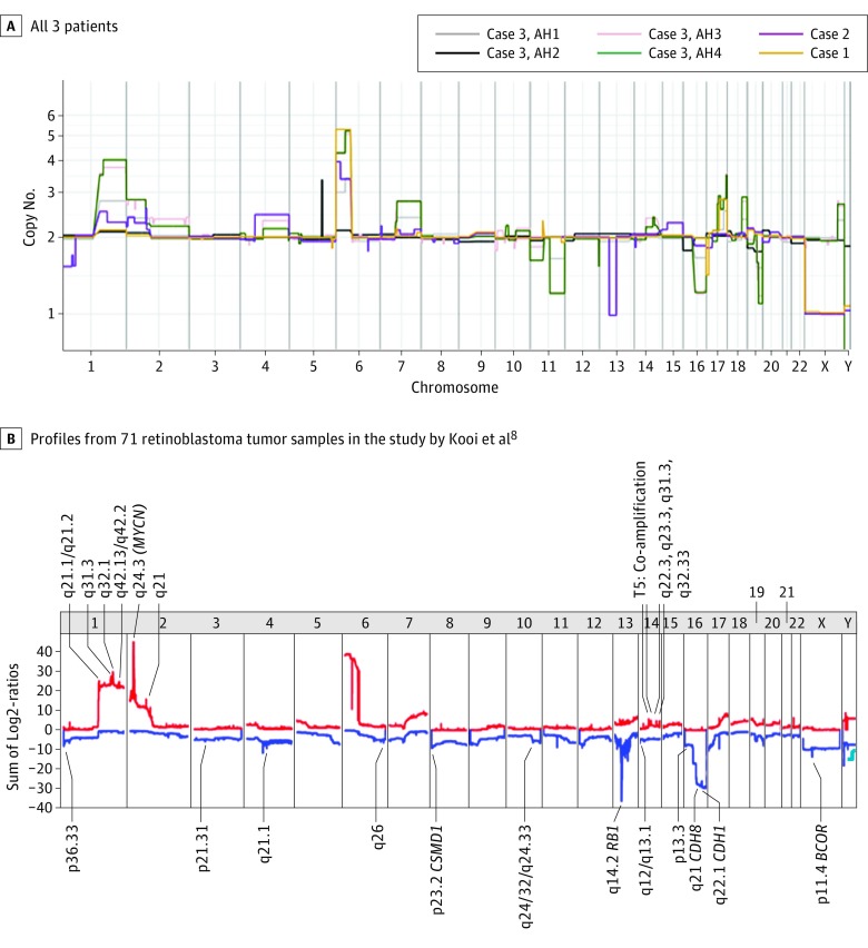
Separate samples demonstrated that there were measurable levels of nucleic acids in the AH of these eyes, and our next aim was to investigate whether the AH DNA was tumor derived. Copy number variation profiling confirmed the presence of somatic chromosomal variations that would be expected from the tumor DNA and have been described in Rb. We were able to demonstrate that copy number variation in cfDNA could be detected in the AH and that the chromosomal copy number profiles in the AH reflected those of the enucleated tumors. In Case 3, evaluation of the AH also provided access to Rb tumor DNA without enucleating the eye. If corroborated in additional patients, this result could establish a minimally invasive, nondestructive method of identifying specific genetic changes at diagnosis or during treatment. In this study, we demonstrated that the DNA in the AH is consistent with expectations for Rb tumor DNA, exhibiting similar known chromosomal changes, including a gain of 1q, 2p, and 6p and a loss of 16q. Comparisons of the chromosomal copy number variation profiles between the AH and the tumor are consistent with the hypothesis that cfDNA from the AH is tumor derived. The AH did not perfectly mimic the chromosomal changes in the tumor in all cases, which may suggest that in a patient with multifocal tumors, there may be variations in the genetic changes of each tumor clone, all of which separately contribute to the AH cfDNA. Further research is needed to test this hypothesis. In addition, evaluation of a greater number of samples of the AH cfDNA, as well as longitudinal sampling, is needed to determine whether the AH continues to mimic the known chromosomal changes that have been described in Rb tumor samples. Kooi et al recently published a report entitled “Somatic Genomic Alterations in Retinoblastoma Beyond RB1 Are Rare and Limited to Copy Number Changes,” in which they evaluated 71 Rb tissue samples for copy number gain and loss. Evaluation of 6 AH samples from our 3 patients is similar in distribution and depth of chromosomal variations found in their compiled tumor samples (Figure 4). Finally, future research is necessary to determine if copy number variation profiles differ with vitreous seeding progression, response or resistance to chemotherapy, more aggressive disease, or tumor anaplasia, which portends a worse prognosis. There is currently no known manner of determining which tumors may be more or less responsive to therapy; therefore, a better understanding of the genomic mechanisms in Rb may aid in the development of personalized therapy for Rb.
Figure 4. Single Aqueous Humor (AH) Chromosomal Copy Number Variation Compared With Summative Tumor Chromosomal Copy Number Variation by Kooi et al.
A and B, Chromosomal copy number variation from the AH cell-free DNA in all 3 patients (A) compared with a summation of the chromosomal copy number variation profiles from 71 retinoblastoma tumor samples in the study by Kooi et al (reproduced with permission) (B).
We identified the specific RB1 mutation in the AH in Case 3, with the benefit of knowing which mutation to query. This suggests that, despite lack of whole-cell genomic DNA, an appropriate library may be created to assay for RB1 mutations in cfDNA from the AH. If this process was successful, it would allow the AH to serve as a true surrogate biopsy not only for chromosomal rearrangements but also for the specific RB1 mutation. It is not known whether there is a clinical influence from knowing the RB1 mutation (or mutations) for every Rb tumor because this information is only available if the eye is enucleated. One scenario in which patient care could be influenced by the AH surrogate biopsy is the rare Rb tumor initiated by MYCN (OMIM 615968) amplification and not RB1 loss. It has been suggested in previously published literature that MYCN-amplified eyes should be enucleated due to the aggressive nature of these tumors. However, clinicians currently do not have this information when making treatment decisions because it is only known by evaluating tumor tissue or DNA. Further research is needed to discern if the AH will be a reliable source to query for RB1 mutations.
Limitations
A limitation of this study is that only 3 advanced Rb eyes were evaluated. Therefore, the findings presented herein need to be validated in more AH samples, particularly in eyes undergoing therapy and in less advanced Rb eyes to determine whether smaller tumors also have detectable amounts of cfDNA in the AH.
Conclusions
This study demonstrates that tumor-specific DNA is present in the AH of Rb eyes and can be safely “biopsied” and evaluated. To our knowledge, the present study is the first description of using the AH as a surrogate tumor biopsy for Rb when tumor tissue is not available. With further research, this novel method may allow clinicians to diagnose Rb in the setting of a diagnostic dilemma and, potentially, to determine whether certain chromosomal copy number variation profile parameters or RB1 mutations correlate with prognosis or therapeutic response. While additional investigation is required, this surrogate biopsy has the potential to revolutionize how we manage this childhood cancer.
eTable. Concentration of Nucleic Acids in the Aqueous Humor
eFigure. RB1 Mutation Isolated From cfDNA in the Aqueous Humor of Case 3 by Sanger Sequencing
References
- 1.Harbour JW. Retinoblastoma: pathogenesis and diagnosis In: Char DH, et al. . Tumors of the Eye and Ocular Adnexa. London, England: BC Decker Inc; 2001;253-265. [Google Scholar]
- 2.Fung YK, Murphree AL, T’Ang A, Qian J, Hinrichs SH, Benedict WF. Structural evidence for the authenticity of the human retinoblastoma gene. Science. 1987;236(4809):1657-1661. [DOI] [PubMed] [Google Scholar]
- 3.Friend SH, Bernards R, Rogelj S, et al. . A human DNA segment with properties of the gene that predisposes to retinoblastoma and osteosarcoma. Nature. 1986;323(6089):643-646. [DOI] [PubMed] [Google Scholar]
- 4.Dryja TP, Friend S, Weinberg RA. Genetic sequences that predispose to retinoblastoma and osteosarcoma. Symp Fundam Cancer Res. 1986;39:115-119. [PubMed] [Google Scholar]
- 5.Goodrich DW. The retinoblastoma tumor suppressor gene: the exception that proves the rule. Oncogene. 2006;25(38):5233-5243. [DOI] [PMC free article] [PubMed] [Google Scholar]
- 6.Xie T, Zhang C, Zhang B, et al. . A survey of cancer cell lines reveals highly structured and hierarchical relationships within and between DNA and mRNA that may be the result of selection. OMICS. 2010;14(1):91-97. [DOI] [PubMed] [Google Scholar]
- 7.Pratt ED, Stepansky A, Hicks J, Kirby BJ. Single-cell copy number analysis of prostate cancer cells captured with geometrically enhanced differential immunocapture microdevices. Anal Chem. 2014;86(22):11013-11017. [DOI] [PubMed] [Google Scholar]
- 8.Kooi IE, Mol BM, Massink MP, et al. . Somatic genomic alterations in retinoblastoma beyond RB1 are rare and limited to copy number changes. Sci Rep. 2016;6:25264. [DOI] [PMC free article] [PubMed] [Google Scholar]
- 9.Kooi IE, Mol BM, Massink MP, et al. . A meta-analysis of retinoblastoma copy numbers refines the list of possible driver genes involved in tumor progression. PLoS One. 2016;11(4):e0153323. [DOI] [PMC free article] [PubMed] [Google Scholar]
- 10.Shields JA, Shields CL, Ehya H, Eagle RC Jr, De Potter P. Fine-needle aspiration biopsy of suspected intraocular tumors: the 1992 Urwick Lecture. Ophthalmology. 1993;100(11):1677-1684. [DOI] [PubMed] [Google Scholar]
- 11.Karcioglu ZA. Fine needle aspiration biopsy (FNAB) for retinoblastoma. Retina. 2002;22(6):707-710. [DOI] [PubMed] [Google Scholar]
- 12.Karcioglu ZA, Gordon RA, Karcioglu GL. Tumor seeding in ocular fine needle aspiration biopsy. Ophthalmology. 1985;92(12):1763-1767. [DOI] [PubMed] [Google Scholar]
- 13.Eide N, Syrdalen P, Walaas L, Hagmar B. Fine needle aspiration biopsy in selecting treatment for inconclusive intraocular disease. Acta Ophthalmol Scand. 1999;77(4):448-452. [DOI] [PubMed] [Google Scholar]
- 14.Eide N, Walaas L. Fine-needle aspiration biopsy and other biopsies in suspected intraocular malignant disease: a review. Acta Ophthalmol. 2009;87(6):588-601. [DOI] [PubMed] [Google Scholar]
- 15.Eriksson O, Hagmar B, Ryd W. Effects of fine-needle aspiration and other biopsy procedures on tumor dissemination in mice. Cancer. 1984;54(1):73-78. [DOI] [PubMed] [Google Scholar]
- 16.Ali MJ, Honavar SG, Vemuganti GK, Singh AD. Fine needle aspiration biopsy of retinal tumors. Monogr Clin Cytol. 2012;21:72-81. [DOI] [PubMed] [Google Scholar]
- 17.Munier FL, Gaillard MC, Balmer A, et al. . Intravitreal chemotherapy for vitreous disease in retinoblastoma revisited: from prohibition to conditional indications. Br J Ophthalmol. 2012;96(8):1078-1083. [DOI] [PubMed] [Google Scholar]
- 18.Munier FL, Soliman S, Moulin AP, Gaillard MC, Balmer A, Beck-Popovic M. Profiling safety of intravitreal injections for retinoblastoma using an anti-reflux procedure and sterilisation of the needle track. Br J Ophthalmol. 2012;96(8):1084-1087. [DOI] [PubMed] [Google Scholar]
- 19.Seregard S, Kock E, af Trampe E. Intravitreal chemotherapy for recurrent retinoblastoma in an only eye. Br J Ophthalmol. 1995;79(2):194-195. [DOI] [PMC free article] [PubMed] [Google Scholar]
- 20.Ghassemi F, Shields CL. Intravitreal melphalan for refractory or recurrent vitreous seeding from retinoblastoma. Arch Ophthalmol. 2012;130(10):1268-1271. [DOI] [PubMed] [Google Scholar]
- 21.Shields CL, Fulco EM, Arias JD, et al. . Retinoblastoma frontiers with intravenous, intra-arterial, periocular, and intravitreal chemotherapy. Eye (Lond). 2013;27(2):253-264. [DOI] [PMC free article] [PubMed] [Google Scholar]
- 22.Ghassemi F, Shields CL, Ghadimi H, Khodabandeh A, Roohipoor R. Combined intravitreal melphalan and topotecan for refractory or recurrent vitreous seeding from retinoblastoma. JAMA Ophthalmol. 2014;132(8):936-941. [DOI] [PubMed] [Google Scholar]
- 23.Lawson BM, Saktanasate J, Say EA, Shields CL. Intravitreal chemotherapy provides control for massive vitreous seeding from retinoblastoma. J Pediatr Ophthalmol Strabismus. 2014;51 Online:e92-e94. [DOI] [PubMed] [Google Scholar]
- 24.Shields CL, Manjandavida FP, Arepalli S, Kaliki S, Lally SE, Shields JA. Intravitreal melphalan for persistent or recurrent retinoblastoma vitreous seeds: preliminary results. JAMA Ophthalmol. 2014;132(3):319-325. [DOI] [PubMed] [Google Scholar]
- 25.Manjandavida FP, Shields CL. The role of intravitreal chemotherapy for retinoblastoma. Indian J Ophthalmol. 2015;63(2):141-145. [DOI] [PMC free article] [PubMed] [Google Scholar]
- 26.Tuncer S, Balcı Ö, Tanyıldız B, Kebudi R, Shields CL. Intravitreal lower-dose (20 µg) melphalan for persistent or recurrent retinoblastoma vitreous seeds. Ophthalmic Surg Lasers Imaging Retina. 2015;46(9):942-948. [DOI] [PubMed] [Google Scholar]
- 27.Francis JH, Marr BP, Brodie SE, Gobin P, Dunkel IJ, Abramson DH. Intravitreal melphalan as salvage therapy for refractory retinal and subretinal retinoblastoma. Retin Cases Brief Rep. 2016;10(4):357-360. [DOI] [PMC free article] [PubMed] [Google Scholar]
- 28.Kivelä T, Eskelin S, Paloheimo M. Intravitreal methotrexate for retinoblastoma. Ophthalmology. 2011;118(8):1689.e1-1689.e6. [DOI] [PubMed] [Google Scholar]
- 29.Sun YB, Hui P, Punyara K, et al. . Intravitreal injection of melphalan in the treatment of retinoblastoma with vitreous cavity seeding. Chin Med J (Engl). 2013;126(8):1587. [PubMed] [Google Scholar]
- 30.De Francesco S, Galluzzi P, Bracco S, Menicacci F, Motolese E, Hadjistilianou T. Alternated intra-arterial and intravitreal chemotherapy for advanced intraocular retinoblastoma: preliminary successful results without systemic chemotherapy. Int Ophthalmol. 2015;35(6):887-895. [DOI] [PubMed] [Google Scholar]
- 31.Hou X, Cheng Y, Zhang Q, Liang J, Li X. Efficacy of intravitreal carboplatin plus bevacizumab in refractory retinoblastoma [in Chinese]. Zhonghua Yan Ke Za Zhi. 2015;51(2):126-129. [PubMed] [Google Scholar]
- 32.Suzuki S, Aihara Y, Fujiwara M, Sano S, Kaneko A. Intravitreal injection of melphalan for intraocular retinoblastoma. Jpn J Ophthalmol. 2015;59(3):164-172. [DOI] [PubMed] [Google Scholar]
- 33.Lee JH, Han JW, Hahn SM, Lyu CJ, Kim DJ, Lee SC. Combined intravitreal melphalan and intravenous/intra-arterial chemotherapy for retinoblastoma with vitreous seeds. Graefes Arch Clin Exp Ophthalmol. 2016;254(2):391-394. [DOI] [PubMed] [Google Scholar]
- 34.Kiratli H, Koç İ, Varan A, Akyüz C. Intravitreal chemotherapy in the management of vitreous disease in retinoblastoma. Eur J Ophthalmol. 2017;27(4):423-427. [DOI] [PubMed] [Google Scholar]
- 35.Francis JH, Schaiquevich P, Buitrago E, et al. . Local and systemic toxicity of intravitreal melphalan for vitreous seeding in retinoblastoma: a preclinical and clinical study. Ophthalmology. 2014;121(9):1810-1817. [DOI] [PubMed] [Google Scholar]
- 36.Munier FL, Gaillard MC, Balmer A, Beck-Popovic M. Intravitreal chemotherapy for vitreous seeding in retinoblastoma: recent advances and perspectives. Saudi J Ophthalmol. 2013;27(3):147-150. [DOI] [PMC free article] [PubMed] [Google Scholar]
- 37.Smith SJ, Smith BD. Evaluating the risk of extraocular tumour spread following intravitreal injection therapy for retinoblastoma: a systematic review. Br J Ophthalmol. 2013;97(10):1231-1236. [DOI] [PubMed] [Google Scholar]
- 38.Smith SJ, Smith BD, Mohney BG. Ocular side effects following intravitreal injection therapy for retinoblastoma: a systematic review. Br J Ophthalmol. 2014;98(3):292-297. [DOI] [PubMed] [Google Scholar]
- 39.Dunmire JJ, Lagouros E, Bouhenni RA, Jones M, Edward DP. MicroRNA in aqueous humor from patients with cataract. Exp Eye Res. 2013;108:68-71. [DOI] [PubMed] [Google Scholar]
- 40.Tanaka Y, Tsuda S, Kunikata H, et al. . Profiles of extracellular miRNAs in the aqueous humor of glaucoma patients assessed with a microarray system. Sci Rep. 2014;4:5089. [DOI] [PMC free article] [PubMed] [Google Scholar]
- 41.Abramson DH, Piro PA, Ellsworth RM, Kitchin FD, McDonald M. Lactate dehydrogenase levels and isozyme patterns: measurements in the aqueous humor and serum of retinoblastoma patients. Arch Ophthalmol. 1979;97(5):870-871. [DOI] [PubMed] [Google Scholar]
- 42.Mukhopadhyay S, Ghosh S, Biswas PN, Dutta SK, Chattopadhyay D. A cross-sectional study on aqueous humour lactate dehydrogenase level in retinoblastoma. J Indian Med Assoc. 2008;106(2):99-100. [PubMed] [Google Scholar]
- 43.Piro PA Jr, Abramson DH, Ellsworth RM, Kitchin D. Aqueous humor lactate dehydrogenase in retinoblastoma patients: clinicopathologic correlations. Arch Ophthalmol. 1978;96(10):1823-1825. [DOI] [PubMed] [Google Scholar]
- 44.Wang CY. Significance of the lactate dehydrogenase level in aqueous humor in the diagnosis of retinoblastoma [in Chinese]. Zhonghua Yan Ke Za Zhi. 1989;25(5):289-291. [PubMed] [Google Scholar]
- 45.Abramson DH, Piro PA, DeCresce R, Ellsworth RM, Kitchin D. Catecholamine metabolites in the aqueous of retinoblastoma. J Pediatr Ophthalmol Strabismus. 1978;15(2):77-78. [DOI] [PubMed] [Google Scholar]
- 46.Abramson DH, Senft SH, Servodidio CA, Ellsworth RM, Gamache PH. Retinoblastoma aqueous humor: aromatic amino acids. Ophthalmic Paediatr Genet. 1993;14(3):127-130. [DOI] [PubMed] [Google Scholar]
- 47.Mendelsohn ME, Abramson DH, Senft S, Servodidio CA, Gamache PH. Uric acid in the aqueous humor and tears of retinoblastoma patients. J AAPOS. 1998;2(6):369-371. [DOI] [PubMed] [Google Scholar]
- 48.Hadjistilianou T, Giglioni S, Micheli L, et al. . Analysis of aqueous humour proteins in patients with retinoblastoma. Clin Exp Ophthalmol. 2012;40(1):e8-e15. [DOI] [PubMed] [Google Scholar]
- 49.Shehata HH, Abou Ghalia AH, Elsayed EK, Ziko OO, Mohamed SS. Detection of survivin protein in aqueous humor and serum of retinoblastoma patients and its clinical significance. Clin Biochem. 2010;43(4-5):362-366. [DOI] [PubMed] [Google Scholar]
- 50.Shehata HH, Abou Ghalia AH, Elsayed EK, Ahmed Said AM, Mahmoud SS. Clinical significance of high levels of survivin and transforming growth factor beta-1 proteins in aqueous humor and serum of retinoblastoma patients. J AAPOS. 2016;20(5):444.e1-444.e9. [DOI] [PubMed] [Google Scholar]
- 51.Baslan T, Kendall J, Rodgers L, et al. . Genome-wide copy number analysis of single cells. Nat Protoc. 2012;7(6):1024-1041. [DOI] [PMC free article] [PubMed] [Google Scholar]
- 52.Baslan T, Kendall J, Rodgers L, et al. . Corrigendum: genome-wide copy number analysis of single cells. Nat Protoc. 2016;11(3):616. [DOI] [PubMed] [Google Scholar]
- 53.Ward JH., Jr Hierarchical grouping to optimize an objective function. J Am Stat Assoc. 1963;58(301):236-244. [Google Scholar]
- 54.Berry JL, Jubran R, Wong K, Lee TC, Murphree AL, Kim JW. Factors predictive of long-term visual outcomes of Group D eyes treated with chemoreduction and low-dose IMRT salvage: the Children’s Hospital Los Angeles experience. Br J Ophthalmol. 2014;98(8):1061-1065. [DOI] [PubMed] [Google Scholar]
- 55.Linn Murphree A. Intraocular retinoblastoma: the case for a new group classification. Ophthalmol Clin North Am. 2005;18(1):41-53, viii. [DOI] [PubMed] [Google Scholar]
- 56.Thériault BL, Dimaras H, Gallie BL, Corson TW. The genomic landscape of retinoblastoma: a review. Clin Exp Ophthalmol. 2014;42(1):33-52. [DOI] [PMC free article] [PubMed] [Google Scholar]
- 57.Berry JL, Jubran R, Kim JW, et al. . Long-term outcomes of Group D eyes in bilateral retinoblastoma patients treated with chemoreduction and low-dose IMRT salvage [published dosage correction appears in Pediatr Blood Cancer. 2014;61(6):1147]. Pediatr Blood Cancer. 2013;60(4):688-693. [DOI] [PubMed] [Google Scholar]
- 58.Berry JL, Jubran R, Lee TC, Murphree AL, Lee D, Kim JW. Low-dose chemoreduction for infants diagnosed with retinoblastoma before 6 months of age. Ocul Oncol Pathol. 2015;1(2):103-110. [DOI] [PMC free article] [PubMed] [Google Scholar]
- 59.Mendoza PR, Specht CS, Hubbard GB, et al. . Histopathologic grading of anaplasia in retinoblastoma. Am J Ophthalmol. 2015;159(4):764-776. [DOI] [PMC free article] [PubMed] [Google Scholar]
- 60.Mendoza PR, Grossniklaus HE. Therapeutic options for retinoblastoma. Cancer Control. 2016;23(2):99-109. [DOI] [PubMed] [Google Scholar]
- 61.Rushlow DE, Mol BM, Kennett JY, et al. . Characterisation of retinoblastomas without RB1 mutations: genomic, gene expression, and clinical studies. Lancet Oncol. 2013;14(4):327-334. [DOI] [PubMed] [Google Scholar]
Associated Data
This section collects any data citations, data availability statements, or supplementary materials included in this article.
Supplementary Materials
eTable. Concentration of Nucleic Acids in the Aqueous Humor
eFigure. RB1 Mutation Isolated From cfDNA in the Aqueous Humor of Case 3 by Sanger Sequencing


