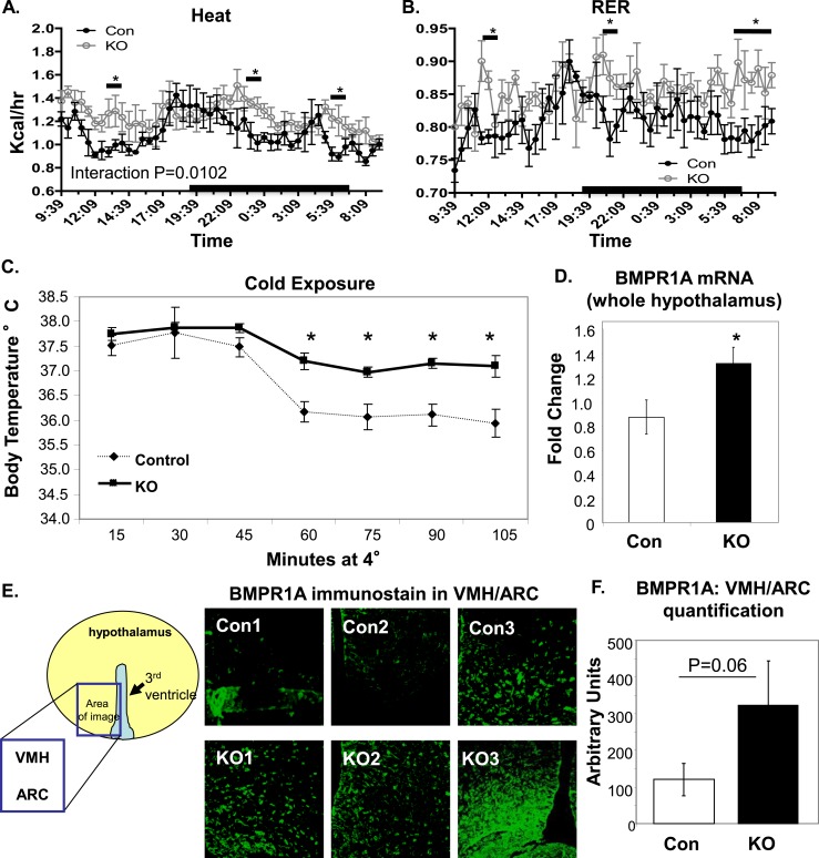
Figure 2.
Increased energy expenditure in POMC/BMPR1A-KO mice. (A and B) POMC/BMPR1A-KO mice had higher energy expenditure as measured in CLAMS while on a 45% HFD, including (A) transiently increased heat production (calculated from Vo2 and Vco2) and (B) RER. Interaction statistics gave a P value for heat of 0.0102 (significant), and for RER, it was 0.0888 (not significant). Mice undergoing CLAMS analysis were at the same body weight, but data were normalized to lean body mass as measured by DEXA (see Supplemental Material (49.5KB, doc) ), which was also unchanged. N = 6 mice per group; data presented as wave-forms (see Supplemental Material (49.5KB, doc) ). (C) During cold exposure after a 45% HFD, POMC/BMPR1A-KO mice were better able to regulate their body temperature than control mice, as measured by rectal temperature taken every 15 minutes at 4°C (N = 6 to 8 mice per group). (D−F) Despite POMC-neuron ablation, POMC/BMPR1A-KO mice expressed more total hypothalamic BMPR1A, as measured at the (D) mRNA and (E and F) protein levels. BMPR1A immunostaining in (E) indicates greater BMPR1A expression in multiple hypothalamic regions, including the ventromedial hypothalamus (VMH) and ARC. Fluorescent BMPR1A signal from (E) is quantified in (F) from three animals. Despite interindividual variation, there was a trend for increased BMPR1A in POMC/BMPR1A-KO hypothalami of all hypothalamic regions analyzed. The cartoons demonstrate where in the hypothalamus the confocal photomicrographs were taken. All animals were on a 45% HFD. *Indicates significance where P < 0.05. Error bars represent standard error of the mean. Con, control; RER, respiratory exchange ratio.

