

Abstract
It has recently been shown that cancer treatments such as radiation and hyperthermia, which have conventionally been viewed to have modest immune based anti-cancer effects, may, if used appropriately stimulate a significant and potentially effective local and systemic anti-cancer immune effect (abscopal effect) and improved prognosis. Using eight spontaneous canine cancers (2 oral melanoma, 3 oral amelioblastomas and 1 carcinomas), we have shown that hypofractionated radiation (6 x 6 Gy) and/or magnetic nanoparticle hyperthermia (2 X 43°C / 45 minutes) and/or an immunogenic virus-like nanoparticle (VLP, 2 x 200 μg) are capable of delivering a highly effective cancer treatment that includes an immunogenic component. Two tumors received all three therapeutic modalities, one tumor received radiation and hyperthermia, two tumors received radiation and VLP, and three tumors received only mNP hyperthermia. The treatment regimen is conducted over a 14-day period. All patients tolerated the treatments without complication and have had local and distant tumor responses that significantly exceed responses observed following conventional therapy (surgery and/or radiation). The results suggest that both hypofractionated radiation and hyperthermia have effective immune responses that are enhanced by the intratumoral VLP treatment. Molecular data from these tumors suggest Heat Shock Protein (HSP) 70/90, calreticulin and CD47 are targets that can be exploited to enhance the local and systemic (abscopal effect) immune potential of radiation and hyperthermia cancer treatment.
Keywords: hypo-fractionated radiation, hyperthermia, viral-like nanoparticle (VLP), cancer therapy, spontaneous canine cancer, abscopal effect
INTRODUCTION
Combinatorial therapy is currently accepted as the optimal approach to treating cancer. This includes modalities such as chemotherapy, surgery, hyperthermia, radiation, molecular targeting, and immunotherapy. The latter is rapidly becoming one of the most promising and important approaches. How these modalities are optimally combined to achieve the best outcome is a central question in translational cancer research. The ability of local cancer treatments to induce an effective systemic/metastatic cancer treatment is known as the abscopal effect. The abscopal effect of radiation therapy is a rare but well documented event. Although the mechanism is unclear most believe the abscopal effect to be immunologically based. The overall goal of this research is that specific radiation or hyperthermia doses/techniques have definable and reproducible antitumor immune responses that can be exploited through combination with immunoreactive agents, such as the viral-like nanoparticles we are using. Another way of looking at this situation is that combining radiation or hyperthermia with a local immunotherapy reagent creates an “in situ vaccination”. Essentially the radiation and immunomodulatory agent, such as VLP, stimulates tumor antigen and neoantigen presentation and systemic anti-tumor immune effect. The in situ vaccination approach is attractive because the cell stress and death caused by the radiation is likely to support stronger antitumor immune responses when the in situ vaccination modifies the tumor microenvironment to reverse local immunosuppression and support local and systemic antitumor immune responses. [1–8]
Radiation and the abscopal effect
The field of radiation therapy is becoming increasingly accepting of hypo-fractionated therapy. The primary reasons are four-fold: 1) few patient treatments, 2) larger fraction size is more cytotoxic to the tumor (but of course carries a greater risk of normal tissue damage), 3) greatly improved tumor imaging, treatment planning and beam management, and importantly 4) larger fraction sizes promoting a tumor cell killing mechanism that induces immune activation – a situation that is not appreciated with conventional small fraction size regimens.
While a combinatorial approach is most effective for cancer treatment, the new insights into tumor immunotherapy have not yet progressed to understanding combinations of immunotherapy with each other. Using spontaneous canine tumors in cancer therapy research is significant because the patients are similar to humans in size and environment, are genetically diverse, and develop cancer at an incidence and type that are similar to humans.
The abscopal effect has recently been well reviewed in multiple places (Grass, 2016). The main concepts are: that the effect is immune based and is due to the tendency of radiation therapy (RT) damage of tumors to create a more immunogenic tumor microenvironment than the immune suppressive environment that characterizes untreated tumors. Clearly, RT by itself is generally not sufficient to create an effective and reliable antitumor immunity (Siva, 2015). Studies report that the damage of RT alone recruits M2 type tissue repair macrophages that suppress adaptive immunity (Crittenden, 2014). The crucial aspect appears to be “immunogenic cell death” (ICD) that occurs when cells die in a manner that stimulates the immune system. ICD is characterized by a grouping of the following danger associated molecular signals (DAMPs): calreticulin expression on the cell surface, release of ATP, release of HMGB1 protein, and expression of type one interferons (IFN alpha and beta) (Grass, 2016). When the tumor environment is sufficiently immunogenic, tumor associated antigens (normal proteins expressed at the wrong level, wrong tissue or wrong time during development) and neoantigens (mutant proteins expressed by tumors due to accumulated mutations) are taken up by antigen presenting cells that go to the lymph nodes, present them to T cells and stimulate an adaptive immune response against tumor cells. That adaptive immune response not only impacts tumors near that lymph node but also can become a systemic response against the same tumor wherever it may occur. [9–15]
Hyperthermia and the abscopal effect
Although less well known and appreciated there is also documentation of therapeutic heat/hyperthermia induced abscopal effect. In one recent paper (Wang et al, 2014) the abscopal effect it was demonstrated in a Walker-256 carcinoma using magnetic seed-mediated hyperthermia at a higher temperatures (42°C–46°C for 30 minutes and 50 – 55°C for 10 minutes). The abscopal endpoint effect was regression of a non-heated tumor (contralateral) following heat treatment of one tumor in rat with bilateral tumor growth. It was also noted that the levels of CD4+ and CD8+ lymphocytes and IFN-γ and IL-2 cytokines were significantly increased in the blood of hyperthermia groups compared with those of the three control groups; the increase in CD8+ cells was higher than that of the CD4+ cells.
It is important to emphasize that although there is clearly a relationship between local immune control of a treated tumor and systemic resistance to untreated tumors, that relationship is not manifested in the best local control manifesting in the best systemic antitumor immune response. We have shown that hyperthermia treatment of an established tumor can generate systemic resistance to rechallenge (Toraya-Brown, 2014), however this was a minimal heat dose that did not eliminate the treated tumor. [16–27]
MATERIALS and METHODS
2.1 Spontaneous Canine Tumor Models
All dogs studied and proposed for study are companion pets that have been referred from regional veterinarians. Owners must consent to the study parameters, follow-up schedule, and a post mortem examination for the patient. Inclusion in the study requires the dog be healthy (no serious medical condition or metastatic disease), have an appropriate biopsy-based diagnosis and CT scan of the tumor site. In this report we include six canine patients; two oral melanoma, three oral amelioblastomas and one non-oral carcinomas. During a 14-day treatment regimen; the dogs received hypofractionated radiation (6 x 6 Gy) and/or magnetic nanoparticle hyperthermia (2 X 43°C / 45 minutes) and/or an immunogenic-VLP (2 x 200 μg). Two tumors received all three therapeutic modalities, one tumor received radiation and hyperthermia, two tumors received radiation and VLP, and three treatments included only mNP hyperthermia. In brief, acanthomatous amelioblastomas (AA) are locally aggressive tumors that arise from periodontal epithelium. Acanthomatous amelioblastomas invade bone and recur following excisional surgery or other therapies, mandibulectomy or hemi-mandibulectomy are the only curative therapies. Oral melanoma is a common and lethal cancer in dogs. Untreated dogs rarely live more than 6-months post diagnosis. Surgery and/or radiation can extend post treatment to 12–18 months. The other tumors treated in this series are invasive carcinomas of the digit and breast. Both tumor types have the potential to be metastatic and rapidly fatal unless treated early with highly aggressive surgery and/or radiation. [28 – 31] The radiation-hyperthermia - immunomodulation study is designed t o monitor tumor response and multiple immune parameters (tumor biopsies and at multiple immune cell, hematology and cytokine markers) over time following treatment. The length of survival and time to metastasis is a critical endpoint for us, however it is the not primary endpoint of this study. Our canine treatments consist of 6 x 6 Gy radiation treatments and 4 x 200 ug intra-tumoral VLP treatments. The treatments are given concurrently, every other day for two weeks. Tumor biopsies and blood samples are taken, before treatment, one week into treatment, immediately following treatment, one week following treatment and at 2 – 4 week intervals thereafter for the first six months post therapy. Dogs are intubated and under surgical plane anesthesia (with continuous intravenous access) for all procedures. All procedures are approved by the Dartmouth College Institutional Animal Care and Use Committee (IACUC) and adhere to all USDA, AAALAC, and institutional guidelines.
2.2 Iron Oxide Nanoparticle (mNP) / Alternating Magnetic Field Hyperthermia (AMF) Technology and Treatment
In these studies, we used bionised nanoferrite (BNF) iron oxide nanoparticles (product number 10-00-801 from Micromod Partikeltechnologie GmBH, Rostock, Germany). These iron oxide nanoparticles (mNP) are composed of a hematite core surrounded with crystals of an average diameter of approximately 20 nm (total core diameter approximately 40 nm) and a hydroxyethyl starch or dextran shell to a final average hydrodynamic diameter of 117 nm. The mNP were concentrated to a final particle concentration of 44 mg/ml and iron concentration of 28 mg/ml. A pancake coil with an inner diameter of 6 cm and an outer diameter of 12 cm composed of hollow copper tubing was used to generate the alternating magnetic field (AMF). The AMF coil was powered at variable kHz by a TIG 10/300 generator (Huttinger Elektronik GmbH, Freiburg, Germany). The AMF coil and generator were cooled by a chiller (Tek-Temp Instruments, Croydon PA) operating at 20°C and four gallons per minute.
In the images above, figure 1a shows the AMF treatment facility, figure 1b is a top down view of the pancake coil used to treat most oral tumors. Figure 1c is a temperature verified model of the alternating magnetic field delivered at 150 kHz and 450 Oe. Figure 1d demonstrates a pig breast being treated on the pancake coil. Clinically each heated tumor received a mNP injection of 5 mg/gm tumor and two 43°C / 60 minutes AMF exposures of 150 kHz/450 Oe (average thermal dose) Tumors are treated twice over a two week period (once per week).
Figure 1.

Figures 2a and 2b, above, demonstrate the clinical appearance and CT scan image of an acanthomatous amelioblastoma, before treatment. Tumor invasion of the bone (degeneration) can be seen in the left anterior mandible on CT scan. Figure 2c demonstrates injection of the mNP into the tumor site (treatment #2). Figure 2d demonstrates a tumor free site, 30 months post-treatment.
Figure 2.

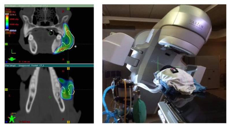
2.3 Clinical Irradiation
All dogs in this series receive a pretreatment CT and/or MRI scan and 3-D radiation treatment plan (figure 3). For a dog receiving tumor irradiation, this scan is used to generate a state-of-the-art 3-D radiation treatment plan for optimal safe and accurate delivery of the radiation. Eclipse RT treatment planning software and Varian 2100C or Truebeam linear accelerator are used with a mean radiation dose of 6 Gy delivered to the tumor margin (360°). Radiation treatments are delivered three times per week (M, W, F) for two weeks (6 total treatments). The images below demonstrate the CT scan and integrated 3-D radiation plan and the dog being irradiated on a Varian Truebeam irradiator.
Figure 3.
2.4 Immunogenic Viral – Like Nanoparticles
The viral-like nanoparticle platform we have used in these treatments is a modified version of the Cowpea Mosaic Virus (CPMV). The RNA has been removed from the VLP rendering it non-infectious and the protein coat engineered to stimulate a greater immune response. This is done through parallel expression of a viral protease (Pro) and the VP60 precursor protein encoding large and small coat proteins. While similar in protein organization, the eCPMV (empty) platform nanotechnology offers key advantages over the naturally occurring Cowpea mosaic virus such as it is not infectious and the empty interior cavity can be engineered to carry medical payload.
Figure 4a is a computer rendition of the CPMV (approximate 30nm diameter). Transmission electron micrographs (taken by the Hoopes laboratory group) in figures 4b and 4c demonstrate the association of 30 nm viral-like nanoparticles (CPMV) and B16 F10 murine melanoma cells. The cells were harvested and processed for TEM six hours following the addition of the VLP to cells/cell culture media. Figure 4b demonstrates VLP aggregate adjacent (possibly attached) to the outer plasma membrane of a B16 melanoma cell. Figure 4c demonstrates an intracellular vesicle containing VLP.
Figure 4.

2.5 Radiation, mNP hyperthermia and VLP
As discussed previously the radiation and mNP hyperthermia treatments are given over a two-week period. Radiation is delivered 3 x 6 Gy / week x 2 weeks (total of 36 Gy), mNP hypertheremia is delivered 1 x 60 CEM x 2 weeks. VLP are delivered intratumorally 2 times/week x 2 weeks (4 treatments). Each 200 μg (500 μl) intratumoral VLP injection is placed in three tumor sites.
Figures 5 a–e below are a sequential demonstration of the radiation-mNP hyperthermia-VLP treatment of an invasive caricinoma encompassing an entire canine digit (Figure 5a). Figures 5b,c demonstrate the intratumoral delivery of iron oxide nanoparticle to the tumor and heat activation using the helical AMF coil (43°C / 60 min x 2 treatment over a two week period). Figure 5d demonstrates intratumoral delivery of the VLP (200 μg x 4 treatment over a two week period). Figure 5e is an H&E photomicrograph of biopsies tissue, from the tumor region, demonstrating post-treatment fibrosis and chronic inflammation, but no tumor cells six months post treatment.
Figure 5.

2.6 Pathology and Immunopathology
All patients/tumors included in this study have a biopsy (pathology) confirmed diagnosis prior to treatment. Although variability in early cases limited the uniformity of mid and post-treatment tumor and peri-tumor tissue acquisition we have consistently acquired and blood sampled at one mid-treatment and three post-treatment time endpoints (two-three week intervals). These samples are used for qualitative and quantitative morphology and immunohistochemistry assessment of the tumor and peri-tumor direct and immune based treatment responses. The photomicrograph series below (Figures 6a–d) demonstrates this situation. Figure 6a is a pretreatment oral melanoma. The tumor is morphologically composed almost entirely of melanin containing cancerous melanocytes and macrophages. There are occasional spindle cells with dense nuclei (fibroblasts / fibrocytes) and small densely nucleated cells / lymphocytes). Figure 6b is biopsy tissue taken 21 days post radiation –VLP treatment (tumor margin). The sample is morphologically characterized by a marked reduction in melanoma cancer cells (these cell may have lethal mitotic damage, but appear morphologically viable) and very a significant increase in the number of tumor macrophages and lymphocytes. Figures 6c and 6d are high magnification H&E and IHC (CD11c) photomicrographs, respectively, showing the preponderance of CD11c tagged lymphocytes. Morphologically, the lymphocyte population is compatible with an abundance of B-cell and a lesser number of T-cells. Although CD11c is typically viewed as a dendritic cell (DC) marker, new information suggests it is also a marker of activated lymphocytes in some immune situations.
Figure 6.

2.7 Canine Tumor Propagation
In an attempt to better understand the translational therapeutic and immunobiology of the canine cancers, we are attempting to propagate, in cell culture, as many canine tumors as possible. Figures 7a and 7b below demonstrate clinical growth, and associated microscopic tumor pathology of a canine melanoma growing in an immunosuppressed (NSG) mouse. The histomorphological appearance of this tumor is very similar to original canine tumor. This allows us to perform wide variety biology, immunology and therapeutic assessments, on canine tumors, that would not otherwise be available.
Figure 7.

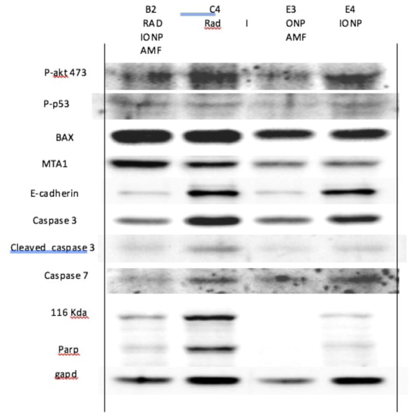
2.8 Assessment of Pathobiological and Therapeutic Markers
In addition to the qualitative and quantitative histopathological and immunohistochemistry assessment described above, western blot assessments of tumor and peri-tumor tissues are used to identify and semi-quantify potentially important therapeutic and immune proteins. Preliminary results, figure 8 below, suggests apoptosis markers (caspases), heat shock proteins, calreticulin, CD47 and MTA1 are potentially important proteins in the radiation-hyperthermia-abscopal/immune-pathway.
Figure 8.
RESULTS
In this preliminary assessment of hypo-fractionated radiation-hyperthermia-viral immunotherapy treatment of spontaneous canine tumors we have shown that the therapeutic regimen we have used not only provides therapeutic benefits that exceed conventional therapies, but strongly suggest a meaningful systemic anti-tumor immune-based activity. Radiation treatments were 6 x 6 Gy, mNP-AMF hyperthermia treatments were based on intratumoral mNP at dose of 5mg/gram of tumor and two mNP-AMF hyperthermia exposures 43C° / 60 min (CEM 60), VLP treatments consisted of four intratumoral injections (200 μg each); all treatment were delivered, concurrently when applicable, over a two week period.
Magnetic nanoparticle hyperthermia (n=3): Three tumors were treated in this group, two amelioblastomas and one oral malignant melanoma. Both amelioblastoma cases have/had complete tumor remission lasting greater than 4 years. One of the amelioblastoma patients remains in remission while the second dog died of age related causes approximately 5 years post treatment. The oral malignant melanoma died of local recurrent and metastatic melanoma 45 months post therapy.
Radiation (6 x 6 Gy) and magnetic nanoparticle hyperthermia (n=1): One amelioblastoma was treated in this group. This dog died of age related causes with complete tumor remission 58 months post treatment.
Radiation (6 x 6 Gy), magnetic nanoparticle hyperthermia, and intratumoral VLP (n=2): One case is a oral malignant melanoma in remission at 18 months, the second case is an invasive squamous cell carcinoma of the digit, this tumor was in remission at the time of age-related death, 22 months post treatment.
CONCLUSIONS
In this preliminary study six spontaneous canine cancers (2 oral melanoma, 3 oral acanthomatous amelioblastoma, 1 non-oral carcinomas), we have shown that hypofractionated radiation (6 x 6 Gy) and/or magnetic nanoparticle hyperthermia (2 X 43°C / 60 minutes) and/or an engineered immunogenic virus-like nanoparticle (2 x 200 μg) are capable of delivering a highly effect cancer treatment, that includes a tumor immunogenic component. While this study is too limited in size, scope and statistics to make conclusive determinations regarding the treatment efficacy and abscopal effect contributions of each modality for individual tumor types. However it can be clearly stated that these cancer treatment modalities all have significant independent therapeutic responses, as proven by prolonged disease free survival, and that this effect is markedly enhanced through their combined use. The study also demonstrates that a clinical anti-tumor immune response, resulting from these treatment modalities, is likely to like to be a major contributor to the overall therapeutic efficacy.
References
- 1.Lizotte PH, Wen AM, Sheen MR, Fields J, Rojanasopondist P, Steinmetz NF, et al. In situ vaccination with cowpea mosaic virus nanoparticles suppresses metastatic cancer. Nat Nanotechnol. 2016;11(3):295–303. doi: 10.1038/nnano.2015.292. [DOI] [PMC free article] [PubMed] [Google Scholar]
- 2.Varn FS, Andrews EH, Mullins DW, Cheng C. Integrative analysis of breast cancer reveals prognostic haematopoietic activity and patient-specific immune response profiles. Nat Commun. 2016;7:10248. doi: 10.1038/ncomms10248. [DOI] [PMC free article] [PubMed] [Google Scholar]
- 3.Whiteside TL. Disarming suppressor cells to improve immunotherapy. Cancer Immunother. 2012;61(2):283–8. doi: 10.1007/s00262-011-1171-7. [DOI] [PMC free article] [PubMed] [Google Scholar]
- 4.Hammerich L, Binder A, Brody JD. In situ vaccination: Cancer immunotherapy both personalized and off-the-shelf. Mol Oncol. 2015;9(10):1966–81. doi: 10.1016/j.molonc.2015.10.016. [DOI] [PMC free article] [PubMed] [Google Scholar]
- 5.Lee KL, Twyman RM, Fiering S, Steinmetz NF. Virus-based nanoparticles as platform technologies for modern vaccines. Wiley Interdiscip Rev Nanomed Nanobiotechnol. 2016;8(4):554–78. doi: 10.1002/wnan.1383. [DOI] [PMC free article] [PubMed] [Google Scholar]
- 6.Lebel ME, Chartrand K, Tarrab E, Savard P, Leclerc D, Lamarre A. Potentiating Cancer Immunotherapy Using Papaya Mosaic Virus-Derived Nanoparticles. Nano Lett. 2016;16(3):1826–32. doi: 10.1021/acs.nanolett.5b04877. [DOI] [PubMed] [Google Scholar]
- 7.Baird JR, Byrne KT, Lizotte PH, Toraya-Brown S, Scarlett UK, Alexander MP, et al. Immune-mediated regression of established B16F10 melanoma by intratumoral injection of attenuated Toxoplasma gondii protects against rechallenge. J Immunol. 2013;190(1):469–78. doi: 10.4049/jimmunol.1201209. [DOI] [PMC free article] [PubMed] [Google Scholar]
- 8.Baird JR, Fox BA, Sanders KL, Lizotte PH, Cubillos-Ruiz JR, Scarlett UK, et al. Avirulent Toxoplagondii generates therapeutic antitumor immunity by reversing immunosuppression in the ovarian cancer microenvironment. Cancer Res. 2013;73(13):3842–51. doi: 10.1158/0008-5472.CAN-12-1974. [DOI] [PMC free article] [PubMed] [Google Scholar]
- 9.Hiniker SM, Chen DS, Reddy S, Chang DT, Jones JC, Mollick JA, et al. A systemic complete responseof metastatic melanoma to local radiation and immunotherapy. Transl Oncol. 2012;5(6):404–7. doi: 10.1593/tlo.12280. [DOI] [PMC free article] [PubMed] [Google Scholar]
- 10.Barker CA, Postow MA. Combinations of radiation therapy and immunotherapy for melanoma: a reviewof clinical outcomes. Int J Radiat Oncol Biol Phys. 2014;88(5):986–975. doi: 10.1016/j.ijrobp.2013.08.035. [DOI] [PMC free article] [PubMed] [Google Scholar]
- 11.Grass GD, Krishna N, Kim S. The immune mechanisms of abscopal effect in radiation therapy. Curr Probl Cancer. 2016;40(1):10–24. doi: 10.1016/j.currproblcancer.2015.10.003. [DOI] [PubMed] [Google Scholar]; Siva S, MacManus MP, Martin RF, Martin OA. Abscopal effects of radiation therapy: a clinical reviewfor the radiobiologist. Cancer Lett. 2015;356(1):82–90. doi: 10.1016/j.canlet.2013.09.018. [DOI] [PubMed] [Google Scholar]
- 12.Crittenden MR, Savage T, Cottam B, Baird J, Rodriguez PC, Newell P, et al. Expression of arginase I in myeloid cells limits control of residual disease after radiation therapy of tumors in mice. Radiat Res. 2014;182(2):182–90. doi: 10.1667/RR13493.1. [DOI] [PubMed] [Google Scholar]
- 13.Reynders K, Illidge T, Siva S, Chang JY, De Ruysscher D. The abscopal effect of local radiotherapy: using immunotherapy to make a rare event clinically relevant. Cancer Treat Rev. 2015;41(6):503–10. doi: 10.1016/j.ctrv.2015.03.011. [DOI] [PMC free article] [PubMed] [Google Scholar]
- 14.Deng L, Liang H, Xu M, Yang X, Burnette B, Arina A, et al. STING-Dependent Cytosolic DNA SensingPromotes Radiation-Induced Type I Interferon-Dependent Antitumor Immunity in Immunogenic Tumors. Immunity. 2014;41(5):843–52. doi: 10.1016/j.immuni.2014.10.019. [DOI] [PMC free article] [PubMed] [Google Scholar]
- 15.Baird JR, Friedman D, Cottam B, Dubensky TW, Jr, Kanne DB, Bambina S, et al. Radiotherapy Combined with Novel STING-Targeting Oligonucleotides Results in Regression of Established Tumors. Cancer Res. 2016;76(1):50–61. doi: 10.1158/0008-5472.CAN-14-3619. [DOI] [PMC free article] [PubMed] [Google Scholar]
- 16.Jordan A, et al. The effect of thermotherapy using magnetic nanoparticles on rat malignant glioma. Journal of Neuro-Oncology. 2006;78(1):7–14. doi: 10.1007/s11060-005-9059-z. [DOI] [PubMed] [Google Scholar]
- 17.Cassim SM, Hoopes PJ, et al. Iron oxide nanoparticle hyperthermia and radiation cancer treatment. Proc SPIE. 2009;7181:71810O. doi: 10.1117/12.810035. [DOI] [PMC free article] [PubMed] [Google Scholar]
- 18.Maier-Hauff K, et al. Intracranial Thermotherapy using Magnetic Nanoparticles Combined with External Beam Radiotherapy: Results of a Feasibility Study on Patients with Glioblastoma Multiforme. Journal of Neuro-Oncology. 2006;81(1):53–60. doi: 10.1007/s11060-006-9195-0. [DOI] [PubMed] [Google Scholar]
- 19.Johannsen M, et al. Morbidity and quality of life during thermotherapy using magnetic nanoparticles in locally recurrent prostate cancer: Results of a prospective phase I trial. International Journal of Hyperthermia. 2007;23(3):315–323. doi: 10.1080/02656730601175479. [DOI] [PubMed] [Google Scholar]
- 20.Giustini AJ, Hoopes PJ, et al. Magnetic Nanoparticle Hyperthermia in Cancer Treatment. Nano LIFE. 2010;1(1–2):17–32. doi: 10.1142/S1793984410000067. [DOI] [PMC free article] [PubMed] [Google Scholar]
- 21.Zhang G, Liao Y, Baker I. Surface engineering of core/shell iron/iron oxide nanoparticles from microemulsions for hyperthermia. Materials Science and Engineering: C. 2010;30(1):92–97. doi: 10.1016/j.msec.2009.09.003. [DOI] [PMC free article] [PubMed] [Google Scholar]
- 22.Dennis CL, et al. Nearly complete regression of tumors via collective behavior of magnetic nanoparticles in hyperthermia. Nanotechnology. 2009;20:395103. doi: 10.1088/0957-4484/20/39/395103. [DOI] [PMC free article] [PubMed] [Google Scholar]
- 23.Sapareto SA, Dewey WC. Thermal dose determination in cancer therapy. International journal of radiation oncology, biology, physics. 1984;10(6):787–800. doi: 10.1016/0360-3016(84)90379-1. [DOI] [PubMed] [Google Scholar]
- 24.Toraya-Brown S, Sheen MR, Baird JR, Demidenko E, Hoopes PJ, Fiering SN, et al. Local hyperthermia treatment of tumors induces CD8(+) T cell-mediated resistance against distal and secondary tumors. Nanomedicine : nanotechnology, biology, and medicine. 2014;10(6):1273–85. doi: 10.1016/j.nano.2014.01.011. [DOI] [PMC free article] [PubMed] [Google Scholar]
- 25.Toraya-Brown S, Fiering S. Local tumour hyperthermia as immunotherapy for metastatic cancer. International journal of hyperthermia : the official journal of European Society for Hyperthermic Oncology, North American Hyperthermia Group. 2014;30(8):531–9. doi: 10.3109/02656736.2014.968640. [DOI] [PMC free article] [PubMed] [Google Scholar]
- 26.Toraya-Brown S, Sheen MR, Baird JR, Barry S, Demidenko E, Turk MJ, et al. Phagocytes mediate targeting of iron oxide nanoparticles to tumors for cancer therapy. Integrative biology : quantitative biosciences from nano to macro. 2013;5(1):159–71. doi: 10.1039/c2ib20180a. [DOI] [PMC free article] [PubMed] [Google Scholar]
- 27.Wang, et al. Abscopal antitumor immune effects of magnet-mediated hyperthermia at a high therapeutic temperature on Walker-256 carcinosarcomas in rats. Oncol Lett. 2014 Mar;7(3):764–770. doi: 10.3892/ol.2014.1803. [DOI] [PMC free article] [PubMed] [Google Scholar]
- 28.Bergman PJ. Canine oral melanoma. Clin Tech Small Anim Pract. 2007;22(2):55–60. doi: 10.1053/j.ctsap.2007.03.004. [DOI] [PubMed] [Google Scholar]
- 29.Gardner HL, Fenger JM, London CA. Dogs as a Model for Cancer. Annu Rev Anim Biosci. 2016;4:199–222. doi: 10.1146/annurev-animal-022114-110911. [DOI] [PMC free article] [PubMed] [Google Scholar]
- 30.Williams LE, Packer RA. Association between lymph node size and metastasis in dogs with oral malignant melanoma: 100 cases (1987–2001) J Am Vet Med Assoc. 2003;222(9):1234–6. doi: 10.2460/javma.2003.222.1234. [DOI] [PubMed] [Google Scholar]
- 31.Proulx DR, Ruslander DM, Dodge RK, Hauck ML, Williams LE, Horn B, et al. A retrospective analysis of 140 dogs with oral melanoma treated with external beam radiation. Vet Radiol Ultrasound. 2003;44(3):352–9. doi: 10.1111/j.1740-8261.2003.tb00468.x. [DOI] [PubMed] [Google Scholar]