Abstract
Background:
Comminuted fractures happen frequently due to traumas. Fixation without opening the fracture site, known as minimally invasive plate osteosynthesis (MIPO), has recently become prevalent. This study has been designed to assess the outcomes of this treatment for tibial and femoral comminuted fractures.
Methods:
A total of 60 patients with comminuted femoral or tibial fractures were operated with MIPO method in this cross-sectional study at Alzahra university hospital in 2015. Eleven patients were excluded due to lack of adequate follow-ups. Patients’data including union time; infection in the fractured site; hip and knee range of motion; and any malunion or deformities like limb length discrepancy were collected after the surgery in every session.
Results:
Among 32 femoral and 17 tibial fractures, union was completed in48 patients, while only one patient with femoral fracture had nonunion. The mean union time was 18.57±2.42 weeks. Femur fractures healed faster than tibia (17.76±2.36 compared to 19±2.37 weeks, respectively, P=0.09). None of the patients suffered from infections or fistula. The range of motion in hip and knee remained intact in approximately all patients. Malunion happened in 3 patients; 100 internal rotation in 1 patient; and 1cm limb shortening in 2 patients.
Conclusion:
According to the result of this study, MIPO is a simple and effective method of fixation with a high rate of union as well as minimal complications for comminuted fractures of long bones. Infection is rare, and malunion or any deformity is infrequent. MIPO appears to be a promising and safe treatment alternative for comminuted fractures.
Keywords: Comminuted fractures, Femur, Minimally invasive plate osteosynthesis, MIPO, Plate osteosynthesis, Tibia
Introduction
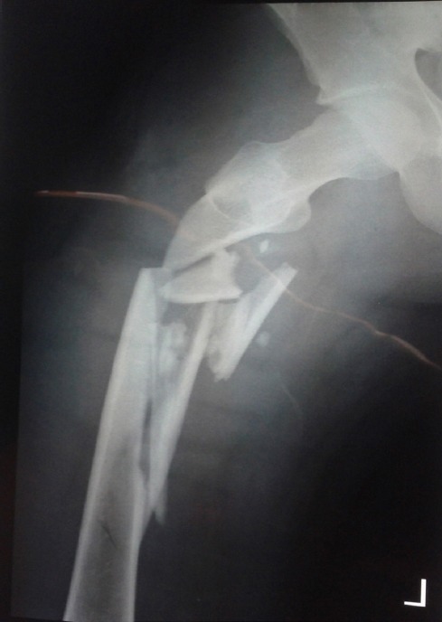
Comminuted fracture is one of the most frequent trauma related fractures in which severe damage to the adjacent soft tissue is accompanying the segmented bone. The long bone breaks into the proximal and distal major pieces along with the small fragments disseminated in the fractured area [Figure 1]. Ischemia may also occur in the region soon after a comminuted fracture, making its management quite challenging. The conventional treatments like internal/external fixation, and pin and plaster have high rate of complications such as infections, malunion, and nonunion (1-3).
Figure 1.

Comminuted femur fracture before the surgery.
Although intramedullary nailing (IMN) is the ideal approach, however, this method is not free of complication either and is not applicable where the fracture is adjacent to joints or when there is intra-articular component with extension to the metaphyseal-diaphyseal junction. This method would not be ideal in circumstances such as: diaphysis fractures extended to the metaphysis or joint, children having epiphyseal growth plates (4-10).
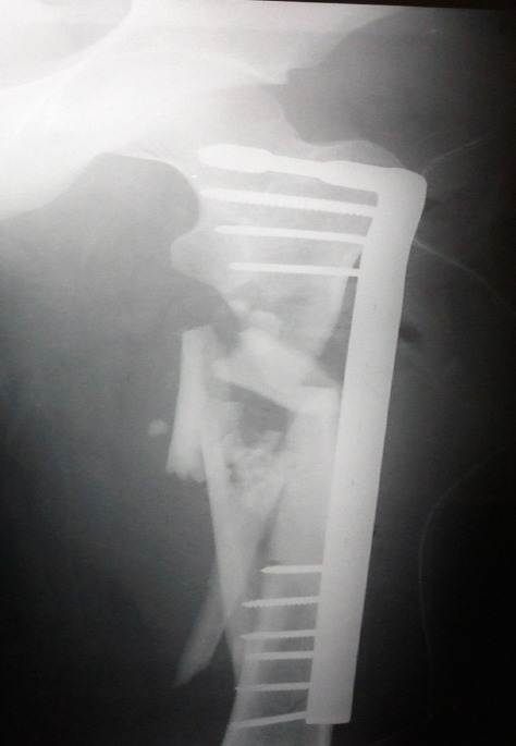
Minimally invasive plate osteosynthesis (MIPO) or biological plating and bridging platehasrecently become more popular but its outcomes for comminuted fractures have not been thoroughly investigated (1-9). The bridging plate method is named due to the action of the plate as a bridge between the proximal and distal bones [Figure 2] (9). This provides stability while preserving the vasculature integrity of the soft tissue (1-3).
Figure 2.

Comminuted femur fracture after the MIPO surgery.
In contrast to conventional methods in which the periosteum, which is enriched with blood supply for bone healing, had to be detached and the hematoma had to be removed to fix the fractured pieces; however both the periosteum and hematoma remain intact in MIPO to enhance union (9, 11, 12). MIPO is the reasonable choice when the fractured site is near the joints (juxtaarticular, periarticular, or paraarticular). In addition, in contrast with intramedullary nailing, MIPO does not interfere with intramedullary blood circulation (1, 2, 13-15).
Shorter surgery time, less blood loss, unharmed soft tissue, restricted inflammation in fractured area, decreased infection rate, accelerated union due to little movements in small bones, possibility of early mobilization of the limb, decreased incidence of other fractures in the same area after plate removal, and decreased need for bone grafting are the advantages of MIPO (16-20).
Higher rates of wound infection with big fracture areas and wound exposures are the potential disadvantages of MIPO. Also, although non-union is rare in this method, bone graft might be needed with severe damages. Plate breaking after MIPO is possible due to delayed union as anatomical obstacles like ruptured muscles in fractured site can pull and separate the fractured bones (15-20).
In this study we have aimed to evaluate the outcomes of MIPO method in tibial and femoral comminuted fractures while there is no clear and documented study available on the long bones.
Materials and Methods
A total of 60 patients were enrolled in this cross-sectional study at Alzahra university hospital between August 2014 and September 2015. The approval to conduct this study was provided by the Ethics Department of Isfahan University of Medical Science. The mean age ofthe patients was 31±6.2 years; 63.3% were men (n=38) and 36.6% were women (n=22). The exclusion criteria were including: metabolic disease leading to fracture; pathological fracture; and refusal to participate in the study. After acquiring the medical history, thorough physical examination was conducted and radiographs were evaluated. Postoperatively, patients were discharged after 3 days unless there were any complications. Patients were asked to come back every 4 weeks for follow-up visitsuntil union was fully achieved. This was done for 6 consecutive months during which the checklists were filled in and the union, range of motion in hip and knee joints, and the leg length were evaluated.
Surgical Technique
A consonant team of surgeons conducted all the procedures. The femoral fractures were fixed by dynamic hip screws (DHSs), dynamic compression screws (DCSs), and dynamic compression plates (DCPs), while the tibial fractures were fixed by DCPs and T-Plates. Traction was applied prior to surgery to reduce the fracture. Two minimal incisions (4-5 cm) were made away from the fractured area at the proximal and distal sites. The plate passed through these incisions, producing a subcutaneous tunnel, going underneath the muscles and above the periosteum while bridging the fractured area with minimal invasion to fractured pieces and adjacent soft tissue. Reaching to the distal part, we reduced the comminuted fracture with traction and no additional damage. A longer skin incision had to be made to avoid disrupting muscles, arteries, or nerves [Figure 2]. Generous traction was needed after fixing the plate in proximal to avoid length discrepancy by assessing the distance between Anterior Superior Iliac Spine (ASIS) and medial malleolus and comparing the two limbs with each other.
Open reduction and internal fixation (ORIF) was used in cases of plateau, metaphysis, and diaphysis fractures; through letting the plate to pass from metaphysis to diaphysis. Gentle approach was crucial in these fractures to minimize the risk of osteoarthritis in future. Knee or hip motion began on the 2nd day postoperatively and the patients were mobilized 5 days later with walker or crutch. A checklist consisting of union time, malunion and/or nonunion, infection, limb shortening, and limitation of movements in proximal and distal joints was filled in for postoperative patients. Foot-Thigh Angles in both legs were compared after the procedure to determine rotations in the fractured leg hip and knee flexion/extension were measured by a goniometer. Data analysis was performed using IBM SPSS version 22.0 (SPSS Inc., Chicago, IL, USA).
Results
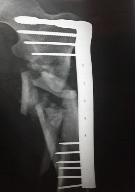
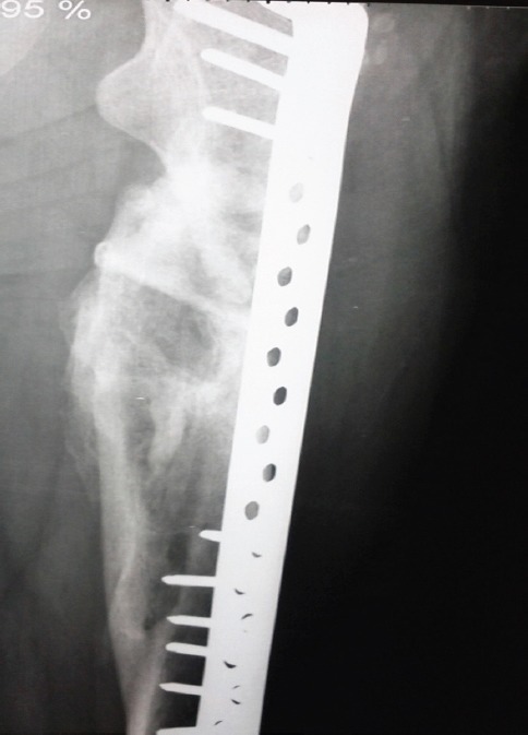
Eleven patients regretted to continue the follow-up visits. Among the rest of 49 tibial and femoral fracture cases, 32 (65.3%) patients were male and 17 (34.7%) were female with an average age of 31.56±6.41 and 30.76±5.89, respectively. The patient groups in terms of age and sex were statistically homogeneous (P=0.67). Seventeen (34.7%) patients had tibial fracture and 32 (65.3%) patients had femoral fracture with average age of 30.41±5.66 and 31.75±6.48, respectively. No significant relationship was found between the age and site of fracture (P=0.48). In tibial fracture group, there were 8 (47.1%) men and 9 (52.9%) women. In addition, 24 (75%) men and 8 (25%) women had femoral fractures, however, no significant relationship was found between the sex and site of fracture (P=0.05). Complete union within the duration of follow-ups was achieved in 48 (98%) patients, however, bone graft had to be used inonly 1 (2%) patient with femur fracture who was complicated with additional surgeries but finally achieved complete union after 9 months. The overall mean union time in these 49 patients was 18.57±2.42 weeks. Union in femoral fractures occurred at 17.76±2.36 weeks and union in tibial fractures occurred at 19±2.37 weeks [Figures 3; 4], however, no significant relationship was found between the union time and the site of fracture (P=0.09) and sex (P=0.93) [Table 1]. None of our patients suffered from wound infections or fistula.
Figure 3.

One month post-surgery, partial union.
Figure 4.

Six months post-surgery, full union.
Table 1.
Relationship between site of fracture, sex and age with mean union time
| Union Time (weeks) | P | ||
|---|---|---|---|
| Site of fracture | Tibia | 17.76±2.36 | 0.09 |
| Femur | 19±2.37 | ||
| Sex | Male | 18.59±2.49 | 0.91 |
| Female | 18.53±2.35 | ||
| Age group (year) | <25 | 18.83±2.4 | 0.83 |
| 25-30 | 18.07±2.58 | ||
| 30-35 | 18.58±2.02 | ||
| 35-40 | 19.15±2.27 | ||
| 40> | 18±4.58 | ||
| Data are presented as mean ± standard deviation. | 18.57±2.42 | ||
The average hip flexion after the procedure was 130.55°±9.44. Three (6.1%) patients with femur fracture had 300 restriction in range of motion. All patients with tibial fractures had intact hip flexion [Table 2]. The average knee flexion was 124.330±8.630. Knee range of motion in femoral fractures was significantly higher than the tibial fractures (P=0.001) [Table 3]. Two (4.1%) patients had restriction in knee flexion that resolved with physiotherapy. None of the patients had restriction in hip or knee extension. Dorsiflexion and plantar flexion of feet remained intact in all patients. Tibial fractures did not end up with malunion in patients treated with MIPO and only one patient with femur fracture showed 100 internal rotation. Two patients with femur fractures had a negligible 1 cm limb shortening, and none of our patients with tibial fractures experienced limb length discrepancy.
Table 2.
Relationship between hip flexion range of motion with site of fracture, sex and age
| Hip flexion range of motion (degrees) | P | ||
|---|---|---|---|
| Site of fracture | Tibia | 130.55±9.44 | <0.001 |
| Femur | 123.53±11.15 | ||
| Sex | Male | 132.09±8.72 | 0.12 |
| Female | 127.65±10.33 | ||
| Age group (year) | <25 | 128.33±14.38 | 0.76 |
| 25-30 | 131.47±9.78 | ||
| 30-35 | 128.33±9.37 | ||
| 35-40 | 131.54±8 | ||
| 40> | 135±0 | ||
| Data are presented as mean ± standard deviation. | 130.55±9.44 | ||
Table 3.
Relationship between knee flexion range of motion with site of fracture, sex and age
| Knee flexion range of motion (degrees) | P | ||
|---|---|---|---|
| Site of fracture | Tibia | 119.12±9.72 | 0.001 |
| Femur | 127.09±6.61 | ||
| Sex | Male | 124.75±8.83 | 0.64 |
| Female | 123.53±8.43 | ||
| Age group(year) | <25 | 124.19±7.36 | 0.96 |
| 25-30 | 123.8±10.17 | ||
| 30-35 | 123.33±9.37 | ||
| 35-40 | 125.38±8.03 | ||
| 40> | 126.67±5.77 | ||
| Data are presented as mean ± standard deviation | 124.33± 8.63 | ||
Discussion
Classic fixation, biological fixation, intramedullary nailing, and use of pins and plaster and various external fixators are well-known methods of treatment for comminuted fractures; however, these methods have high rate of complications, such as infections and delayed union (1, 2, 10). In this study, we showed that using this method brings shorter healing time and higher success rate in comminuted fractures of the long bones. The mean union time was acceptable in our patients underwent MIPO. The infection and fistula formation rate was zero and the rate of any malunion or deformities like limb length discrepancy, rotations or restriction in movements was very low.
The limitations in our study were: the cross-sectional design; relatively small sample size; data collection from one center; and hard follow. To be further tested, randomized clinical trials should be designed for this method to compare the results thoroughly.
Many of the previous studies also showed the effectiveness of MIPO in tibial and femoral fractures. According to the radiographic imaging, MIPO or bridging plate as an indirect method has shown to make callous formation in long bone fractures happen 3-4 weeks earlier than direct methods (4-6). The mean femur union time in children has also been shown to be 12.4 weeks with MIPO (21). Keeping the vessels safe in MIPO has been demonstrated as one of the advantages that result in afaster union in contrast with the conventional fixation methods (20-25).
Severe fractures like comminuted long bones are susceptible to infection, but, the rate of infection in the site of injuries would be decreased with MIPO method (23-30).
If the impact force hit the bone horizontally or obliquely, the possibility of deformity after treatment is higher than when the bone is hit directly and make a comminuted fracture, because callous formation in second events happens faster and limb shortening become less probable (4, 15, 24, 30-33). As noted previously, the mean union time can be decreased as low as 4 weeks in MIPO in contrast with the conventional methods like intramedullary nailing (25).
In a previous study by Wisniewski, 6 out of 56 patients had infections in the site of screws and 4 patients had pin loosening, but no additional movements or plate breaking were observe as in our study (26).
The results of a different study on the application of MIPO in 14 comminuted tibial shaft fractures were similar to our findings. Although they did not declare any malunion, however, they did announce that MIPO requires an expert orthopedic surgeon (3).
Our findings reconfirm the results of the previous studies on patients with lower extremity fractures and tibial fractures (14, 17, 27-29). According to all of these studies, MIPO is a new worthy method in treating comminuted tibial and femoral fractures.
MIPO appears to be a promising and safe treatment alternative for comminuted fractures.
Compared with the traditional approaches, minimally invasive plate osteosynthesis (MIPO) decreases the union time by minimizing the disruption of the soft tissues (including periosteum), lowers the incidence of bone grafting as well as the rates of postoperative complications, and preserves vascular supply to the fracture site.
Though further studies are needed to improve the technique, this method has been proven to be a promising one.
The authors declare no conflicts of interest regarding this manuscript.
Acknowledgment
The authors would sincerely like to thank the heads of the departments of general and orthopedic surgeries at ALZAHRA University Hospital.
All of this work has been done by: Ali Andalib (initial idea, study design, data collection, and figure preparation), Zeynab Andalib (data analysis, drafting the manuscript), Erfan Sheikhbahaei (drafting and editing the manuscript), and Mohammad Ali Tahririan (data collection, figure preparation, and editing the manuscript).
References
- 1.Paige WA, Wood GW., II . Fractures of lower extremity. In: Terry Canale S, Beaty J, editors. Campbell's operative orthopedics. New York: Mosby; 2003. [Google Scholar]
- 2.Charles M. Court Brown Fractures of the tibia and fibula. In: Robert WB, Charles CB, editors. Rockwood & Wilkins fractures in adults. Philadelphia: Lippincott Williams & Wilkins; 2006. [Google Scholar]
- 3.Collinge C, Sanders R, DiPasquale T. Treatment of complex tibial periarticular fractures using percutaneous techniques. Clin Orthop Relat Res. 2000;375(1):69–77. doi: 10.1097/00003086-200006000-00009. [DOI] [PubMed] [Google Scholar]
- 4.Rüedi TP, Murphy WM. AO principles of fracture management. Davos: AO Publishing & Stuttgart; 2000. [Google Scholar]
- 5.Perren SM. Minimally invasive internal fixation history, essence and potential of a new approach. Injury. 2001;32(Suppl 1):SA1–3. [PubMed] [Google Scholar]
- 6.Baumgaertel F, Buhl M, Rahn BA. Fracture healing in biological plate osteosynthesis. Injury. 1998;29(Suppl 3):C3–6. doi: 10.1016/s0020-1383(98)95002-1. [DOI] [PubMed] [Google Scholar]
- 7.Krettek C. Foreword: concepts of minimally invasive plate osteosynthesis. Injury. 1997;28(Suppl 1):A1–2. doi: 10.1016/s0020-1383(97)90108-x. [DOI] [PubMed] [Google Scholar]
- 8.Miclau T, Martin RE. The evolution of modern plate osteosynthesis. Injury. 1997;28(Suppl 1):A3–6. doi: 10.1016/s0020-1383(97)90109-1. [DOI] [PubMed] [Google Scholar]
- 9.Farouk O, Krettek C, Miclau T, Schandelmaier P, Guy P, Tscherne H. Minimally invasive plate osteosynthesis and vascularity: preliminary results of a cadaver injection study. Injury. 1997;28(Suppl 1):A7–12. doi: 10.1016/s0020-1383(97)90110-8. [DOI] [PubMed] [Google Scholar]
- 10.Javdan M, Andalib A, Fattahi F. Biological plating of comminuted fractures of femur and tibia. J Res Med Sci. 2007;12(4):186–9. [Google Scholar]
- 11.Leunig M, Hertel R, Siebenrock KA, Ballmer FT, Mast JW, Ganz R. The evolution of indirect reduction techniques for the treatment of fractures. Clin Orthop Relat Res. 2000;375(1):7–14. doi: 10.1097/00003086-200006000-00003. [DOI] [PubMed] [Google Scholar]
- 12.Burgess AR, Poka A, Brumback RJ, Flagle CL, Loeb PE, Ebraheim NA. Pedestrian tibial injuries. J Trauma. 1987;27(6):596–601. doi: 10.1097/00005373-198706000-00002. [DOI] [PubMed] [Google Scholar]
- 13.Krettek C, Gerich T, Miclau TH. A minimally invasive medial approach for proximal tibial fractures. Injury. 2001;32(Suppl 1):SA4–13. doi: 10.1016/s0020-1383(01)00056-0. [DOI] [PubMed] [Google Scholar]
- 14.Helfet DL, Shonnard PY, Levine D, Borrelli J., Jr Minimally invasive plate osteosynthesis of distal fractures of the tibia. Injury. 1997;28(Suppl 1):A42–7. doi: 10.1016/s0020-1383(97)90114-5. [DOI] [PubMed] [Google Scholar]
- 15.Rüedi TP, Sommer C, Leutenegger A. New techniques in indirect reduction of long bone fractures. Clin Orthop Relat Res. 1998;347(1):27–34. [PubMed] [Google Scholar]
- 16.Vengsarkar N, Goregaonkar AB. Biological plating of comminuted femoral fractures. Bombay Hosp J. 2001;43(1):169–74. [Google Scholar]
- 17.Krettek C, Schandelmaier P, Nliclau T, Tscherne H. Minimally invasive percutaneous plate osteosynthesis (MIPPO) using the DCS in proximal and distal femoral fractures. Injury. 1997;28(Suppl 1):A20–30. doi: 10.1016/s0020-1383(97)90112-1. [DOI] [PubMed] [Google Scholar]
- 18.Johnson EE. Combined direct and indirect reduction of comminuted four-part intraarticular T-type fractures of the distal femur. Clin Orthop Relat Res. 1988;231(1):154–62. [PubMed] [Google Scholar]
- 19.Ostrum RF, Geel C. Indirect reduction and internal fixation of supracondylar femur fractures without bone graft. J Orthop Trauma. 1995;9(4):278–84. doi: 10.1097/00005131-199509040-00002. [DOI] [PubMed] [Google Scholar]
- 20.Bolhofner BR, Carmen B, Clifford P. The results of open reduction and internal fixation of distal femur fractures using a biologic (indirect) reduction technique. J Orthop Trauma. 1996;10(6):372–7. doi: 10.1097/00005131-199608000-00002. [DOI] [PubMed] [Google Scholar]
- 21.Agus H, Kalenderer Ö, Eryanilmaz G, Ömeroglu H. Biological internal fixation of comminuted femur shaft fractures by bridge plating in children. J Pediatr Orthop. 2003;23(2):184–9. [PubMed] [Google Scholar]
- 22.Baumgaertel F, Gotzen L. The“biological” plate osteosynthesis in multi-fragment fractures of the para-articular femur. A prospective study. Unfallchirurg. 1994;97(2):78–84. [PubMed] [Google Scholar]
- 23.Sarafan N, Marashinezhad SA, Mahdinasab SA, Sarami A. Biological plating of comminuted femoral and tibial fractures. Iran J Orthop Surg. 2005;3(11):40–5. [Google Scholar]
- 24.Perren SM. Evolution of the internal fixation of long bone fractures. The scientific basis of biological internal fixation: choosing a new balance between stability and biology. J Bone Joint Surg Br. 2002;84(8):1093–110. doi: 10.1302/0301-620x.84b8.13752. [DOI] [PubMed] [Google Scholar]
- 25.Fernandes HJ, Sakaki MH, Silva Jdos S, Reis FB, Zumiotti AV. Comparative multicenter study of treatment of multi-fragmented tibial diaphyseal fractures with nonreamed interlocking nails and with bridging plates. Clinics. 2006;61(4):333–8. doi: 10.1590/s1807-59322006000400010. [DOI] [PubMed] [Google Scholar]
- 26.Wisniewski TF, Radziejowski MJ. Minimally invasive plating of high proximal tibial fractures unsuitable for nailing. Program and abstracts of the 18th Annual Meeting of the Orthopaedic Trauma Association, Toronto, Ontario, Canada. 2002 [Google Scholar]
- 27.Wenda K, Runkel M, Degreif J, Rudig L. Minimally invasive plate fixation in femoral shaft fractures. Injury. 1997;28(Suppl 1):A13–9. doi: 10.1016/s0020-1383(97)90111-x. [DOI] [PubMed] [Google Scholar]
- 28.Oh CW, Park BC, Kyung HS, Kim SJ, Kim HS, Lee SM, et al. Percutaneous plating for unstable tibial fractures. J Orthop Sci. 2003;8(2):166–9. doi: 10.1007/s007760300028. [DOI] [PubMed] [Google Scholar]
- 29.Wong EW, Lee EW. Percutaneous plating of lower limb long bone fractures. Injury. 2006;37(6):543–53. doi: 10.1016/j.injury.2006.01.020. [DOI] [PubMed] [Google Scholar]
- 30.Xu H, Xue Z, Ding H, Qin H, An Z. Callus formation and mineralization after fracture with different fixation techniques: minimally invasive plate osteosynthesis versus open reduction internal fixation. PloS One. 2015;10(10):e0140037. doi: 10.1371/journal.pone.0140037. [DOI] [PMC free article] [PubMed] [Google Scholar]
- 31.Jiamton C, Apivatthakakul T. The safety and feasibility of minimally invasive plate osteosynthesis (MIPO) on the medial side of the femur: a cadaveric injection study. Injury. 2015;46(11):2170–6. doi: 10.1016/j.injury.2015.08.032. [DOI] [PubMed] [Google Scholar]
- 32.Bhat R, Wani MM, Rashid S, Akhter N. Minimally invasive percutaneous plate osteosynthesis for closed distal tibial fractures: a consecutive study based on 25 patients. Eur J Orthop Surg Traumatol. 2015;25(3):563–8. doi: 10.1007/s00590-014-1539-4. [DOI] [PubMed] [Google Scholar]
- 33.Lill M, Attal R, Rudisch A, Wick MC, Blauth M, Lutz M. Does MIPO of fractures of the distal femur result in more rotational malalignment than ORIF?A retrospective study. Eur J Trauma Emerg Surg. 2015;42(6):733–740. doi: 10.1007/s00068-015-0595-8. [DOI] [PubMed] [Google Scholar]


