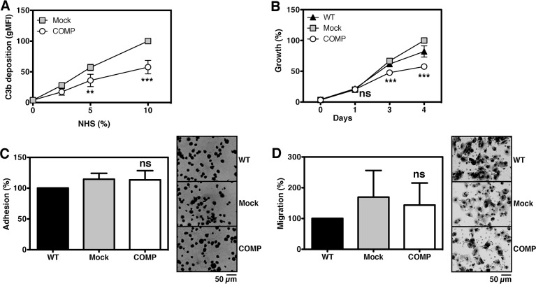
Figure 3. Expression of COMP protects cancer cells against complement.
Complement was activated on the surface of DU145 cells and C3b deposition was measured. Expression of COMP led to a significantly inhibited complement attack as compared to mock (A). In addition, functional assays revealed that DU145 cells expressing COMP have a decreased growth rate in vitro (B), while adhesion (C) and migration (D) was not affected. The graphs represent data from three independent experiments ±SD and was compared to mock by 2-way ANOVA with Bonferroni post-test (A-B) or 1-way ANOVA with Dunnett's multiple comparison, ns, not significant; **, p < 0.005; ***, p < 0.001.

