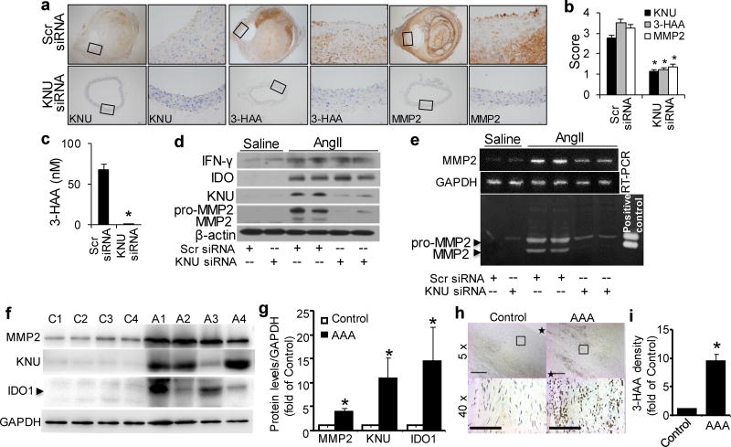
Figure 8. AngII-mediated MMP2 expression abolished by inhibition of 3-HAA formation in mice and upregulated kynurenine pathway in human AAA samples.
After transfections with scrambled (Scr) siRNA or kynureninase (KNU) siRNA, Apoe−/− mice were infused with saline or AngII (1000 ng/min per kg) for 4 weeks. (a–b) Representative immunohistochemical staining (a) and quantification for KNU, 3-HAA, and MMP2 (b) in the suprarenal aortas of AngII-infused mice with the indicated siRNA transfections. (c) Plasma concentrations of 3-HAA detected by HPLC in mice with the indicated siRNA transfections after AngII infusion.(d, e) The protein expression levels of IFN-γ, IDO, KNU, MMP2, and β-actin (d), as well as MMP2 mRNA (e) and activity (by zymography, e), in the suprarenal aortas of saline- or AngII-infused mice with the indicated siRNA transfections. *P<0.01 vs. scrambled siRNA-transfected Apoe−/− mice. All results were obtained from 6–10 mice in each group. (f) IDO1, KNU, and MMP2 increased in patient AAA samples. Representative Western blots were shown. C1~C4 indicates 4 control adjacent nonaneurysmal aortic sections; A1~A4 indicates 4 human AAA samples. (g) Quantification data for (f). n=4, *P<0.01 vs respective control group. (h) Anti-3-HAA staining in patient AAA area. *in control group indicates aortic lumen, * in AAA sample indicates aortic lumen side. (i) Quantification data for (h) (40X). *P<0.01 vs control group. P values in b and c, were obtained by a t test. P values in g and i were obtained by a paired t test. The error bars in b, c, g andi are s.e.m.

