Abstract
Stretch of myocardium, such as occurs upon increased filling of the cardiac chamber, induces two distinct responses: an immediate increase in twitch force followed by a slower increase in twitch force that develops over the course of several minutes. The immediate response is due, in part, to modulation of myofilament Ca2+ sensitivity by sarcomere length (SL). The slowly developing force response, termed the Slow Force Response (SFR), is caused by a slowly developing increase in intracellular Ca2+ upon sustained stretch. A blunted immediate force response was recently reported for myocardium isolated from homozygous giant titin mutant rats (HM) compared to muscle from wild-type littermates (WT). Here, we examined the impact of titin isoform on the SFR. Right ventricular trabeculae were isolated and mounted in an experimental chamber. SL was measured by laser diffraction. The SFR was recorded in response to a 0.2 μm SL stretch in the presence of [Ca2+]o=0.4 mM, a bathing concentration reflecting ~50% of maximum twitch force development at 25 °C. Presence of the giant titin isoform (HM) was associated with a significant reduction in diastolic passive force upon stretch, and ~50% reduction of the magnitude of the SFR; the rate of SFR development was unaffected. The sustained SL stretch was identical in both muscle groups. Therefore, our data suggest that cytoskeletal strain may underlie directly the cellular mechanisms that lead to the increased intracellular [Ca2+]i that causes the SFR, possibly by involving cardiac myocyte integrin signaling pathways.
Keywords: stretch, isolated cardiac trabeculae, rat, passive force, twitch force
INTRODUCTION
Cardiac output is a tightly regulated physiological parameter whereby the volume pumped by the heart matches venous return. Venous return is determined by the body’s requirements for nutrient/oxygen delivery and the concomitant removal of waste products. The main short-term mechanisms (seconds to minutes) by which circulatory homeostasis is achieved are heart rate, contractility, and stroke volume. Long-term mechanisms (hours to weeks) include renal fluid regulation and cardiac chamber remodeling(17).
Stroke volume is defined as the difference between the extent of cardiac chamber filling during diastole and the volume remaining at end-systole following the ejection phase of the cardiac cycle. Contractility (the intrinsic strength of the heart as a muscular pump) and the afterload determine end-systolic volume. Afterload is the mechanical load under which the heart operates that, to a large extent, is determined by systolic arterial blood pressure. End-diastolic volume constitutes the preload of the heart and it is determined by diastolic myocardial stiffness and the pressure gradient between the atrium and the relaxed cardiac chamber during the filling phase of the cardiac cycle.
Increased end-diastolic chamber volume upon augmented venous return results in stretch of the myocardial muscle fibers in the wall of the heart. Stretch of isolated myocardium results in an immediate increase in twitch force that is subsequently followed by a further, slowly developing, increase in twitch force that reaches a steady state after several minutes of sustained stretch. The immediate response reflects the well-known Frank-Starling mechanism(20); for recent reviews see (28)(27). The cellular basis for the Frank-Starling phenomenon is, in part, an immediate increase in myofilament responsiveness towards activating Ca2+ ions(28)(11)(1). Although the molecular mechanisms underlying myofilament length dependent activation are incompletely understood, recent evidence suggest a pivotal role for the cardiac elastic protein titin in modulating cardiac myofilament length dependent activation (1). The slower developing secondary phase takes place during a sustained increase in diastolic volume. As was the Frank-Starling mechanism, the slower response was first described over a century ago by von Anrep(4) and has been termed the “Anrep” effect. The phenomenon can be demonstrated at the whole heart level, both in-situ and ex-vivo(17). The classical manifestation of the Anrep phenomenon is the response to a sudden increase in cardiac afterload that initially induces increases in both end-diastolic and end-systolic volume; this allows for immediate increased cardiac pressure generation by virtue of the Frank-Starling mechanism. With time, however, cardiac contractility slowly increases to reach a steady-state level over the course of several minutes while cardiac volumes are partly restored. The increase in cardiac contractility that underlies the Anrep phenomenon was found not to be caused by circulating catecholamines(21), prompting Sarnoff to coin the phrase “homeometric autoregulation” to describe a phenomenon that develops within the heart in the absence of changes in preload, heart rate, or cardiac muscle fiber length. The immediate response to increased pre-load, in contrast, he termed “heterometric autoregulation” to reflect the immediate response is due to factors that are extrinsic to the heart (that is, stretch of sarcomeres in response to increased preload).
Parmley and Chuck, employing an isolated cat right ventricular papillary muscle preparation, demonstrated a similar phenomenon at the isolated cardiac papillary muscle level(18). They showed that stretch of the isolated muscle induced both an immediate increase in diastolic passive force as well as peak twitch force. This was followed by a slower increase in twitch force that reached steady-state over the course of several minutes, a phenomenon termed the slow force response (SFR). The SFR has also been demonstrated at the single cell level(24). Later work by Allen et al(3) established that the increase in twitch tension during the Slow Force Response (SFR) is caused by an increase in amplitude of the intracellular calcium transient, induced by cellular mechanisms that operate mainly during diastole(12). Thus, the Anrep phenomenon has at its basis a modulation of the cardiac excitation-contraction (EC) coupling processes in response to a sustained increase in (diastolic) cardiac muscle length(3)(12).
The cellular mechanisms that underlie altered calcium homeostasis during the SFR are unknown (for a recent reviews see(17)(6)(5)). One proposed mechanism involves activation of transmembrane stretch sensitive ion channels(23). Opened stretch activated channels either conduct Ca2+ ions directly, or conduct Na+ ions that, in turn, modulate the Na+/Ca2+ exchanger to indirectly enhance cellular Ca2+ loading. Another proposed mechanism involves local release of Angiotensin-II that, following several proposed signal transduction steps, modulates the activity of the Na+/H+ exchanger to, likewise, induce an increase in cellular Ca2+ load via the sarcolemmal Na+/Ca2+ exchange membrane carrier, although this hypothesis is not universally supported(22). Finally, stretch has also been proposed to increase protein kinase activity that may alter cardiac EC-coupling in addition to alterations of contractile protein phosphorylation (16)(2).
We recently demonstrated a blunted immediate twitch force increase in myocardium isolated from the hearts of a homozygous mutant rat strain (HM) that express an unusually long isoform of titin well into adulthood(8), compared to the wild-type (WT) littermates(1). Titin, a very large elastic protein, is located between the Z-disk and the center of the A-band in the striated muscle sarcomere(13)(14). Titin strain is a major determinant of myocardial diastolic stiffness, at least within the physiological range of sarcomere length encountered in the intact heart. Presence of the giant titin isoform reduces passive diastolic force, blunts myofilament length dependent activation, and eliminates the alterations in thick- and thin-filament protein structure that normally accompany diastolic sarcomere stretch(1). Because the sarcomere length change was identical between the HM and WT muscles, these data suggest that stretch induced strain in titin, and the resulting diastolic passive force, directly induces these immediate response molecular events.
Here, we employ the same giant titin mutant and wild-type rat strains to examine the impact of titin strain on the SFR. Isolated electrically stimulated right ventricular trabeculae were studied under conditions of intermediate contractility by reduced extracellular [Ca2+]o. A sustained increase in sarcomere length (SL) was employed to record the stretch induced SFR. Reduced titin strain and passive force in HM myocardium was associated with a ~50% reduction in the SFR. In contrast, neither the responsiveness to [Ca2+]o nor the time-course of the SFR was affected by titin strain. Because SL stretch was identical in both muscle groups, our data suggest that cytoskeletal strain may directly underlie the cellular mechanisms that lead to the increased intracellular [Ca2+]i that causes the SFR, possibly by involving cardiac myocyte integrin signaling pathways.
MATERIALS AND METHODS
Muscle Preparation and Solutions
All experimental procedures involving live rats were performed according to institutional guidelines concerning the care and use of experimental animals, and the Institutional Animal Care and Use Committee of the Loyola University Stritch School of Medicine approved all protocols. Homozygous autosomal titin mutant rats (HM) and wild-type littermate (WT) rats have been described previously(1)(10)(8)(19). Rats were deeply anesthetized with 50 mg/kg pentobarbital sodium in combination with 200U heparin. Hearts were rapidly excised from the thorax and perfused via the aorta with a modified Krebs-Henseleit (KH) solution containing 118.5mM NaCl, 5 mM KCl, 2 mM NaH2PO4, 1.2 mM MgSO4, 10 mM glucose, 25 mM NaHCO3 and 0.1 mM CaCl2 and 20 mM 2,3-butanedione monoxime (BDM) to inhibit spontaneous contractions during dissection; pH was adjusted to 7.4 (at 25 °C) by vigorous bubbling with 95/5 % O2/CO2 (25). Free running unbranched cell membrane intact trabeculae were dissected from the right ventricle(25); average dimension of the muscles included in the study are summarized in Table 1. Dissected muscles were moved from the dissection dish into a 250 μl glass bottomed temperature controlled bath (Aurora, model 1500A) perfused with the same oxygenated KH solution, but without BDM, at a flow rate of ~2.5 ml/min; bath temperature was controlled at 25±0.1 °C. The muscle was mounted horizontally in this bath by positioning its muscular end in a platinum cradle (25) attached to a force transducer (Aurora, model 400A), and by attaching its valvular end to a hook attached to a motor (Aurora, model 322C). Both attaching devices were controlled by micromanipulators. Finally, the bath was covered with a glass slide. Sarcomere length was continuously monitored (4kHz) (27)(1) using He-Ne laser diffraction, where the first order diffraction band was captured on a linear CCD-based FPGA analyzer that outputs calibrated sarcomere lengths (PLIN-2605-2, Dexela Inc). Following dissection and mounting of the trabeculae in the experimental set-up, the preparation was stimulated through two platinum electrodes running parallel to the muscle at a rate of 1.0 Hz. Stimulus intensity was 50% above threshold, and stimulus duration was 2–5 ms. Muscles were stretched to a resting sarcomere length of about 2.2 μm, a length at which passive force was usually 5–10% of peak active twitch force, and left to equilibrate for at least one hour in presence of 1.5 mM extracellular [Ca2+]o). After this equilibration period, the muscle was restretched to a diastolic sarcomere length of 2.2 μm. If peak twitch force production had decreased to <70% of control at this time, the preparation was discarded
Table 1.
Average muscle dimensions, twitch kinetics, twitch force, and [Ca2+]o responsiveness.
| WT (n=7) | HM (n=8) | |
|---|---|---|
| Length (mm) | 2.9 ± 0.3 | 2.6 ± 0.1 |
| Width (μm) | 329 ± 61 | 333 ± 55 |
| Thickness (μm) | 147 ± 36 | 101 ± 9 |
| Activation time (ms) | 86 ± 4 | 74 ± 4 |
| Relaxation time (ms) | 139 ± 9 | 80 ± 5* |
| Twitch duration (ms) | 225 ± 11 | 154 ± 7* |
| Fmax (mN/mm2) | 92.7 ± 7.3 | 44.1 ± 5.5* |
| Passive Diastolic Stress (mN/mm2) | 3.68 ± 0.45 | 0.67 ± 0.08* |
| EC50 (mM) | 0.42 ± 0.06 | 0.51 ± 0.05 |
| Hill coefficient | 2.5 ± 0.1 | 2.4 ± 0.2 |
Data were collected from electrically stimulated right ventricular cardiac trabeculae isolated from wild-type littermate (WT) or giant titin homozygous mutant (HM) rats. Systolic SL=2.2 μm; stimulus frequency=1 Hz; temperature 25 °C.
p<0.05.
Experimental protocols
The study protocol started with the assessment of the active developed twitch stress–[Ca2+]o relationships at 25 °C. [Ca2+]o was randomly varied between 0.1 and 2.0 mM(25). Upon steady state (~3–5 minutes) at each [Ca2+]o, twitch stress was measured at steady-state and systolic sarcomere shortening to SL=2.2 μm. A final solution containing [Ca2+]o=2.0 mM was applied to assess twitch kinetics (Table 1), that is, time between 50% twitch force and peak twitch force, and time between peak twitch force tension and decline to 50% of peak force (a measure of relaxation velocity).
Next, the Slow Force Response (SFR) of the muscle was determined. [Ca2+]o was reduced to 0.4 mM, which in isolated rat myocardium reflects ~50% maximum activation at diastolic SL=2.1 μm at 25 °C (25). When a steady-state was attained (~5 minutes), twitch force was continuously recorded for the next 7 minutes at 30 second intervals; after 1.5 minutes, diastolic muscle length was increased so as to attain SL=2.3 μm and kept constant thereafter for the next 5.5 minutes.
Data Analysis
Muscle mechanical stress was calculated as passive and active twitch force divided by the cross-sectional area, calculated from the muscle dimensions.
Sigmoidal stress-[Ca2+]o relationships were fit by non-linear regression to a modified Hill equation:
| (1) |
where Fmax is the maximum, [Ca2+]o saturated peak twitch stress development; nH is the Hill coefficient, a measure of cooperativity; EC50 is the [Ca2+]o at which peak twitch stress is half-maximal.
Twitch kinetics were assessed by measuring the time elapsed between a particular fractional force level (10%, 50%, or 75%) to peak twitch force (activation time), and from the time of peak twitch force down to that fractional twitch force (relaxation time). Total duration is defined as activation time plus relaxation time.
The rate at which twitch force increases during the SFR response was assessed by non-linear exponential fit of the peak twitch force data as function of time following the stretch to SL=2.3 μm.
Statistical analysis
All data are represented as mean ± SEM. Statistical analysis was performed using Systat 13 (Systat Software, Inc) using Student’s t-test. Statistical significance was defined as P < 0.05.
RESULTS
Steady state twitches
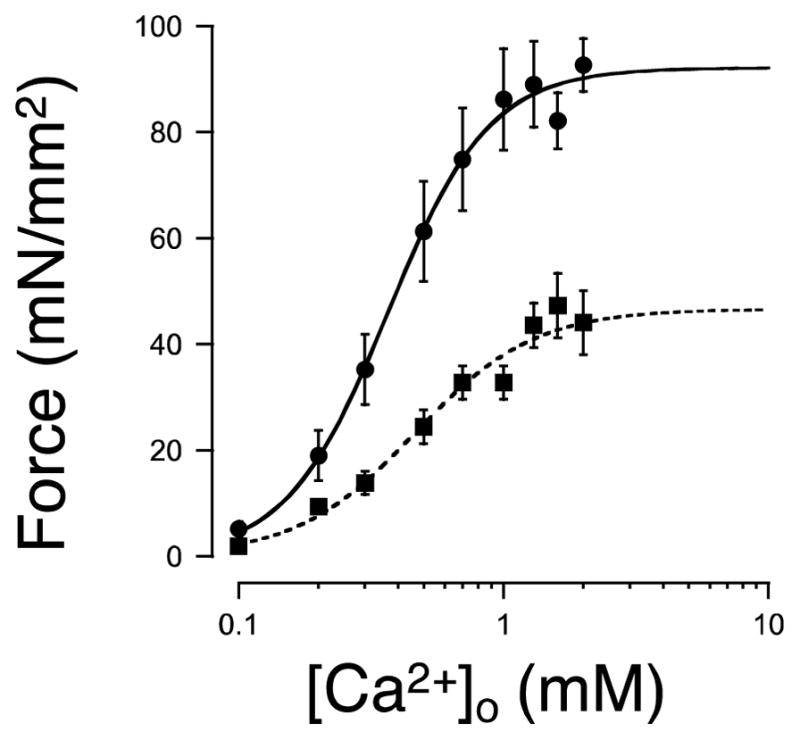
Figure 1 summarizes the impact of varying extracellular [Ca2+]o on twitch force development in the isolated right ventricular cardiac trabeculae. The average modified Hill fit parameters derived from individual muscles is summarized in Table 1. SL in these series of experiments was set at SL=2.2 μm, halfway in between the SL range used for determination of the SFR (see below). To allow for comparison between muscles, force is expressed as stress (force divided by cross-sectional area of the muscle). Twitch force depended in a sigmoidal fashion on [Ca2+]o in the bathing solution, reaching half maximum values at [Ca2+]o=~0.5 mM in either WT or HM muscles. This result is consistent with previous reports on isolated WT rat myocardium at 25 °C (25). Moreover, presence of the giant isoform in HM muscles did not affect the apparent cooperativity of the response to [Ca2+]o (see Table 1). Maximum twitch stress at saturating [Ca2+]o, in contrast, was significantly lower in HM compared to WT muscles, as was passive diastolic stress at this SL.
Figure 1. Average twitch force – extracellular [Ca2+]o relationships.

Twitch stress (force divided by cross-sectional area) was measured in electrically stimulated right ventricular trabeculae isolated from wild-type (WT; closed circles; n=7), and giant titin homozygous mutant (HM; closed squares; n=8) rats at bath [Ca2+]o ranging between 0.1 and 2.0 mM. Data were fit to a modified Hill equation (see also Table 1). Presence of the giant isoform in the HM muscles was associated with lower twitch tress (at all [Ca2+]o). In contrast, neither the apparent sensitivity to extracellular Ca2+ (EC50), nor the level of cooperativity (Hill coefficient) was affected by titin isoform (see Table 1). Systolic SL=2.2 μm; stimulus frequency=1 Hz; temperature 25 °C.
Analysis of twitch kinetics at saturating [Ca2+]o revealed a 14% accelerated activation time, 42% accelerated relaxation time, and 32% shortening of overall twitch duration at the 50% twitch force level in HM compared to WT muscles at this SL (Table 1).
Slow force response
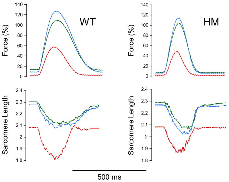
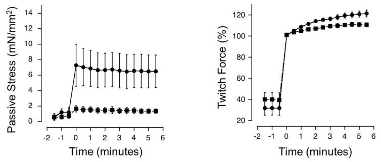
The SFR phenomenon is reportedly caused by a slowly developing increase in the magnitude of the intracellular calcium transient. Therefore, to assess the SFR, it is important to perform the experiment at a sub-maximal level of contractility when twitch force is not Ca2+ saturated. This was achieved by reducing [Ca2+]o to 0.4 mM, a level slightly below the EC50 value in either muscle group at 25 °C (Table 1). Figure 2 shows typical recordings in WT and HM muscles of twitch force and sarcomere length recorded at the steady-state pre-stretch diastolic SL (2.1 μm), immediately following muscle stretch (diastolic SL=2.3 μm), and at 5.5 minutes following stretch. To allow for comparisons between WT and HM muscles, force was normalized to peak developed twitch force of the first twitch following the stretch. The average time course of the SFR is shown in Figure 3, while average SFR parameters are summarized in Table 2. Stretch resulted in an immediate increase in both passive and active developed twitch force. The diastolic passive force increase upon stretch was significantly blunted in HM compared WT muscles (~6% vs. 23%), as was the increase in active twitch force development, albeit to a lesser extent (~60% vs. 68%), consistent with previously reported data(1). Sustained muscle stretch resulted in a slower increase in twitch force that was nearly complete at time=5.5 minutes. The magnitude of the normalized slow increase in twitch force was ~50% smaller in HM compared to WT muscles (~12% vs. 22%). In contrast, the rate at which the SFR developed was comparable between WT and HM muscles (~0.6 min−1). Note that sustained muscle length stretch resulted in a small decline in diastolic SL over the course of the 5.5 minutes in which we measured the SFR (compare the green and blue traces in Figure 2). This likely reflects stretch-relaxation of the passive elastic structures within the sarcomere, a phenomenon that is commonly observed in muscle(13)(14).
Figure 2. Typical Slow Force Response recordings in wild-type (WT) and giant titin homozygous mutant (HM) isolated rat cardiac trabeculae.
Sarcomere stretch (ΔSL=0.2 μm) from baseline diastolic SL (red traces) led to an immediate increase in twitch force (green traces); maintaining diastolic SL at the longer length for a further 5.5 minutes induced a further increase in twitch force (blue traces), reflecting the Slow Force Response (SFR) phenomenon (see also Figure 3 and Table 2). The SFR was smaller in HM compared to WT muscles; twitch kinetics were, overall, faster in HM compared to WT muscles, consistent with the average twitch kinetics recorded at saturating bath [Ca2+]o (2 mM; Table 1). [Ca2+]o=0.4 mM; stimulus frequency=1 Hz; temperature=25 °C.
Figure 3. Average passive stress and normalized active twitch force response to stretch.
The Slow Force Response (SFR) was recorded in WT (closed circles; n=5) and HM (closed squares; n=8) at [Ca2+]o=0.4 mM (which results in each muscle group ~50% of maximum twitch force). Twitch force was first recorded at the baseline diastolic sarcomere length (SL=2.1 μm) for 1.5 mins; at time=0 min, the muscles were quickly stretched during diastole to increase diastolic sarcomere length to SL=2.3 μm (ΔSL=0.2 μm), resulting in an immediate increase in both passive (left panel) and active twitch force (right panel). In the right panel, active twitch force is normalized to peak twitch force of the first elicited twitch following the stretch. Presence of the giant isoform of titin in the HM muscles was associated with significant lower passive stress development following stretch and a ~50% lower final magnitude of the SFR in HM muscles. The kinetics of the SFR, however, were not affected (see also Table 2). [Ca2+]o=0.4 mM; stimulus frequency=1 Hz; temperature=25 °C.
Table 2.
Average Slow Force Response parameters.
| WT (n=5) | HM (n=8) | |
|---|---|---|
| Slow Force Response immediate active twitch force increase (%) | 68.2 + 6.2 | 60.6 + 6.6 |
| Slow Force Response immediate passive force increase (%) | 23.1 + 7.0 | 6.0 + 0.8* |
| Slow Force Response final twitch force increase (%) | 22.1 + 3.1 | 11.8 + 2.7* |
| Slow Force Response twitch force increase rate (min−1) | 0.54 ± 0.09 | 0.69 + 0.20 |
Slow Force Response (SFR) data in response to a quick increase in diastolic sarcomere length from SL=2.1 μm to SL=2.3 μm (ΔSL=0.2 μm) recorded on right ventricular cardiac trabeculae isolated from wild-type littermate (WT) or giant titin homozygous mutant (HM) rats. [Ca2+]o=0.4 mM; stimulus frequency=1 Hz; temperature 25 °C.
p<0.05.
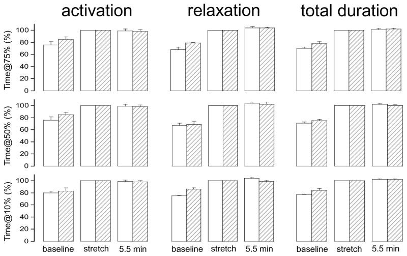
Sustained stretch of cardiac muscle may lead to altered protein kinase activity(16)(2), which could alter twitch kinetics. To address this, we examined twitch kinetic parameters (activation time, relaxation time, and total duration) at the 10%, 50%, and 75% twitch force level during the SFR protocol. The average results are summarized in Figure 4. Twitch timing kinetics were normalized to those of the first twitch immediately following the stretch (middle pair of bars). Diastolic stretch resulted in an immediate relative prolongation of the twitch to a similar extent in either WT and HM muscles. However, sustained stretch in either WT or HM muscles did not further impact twitch timing kinetics, regardless of the force level at which it was examined. Note that peak twitch force increased during that same time period of sustained stretch reflecting the SFR (Figures 2&3, Table 1).
Figure 4. Impact of stretch on twitch kinetics.
Average twitch kinetics at the 75% (top), 50% (middle) and 10% (bottom) twitch force level at: baseline sarcomere length (left pair of bars), immediately following stretch (middle pair of bars), and 5.5 min following the stretch when the Slow Force Response is fully developed (right pair of bars). Data were recorded in wild-type (WT; open bars; n=5) and homozygous giant titin mutant (HM; hatched bars; n=8) muscles. Time durations were normalized to the first twitch immediately following the stretch (i.e. the middle pair of bars representing the immediate response to SL stretch was set to 100%). In both muscle groups and at all three force levels, stretch per se induced an increase in time to reach peak twitch force (activation, left panels) and between the peak of the contraction down to a given twitch force level (relaxation, middle panels), and the overall duration of the twitch (total duration, right panels); prolonged maintained stretch (5.5 minutes), in contrast, did not affect twitch kinetics. [Ca2+]o=0.4 mM; temperature=25 °C; stimulus frequency=1.0 Hz.
DISCUSSION
The main finding of our study is that myocardium containing the giant isoform of titin(8) displays a significantly blunted Slow Force Response (SFR).
It is well established that the SFR is the result of a slowly developing increase in the magnitude of the intracellular calcium transient(17)(6)(5)(12)(3). Therefore, to properly asses the SFR and compare this parameter between groups, it was important to first establish the sensitivity of HM rat myocardium to extracellular [Ca2+]o at 25 °C. Consistent with previous reports that employed either this rat strain(1) or a genetic giant titin mouse model(15), presence of a compliant isoform of titin within the cardiac sarcomere resulted in blunted peak twitch force. This phenomenon likely resides within the myofilaments themselves: First, we observed blunted twitch force at all levels of extracellular [Ca2+]o (Figure 1). Therefore, merely increasing extracellular [Ca2+]o was not sufficient to overcome blunted peak twitch force in the HM muscles. Second, the responsiveness towards extracellular [Ca2+]o was comparable between WT and HM muscles, both in terms of EC50 and the Hill coefficient that indexes cooperativity. It has been reported that the RNA Binding Motif Protein 20 (RBM20) mutation that causes the alteration in titin splicing also affects expression of several calcium homeostasis regulatory proteins within the cardiac myocyte(1)(10). It appears, however, that the impact of these proteins, if any, excludes steps in the cardiac excitation-contraction coupling pathways that determine overall responsiveness of the cardiac myocyte to extracellular calcium ions. This notion is also consistent with the unaltered intracellular calcium transient reported in the murine genetic giant titin model(15). Third, a blunting of maximum Ca2+ saturated force development is also observed at the myofilament level in HM compared to WT muscle. This was the case in both chemically permeabilized (skinned) muscle preparations from the spontaneous rat mutant strain(1)(19), as well as in skinned cardiac muscles prepared from the giant titin genetic mouse model(15). The mechanisms underlying the blunted maximum force development are not clear. Tension-cost, the rate of ATP consumption to sustain a certain level of Ca2+ activated force(29), has been reported to be increased in isolated skinned giant titin mouse myocardium(15). Tension-cost is proportional to cross-bridge detachment rate, at least in a simple two-state model of actin-myosin activation(29). Such an increase in tension-cost is consistent with the accelerated twitch kinetics we observed here in HM compared to WT muscles, particularly accelerated relaxation kinetics (see Table 1). Indeed, the two-state cross-bridge model predicts a reduction in maximum force development upon increased cross-bridge detachment rate due to a reduction of the fraction of cross-bridges that are attached to actin in the strongly bound force generating state at any given time. Such a myofilament based mechanism would cause a reduction in twitch force for a similar magnitude of the intracellular calcium transient in HM muscles. The molecular mechanisms underlying increased cross-bridge detachment rate in HM myocardium, however, cannot be determined from our study and further investigation will be required to elucidate the cause of this phenomenon.
A novel finding in the present study is blunting of the Anrep effect (the SFR) in HM mutant giant titin isoform myocardium. As mentioned above, it is now well established that the cellular mechanisms that underlie the SFR are centered on alterations in the intracellular calcium transient. The blunted immediate response to stretch that is seen in HM myocardium is caused, to a large extent, only to changes in myofilament calcium sensitivity at the short sarcomere length(1)(19). Thus, myofilament calcium sensitivity during the sustained stretch is expected to be similar between the WT and HM muscles. Therefore, the reduction in the SFR in HM myocardium seen in the present study is unlikely the result of a reduced myofilament response to the increased cellular calcium transient. Rather, our data suggest a blunted increase in the intracellular calcium transient upon sustained stretch. The central question remaining, however, is how muscle stretch induces altered cellular calcium homeostasis. Several theories for this phenomenon have been proposed that are not, a priori, mutually exclusive (22)(24)(23)(17)(6)(5):
The SFR response may depend on release of angiotensin-II (Ang-II) by the cardiac myocyte upon muscle stretch. The mechanisms underlying such stress response are not fully understood. The paracrine release of Ang-II, in turn, is believed to induce an increase in myocyte contractility that may involve activation of the AT-1 receptor. However, how AT-1 receptor activation induces an increase in cellular calcium load and contractility is not fully understood. Cingolani et al. have proposed an elaborate signal transduction pathway that includes release of Ang-II and endothelin, activation of the mineralocorticoid receptor and epidermal growth factor receptor, formation of mitochondria reactive oxygen species and activation of the Na+/H+ exchanger leading to increased cellular calcium load via Na+/Ca2+ exchange modulation(6)(5). Of interest, stretch induced release of Ang-II is blunted in myocardium isolated from mice that lack the thrombospondin receptor(7). This protein is an integral component of the focal adhesion-integrin receptor complex; this is the mechanical load bearing structure through which the mechanical force generated by the cross-bridges is transmitted out of the cardiac cell and into the extracellular matrix.
Surface membrane stretch may induce opening of stretch activated channels (SACs) (9). SACs appear to be relatively non-selective ion channels, potentially allowing both mono- and divalent ions to enter the cell upon opening. Thus, stretch induced opening of those channels could allow for direct entry of Ca2+ ions to increase myocyte calcium load. Alternatively, opening of SACs could allow entry of Na+ (over K+ due to the favorable electrochemical gradient that prevails in diastole); the resulting increase in cytosolic Na+ is then expected to indirectly increase cellular calcium load by modulation of the Na+/Ca2+ exchange equilibrium. Evidence, both for and against, this theory has been advanced in the literature (23)(17). One potential problem with the SAC theory, however, is that muscle stretch would have to be associated with altered membrane strain that, although attractive as a theory, may not be consistent with the known cellular anatomy and the load bearing mechanical connections between the cytoskeleton and the extracellular matrix in the heart(13)(14).
Stretch of isolated myocardium has been shown to increase kinase mediated contractile protein phosphorylation(16)(2). The mechanisms underlying increased protein kinase activity upon stretch are not known, but could conceivably be the result of either AT-1 receptor activation or opening of SACs. In addition, the increase in the magnitude of the cytoplasmic calcium transient that causes the SFR could activate Ca2+ sensitive protein kinases, such as protein kinase C or calmodulin kinase. As such, increased kinase activity may merely be the result rather than the cause of the SFR. In the present study, prolonged stretch did not affect twitch timing kinetics, either in WT or HM muscles, at any level of twitch force development (Figure 4). It is well established that contractile protein phosphorylation profoundly affects twitch timing kinetics(28)(30)(26). Moreover, the responsiveness towards extracellular [Ca2+]o was comparable between WT and HM muscle (Figure 1 & Table 1). Hence, although we cannot exclude altered protein kinase activity in our study, our data suggest that the impact, if any, of such activity must have been minor.
Several limitations need to be considered in interpretation of our data. First, we did not measure the intracellular calcium transient. Although it is well established that an increased magnitude of the calcium transient causes the SFR, we can only indirectly deduce that stretch in HM myocardium is caused by a blunted slow increase in the calcium transient. Note that presence of a giant isoform of Titin in a murine genetic model is reportedly not associated with an alteration of the intracellular calcium transient(15). Second, although relative twitch kinetics were not altered upon sustained stretch in either muscle group, we did not directly biochemically assess the contractile protein or calcium homeostasis protein phosphorylation status. It is therefore possible, albeit unlikely, that such a change did occur but that opposing down-stream functional impacts were balanced to maintain overall twitch kinetics.
In conclusion, we measured the Slow Force Response (SFR) in isolated myocardium in which sarcomere compliance was altered due to presence of a mutant giant isoform of titin. Presence of the giant titin isoform was associated with a significant reduction is diastolic passive force and marked blunting of the SFR compared to wild-type titin myocardium. Sarcomere strain and therefore, by inference, membrane strain was comparable in the two groups. Hence, differential activation of stretch activated ion channels in HM and WT myocardium appears unlikely. Rather, our data suggest that cytoskeletal strain underlies the cellular mechanisms that lead to the increased intracellular [Ca2+]i that causes the SFR, possibly by involving strain sensitive integrin signaling pathways.
Acknowledgments
This work was supported, in part, by NIH HL75494, HL62426 (PDT), and NIH HL HL77196 (MLG).
Footnotes
Author contributions: YAM, MZ, JM and PPdT designed and performed research; MG contributed animal models and reagents; YAM, MZ, JM, MG, and PPdT wrote the paper.
The authors declare no conflict of interest.
Publisher's Disclaimer: This is a PDF file of an unedited manuscript that has been accepted for publication. As a service to our customers we are providing this early version of the manuscript. The manuscript will undergo copyediting, typesetting, and review of the resulting proof before it is published in its final citable form. Please note that during the production process errors may be discovered which could affect the content, and all legal disclaimers that apply to the journal pertain.
References
- 1.Ait Mou Y, Hsu K, Farman GP, Kumar M, Greaser ML, Irving TC, de Tombe P. Titin strain contributes to the Frank-Starling law of the heart by structural rearrangements of both thin- and thick-filament proteins. Proc Natl Acad Sci USA. 2016;113:2306–2311. doi: 10.1073/pnas.1516732113. [DOI] [PMC free article] [PubMed] [Google Scholar]
- 2.Ait Mou Y, le Guennec J-Y, Mosca E, de Tombe PP, Cazorla O. Differential contribution of cardiac sarcomeric proteins in the myofibrillar force response to stretch. Pflugers Arch. 2008;457:25–36. doi: 10.1007/s00424-008-0501-x. [DOI] [PMC free article] [PubMed] [Google Scholar]
- 3.Allen DG, Kurihara S. The effects of muscle length on intracellular calcium transients in mammalian cardiac muscle. J Physiol. 1982;327:79–94. doi: 10.1113/jphysiol.1982.sp014221. [DOI] [PMC free article] [PubMed] [Google Scholar]
- 4.Anrep von G. On the part played by the suprarenals in the normal vascular reactions of the body. J Physiol. 1912;45:307–317. doi: 10.1113/jphysiol.1912.sp001553. [DOI] [PMC free article] [PubMed] [Google Scholar]
- 5.Cingolani HE, Ennis IL, Aiello EA, Pérez NG. Role of autocrine/paracrine mechanisms in response to myocardial strain. Pflugers Arch. 2011;462:29–38. doi: 10.1007/s00424-011-0930-9. [DOI] [PubMed] [Google Scholar]
- 6.Cingolani HE, Pérez NG, Cingolani OH, Ennis IL. The Anrep effect: 100 years later. AJP: Heart and Circulatory Physiology. 2013;304:H175–82. doi: 10.1152/ajpheart.00508.2012. [DOI] [PubMed] [Google Scholar]
- 7.Cingolani OH, Kirk JA, Seo K, Koitabashi N, Lee D-I, Ramirez-Correa G, Bedja D, Barth AS, Moens AL, Kass DA. Thrombospondin-4 is required for stretch-mediated contractility augmentation in cardiac muscle. Circ Res. 2011;109:1410–1414. doi: 10.1161/CIRCRESAHA.111.256743. [DOI] [PMC free article] [PubMed] [Google Scholar]
- 8.Greaser ML, Warren CM, Esbona K, Guo W, Duan Y, Parrish AM, Krzesinski PR, Norman HS, Dunning S, Fitzsimons DP, Moss RL. Mutation that dramatically alters rat titin isoform expression and cardiomyocyte passive tension. J Mol Cell Cardiol. 2008;44:983–991. doi: 10.1016/j.yjmcc.2008.02.272. [DOI] [PMC free article] [PubMed] [Google Scholar]
- 9.Guharay F. Stretch-activated single ion channel currents in tissue-cultured embryonic chick skeletal muscle. J Physiol. 1984;352:685–701. doi: 10.1113/jphysiol.1984.sp015317. [DOI] [PMC free article] [PubMed] [Google Scholar]
- 10.Guo W, Schafer S, Greaser ML, Radke MH, Liss M, Govindarajan T, Maatz H, Schulz H, Li S, Parrish AM, Dauksaite V, Vakeel P, Klaassen S, Gerull B, Thierfelder L, Regitz-Zagrosek V, Hacker TA, Saupe KW, Dec GW, Ellinor PT, MacRae CA, Spallek B, Fischer R, Perrot A, Özcelik C, Saar K, Hubner N, Gotthardt M. RBM20, a gene for hereditary cardiomyopathy, regulates titin splicing. Nat Med. 2012;18:766–773. doi: 10.1038/nm.2693. [DOI] [PMC free article] [PubMed] [Google Scholar]
- 11.Kentish JC, Keurs ter HE, Ricciardi L, Bucx JJ, Noble MI. Comparison between the sarcomere length-force relations of intact and skinned trabeculae from rat right ventricle. Influence of calcium concentrations on these relations. Circ Res. 1986;58:755–768. doi: 10.1161/01.res.58.6.755. [DOI] [PubMed] [Google Scholar]
- 12.Kentish JC, Wrzosek A. Changes in force and cytosolic Ca2+ concentration after length changes in isolated rat ventricular trabeculae. J Physiol (Lond) 1998;506(Pt 2):431–444. doi: 10.1111/j.1469-7793.1998.431bw.x. [DOI] [PMC free article] [PubMed] [Google Scholar]
- 13.Labeit S, Kolmerer B, Linke WA. The giant protein titin. Emerging roles in physiology and pathophysiology. Circ Res. 1997;80:290–294. doi: 10.1161/01.res.80.2.290. [DOI] [PubMed] [Google Scholar]
- 14.LeWinter MM, Granzier H. Cardiac titin: a multifunctional giant. Circulation. 2010;121:2137–2145. doi: 10.1161/CIRCULATIONAHA.109.860171. [DOI] [PMC free article] [PubMed] [Google Scholar]
- 15.Methawasin M, Hutchinson KR, Lee E-J, Smith JE, Saripalli C, Hidalgo CG, Ottenheijm CAC, Granzier H. Experimentally increasing titin compliance in a novel mouse model attenuates the Frank-Starling mechanism but has a beneficial effect on diastole. Circulation. 2014;129:1924–1936. doi: 10.1161/CIRCULATIONAHA.113.005610. [DOI] [PMC free article] [PubMed] [Google Scholar]
- 16.Monasky MM, Biesiadecki BJ, Janssen PML. Increased phosphorylation of tropomyosin, troponin I, and myosin light chain-2 after stretch in rabbit ventricular myocardium under physiological conditions. J Mol Cell Cardiol. 2010;48:1023–1028. doi: 10.1016/j.yjmcc.2010.03.004. [DOI] [PMC free article] [PubMed] [Google Scholar]
- 17.Neves JS, Leite-Moreira AM, Neiva-Sousa M, Almeida-Coelho J, Castro-Ferreira R, Leite-Moreira AF. Acute Myocardial Response to Stretch: What We (don’t) Know. Front Physiol. 2015;6:408. doi: 10.3389/fphys.2015.00408. [DOI] [PMC free article] [PubMed] [Google Scholar]
- 18.Parmley WW, Chuck L. Length-dependent changes in myocardial contractile state. Am J Physio Heart. 1973;224:1195–1199. doi: 10.1152/ajplegacy.1973.224.5.1195. [DOI] [PubMed] [Google Scholar]
- 19.Patel JR, Pleitner JM, Moss RL, Greaser ML. Magnitude of length-dependent changes in contractile properties varies with titin isoform in rat ventricles. AJP: Heart and Circulatory Physiology. 2012;302:H697–708. doi: 10.1152/ajpheart.00800.2011. [DOI] [PMC free article] [PubMed] [Google Scholar]
- 20.Patterson SW, Piper H, Starling EH. The regulation of the heart beat. J Physiol. 1914;48:465–513. doi: 10.1113/jphysiol.1914.sp001676. [DOI] [PMC free article] [PubMed] [Google Scholar]
- 21.Sarnoff SJ, GILMORE JP, REMENSNYDER JP, MITCHELL JH. Homeometric autoregulation in the heart. Circ Res. 1960;8:1077–1091. doi: 10.1161/01.res.8.5.1077. [DOI] [PubMed] [Google Scholar]
- 22.Shen X, Cannell MB, Ward M-L. Effect of SR load and pH regulatory mechanisms on stretch-dependent Ca(2+) entry during the slow force response. J Mol Cell Cardiol. 2013;63:37–46. doi: 10.1016/j.yjmcc.2013.07.008. [DOI] [PubMed] [Google Scholar]
- 23.Ward M-L, Williams IA, Chu Y, Cooper PJ, Ju Y-K, Allen DG. Stretch-activated channels in the heart: contributions to length-dependence and to cardiomyopathy. Prog Biophys Mol Biol. 2008;97:232–249. doi: 10.1016/j.pbiomolbio.2008.02.009. [DOI] [PubMed] [Google Scholar]
- 24.Yamaguchi Y, Iribe G, Kaneko T, Takahashi K, Numaga-Tomita T, Nishida M, Birnbaumer L, Naruse K. TRPC3 participates in angiotensin II type 1 receptor-dependent stress-induced slow increase in intracellular Ca(2+) concentration in mouse cardiomyocytes. J Physiol Sci. 2017 Jan 19; doi: 10.1007/s12576-016-0519-3. [DOI] [PMC free article] [PubMed] [Google Scholar]
- 25.de Tombe PP, Keurs ter HE. Force and velocity of sarcomere shortening in trabeculae from rat heart. Effects of temperature. Circ Res. 1990;66:1239–1254. doi: 10.1161/01.res.66.5.1239. [DOI] [PubMed] [Google Scholar]
- 26.de Tombe PP, Keurs ter HE. Lack of effect of isoproterenol on unloaded velocity of sarcomere shortening in rat cardiac trabeculae. Circ Res. 1991;68:382–391. doi: 10.1161/01.res.68.2.382. [DOI] [PubMed] [Google Scholar]
- 27.de Tombe PP, Keurs ter HEDJ. Cardiac muscle mechanics: Sarcomere length matters. J Mol Cell Cardiol. 2015;91:148–150. doi: 10.1016/j.yjmcc.2015.12.006. [DOI] [PMC free article] [PubMed] [Google Scholar]
- 28.de Tombe PP, Mateja RD, Tachampa K, Ait Mou Y, Farman GP, Irving TC. Myofilament length dependent activation. J Mol Cell Cardiol. 2010;48:851–858. doi: 10.1016/j.yjmcc.2009.12.017. [DOI] [PMC free article] [PubMed] [Google Scholar]
- 29.de Tombe PP, Stienen GJM. Impact of temperature on cross-bridge cycling kinetics in rat myocardium. The Journal of Physiology. 2007;584:591–600. doi: 10.1113/jphysiol.2007.138693. [DOI] [PMC free article] [PubMed] [Google Scholar]
- 30.de Tombe PP. Cardiac myofilaments: mechanics and regulation. J Biomech. 2003;36:721–730. doi: 10.1016/s0021-9290(02)00450-5. [DOI] [PubMed] [Google Scholar]


