Abstract
Background:
Sexual identification of immature skeletal remains is still a difficult problem to solve in forensic anthropology. In such situations, the odontometric features of the teeth can be of immense help. Teeth, being the hardest and chemically the most stable tissue in the body, are an excellent material in living and nonliving populations for anthropological, genetic, odontologic, and forensic investigations. Using tooth size standards, whenever it is possible to predict the sex, identification is made easier because then only missing persons of one sex need to be considered.
Aim:
To determine sex from the odontometric data using maxillary canine index and maxillary first molar dimensions and to determine which index gives higher accuracy rate for sex determination using only maxillary cast.
Materials and Methods:
In a sample size of 200 population (100 male and 100 female), alginate impression was taken of maxillary arch and poured with dental stone. Using Vernier caliper, the dimension of maxillary first molar (buccolingual [BL] and mesiodistal [MD]), canine (MD), and intercanine distance was measured on the cast. The obtained data were analyzed using discriminant statistical analysis.
Result and Conclusion:
This study concludes that BL dimension of maxillary first molar is a more reliable indicator for gender determination than other molar and canine dimensions in maxilla.
Key words: Forensic anthropology, forensic science, intercanine width, molar dimension, sex identification canine dimension
Introduction
The teeth being the most durable tissue of our body exhibits the least turnover rate because of its intense resistance to destruction. Hence, they can be considered for gender determination.[1]
Prediction of gender makes the task simpler since the missing person of only one gender is to be evaluated. Measurement of long bones particularly humerus and femur, pelvis, or skull are often used for sex determination. However, odontometric methods are more reliable in case of pediatric cases as teeth complete their development before skeletal maturation.[1] Hence, today, dentist's opinion is often sought to answer queries that arise during a postmortem investigation.[2]
Various odontometric parameters have been used for gender determination such as mandibular and maxillary canine indices, mandibular canine dimensions, maxillary canine dimension, maxillary first molar dimensions, and cumulative dimension of all teeth. Despite being reliable, the mandibular canine index (CI) has its limitations. Mandible being a single bone that is not directly attached to skull poses increased chances of trauma or damage. In cases where only part of the skull with maxilla is obtained, maxillary indices may have to be used for sex determination.
The purpose of this study was to evaluate the probability of determining gender using maxillary CI, buccolingual (BL), and mesiodistal (MD) dimensions of maxillary first molar and to compare the efficacy of these parameters with each other.
Materials and Methods
The study was conducted in the Department of Oral and Maxillofacial Pathology, after obtaining the institutional research committee and university ethical clearance. The study sample comprised population of dental students from the institute.
A base sample of 200 (100 male and 100 female) was chosen using cluster sampling. To detect percentage sexual dimorphism in maxillary first molars about 5.34% with 5% absolute error and 99% confidence interval, minimum observations required for the proposed study was 182. To increase precision (power), we have taken 200 samples.
The age of study population ranged from 15 to 25 years. This age group was particularly selected as dimensional changes due to attrition and abrasion are minimal.
Other inclusion criteria were as follows:
Complete set of fully erupted teeth excluding the third molars
Periodontally healthy teeth
Noncarious teeth
Nonattrited and intact teeth
Satisfactorily aligned maxillary teeth.
Following written consent from the participants, impression of maxillary arch was made using irreversible hydrocolloid – alginate (Finndent™, India) and poured using Type II dental stone (Neelkanth™, India) immediately to avoid any distortion. The maxillary arch study models were then used for analysis. All the measurements were done on the casts for easy reproducibility using digital calipers with a resolution of 0.01 mm by a single observer.
The mesiodistal dimension of maxillary canine (CMD) was measured as the distance between the mesial and distal contact points as shown in Figure 1.[3] The MD width of both left and right canine was measured, and the average value was taken for calculations.
Figure 1.
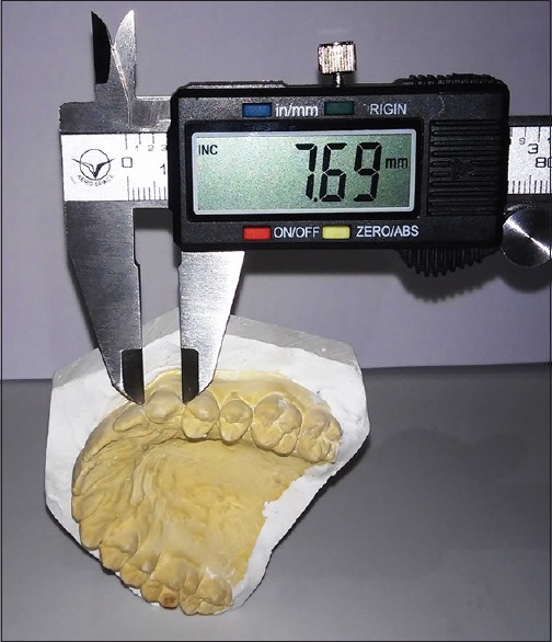
Measurement of mesiodistal dimension of maxillary canine on study cast
The maxillary CI was calculated using the formula:

The intercanine width was measured by placing the beaks of digital Vernier caliper at the cusp tips, and the linear distance between the left and right canine was measured as shown in Figure 2.[3]
Figure 2.
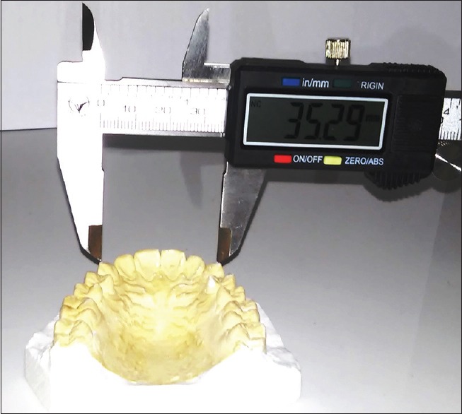
Measurement of intercanine width on study cast
The mesiodistal width of maxillary first molar (MMD) is defined as the greatest distance between the labial surface and the lingual surface of the tooth crown as shown in Figure 3. Both left and right molars were measured, and the average value was taken for calculations.[4]
Figure 3.
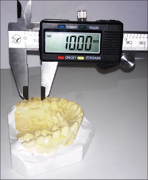
Measurement of mesiodistal width of maxillary first molar of study cast
The BL width of maxillary first molar (MBL) is defined as the greatest distance between the contact points on approximate surface of tooth crown [Figure 4]. Both left and right molars were measured, and the average value was taken for calculations.[4]
Figure 4.
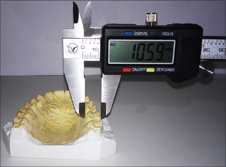
Measurement of buccolingual width of maxillary first molar on study cast
These measurements were then subjected to statistical analyses including descriptive analysis (mean and standard deviation), independent t-test (sexual dimorphism), and linear discriminant analysis using SPSS software version 11 (IBM, Chicago, SPSS Inc., 2009) [p value <0.005].
To assess the gender (y) using tooth dimensions, “discriminant formula” was applied that is:
y = a + b (p)
Where a is the canonical discriminant constant, b is the unstandardized coefficient, and P is the parameter. The constants are obtained from the data given in Table 1. After obtaining the constants, we have generated the formula given below.
Table 1.
Constants used in the formulae

Gender (y) = −0.367 + 0.391 × CMD
Gender (y) = −0.379 + 1.371 × CI
Gender (y) = −18.122 + 18.018 × MMD
Gender (y) = −16.678 + 15.835 × MBL.
The above-mentioned formulae are in format of the discriminant formula y = a + b (p) as mentioned earlier.
Sexual dimorphism is defined as the percentage by which tooth size of males exceeds that of females. The percentage of dimorphism was calculated using the formula.
Sexual dimorphism = (Xm/Xf) − 1 × 100
Where Xm is mean male tooth dimension and Xf is mean female tooth dimension. Sexual dimorphism gives us the percentage value by which the male tooth dimension is greater than the female tooth dimension.
Results
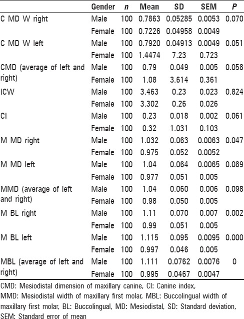
The mean, standard deviation, standard error, and P values of various measurements are tabulated in Table 2. It was observed that the mean value of MD width of canine and CI was greater in females. On the contrary, the mean molar tooth dimensions are greater in males as compared to females (MBL-female = 0.995 cm, MBL-male = 1.11 cm; MMD-female = 0.98 cm, and MMD-male = 1.04 cm). It was observed that P value was highly significant for molar BL diameter (<0.001) and not significant for canine dimensions (CMD = 0.058, CI = 0.061).
Table 2.
Mean, standard deviation, standard error, and P value of all observations were calculated using descriptive analysis
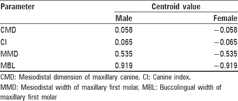
The “y” value that is obtained after applying the discriminant formula for various parameters is compared with group centroids as given in Table 3. The approximate of the “y” value to a particular group centroid value helps us determine the gender of the person.
Table 3.
Centroid value for male and female
The accuracy of gender prediction of each parameter is given in Figure 5. The graph shows that maximum accuracy rate is obtained by molar BL dimension (82.5%) followed by molar MD dimension (69%). Whereas the accuracy rate of CI (51%) and MD width of canine is comparable (50.5%) and is least accurate.
Figure 5.
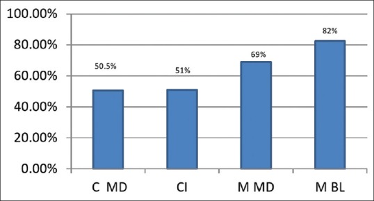
Graphical representation of accuracy of all the parameters of this study
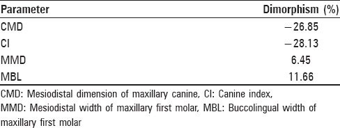
The result of sexual dimorphism for each parameter is given in Table 4. The table shows maximum positive dimorphism for BL width of molar and maximum negative dimension for CI.
Table 4.
Dimorphism of various parameters obtained using independent t-test
Discussion
Determination of sex is of immense importance in forensic investigations. Although DNA analysis is the most precise technique to determine the sex, sometimes lack of facilities and the cost factor may be a hindrance.[5] In such cases, the teeth form an important material as they are hardest and chemically most stable tissues. Their availability even in severe disasters and decomposed bodies makes them invaluable for identification.[6]
Several indices are available for determination of gender such as maxillary incisor width, maxillary CI, maxillary canine width, mandibular CI mandibular canine width, molar width, molar cusp diameter, and cumulative width of all teeth.[7,8,9,10,11,12,13,14] Although some studies[12] employed evaluation of indices on both study models and intraoral measurements, we have not recorded any value clinically to avoid discomfort to the patient and for easy reproducibility of the measurements. We have also encountered studies where right and left side was compared for efficiency, but we have taken average of both side and applied in the formulae.[14] In our study, we have used only maxillary odontometric indices to simulate crime scenes or other scenarios where only skull with maxilla is available, thereby establishing dimorphism. The reason being the possibility in some cases that only skull of the diseased is available. In such case, we recede back to using maxillary odontometric indices such as CI and molar dimensions.
Various theories of teeth dimorphism have been proposed.[14] According to Moss, it is because of the greater thickness of enamel in males due to long period of amelogenesis compared to females and the Y chromosome producing slower male maturation.[14]
The name “canine” is derived from the Latin word for dog canis, as the corresponding teeth are very prominent members of the dentition in these animals. The canine teeth are prominent in other carnivores and also in primates (gorilla, chimpanzee, etc.). It has been postulated that during the evolution of primates, the canines were functionally not masticatory but served the purpose of conveying threat of violence and for prehension of the pray.[15]
A gradual relocation of this aggressive function from the teeth to the fingers took place, but until this transfer was complete, survival was dependent on canines, especially in males. Thus, in humans, sexual dimorphism in the mandibular CI is not merely a coincidence but is the remnant of the past functional activity.[15]
In our present study, we predicted gender using MD width of canine and maxillary CI. It was observed that only 50.5% of cases gave accurate results when MD width of canine was used as a parameter and 51% accuracy was obtained when maxillary CI was used. This proves that no significant difference exists between the dimension of canine teeth for males and females.
Several studies have been done to establish dimorphism of canine teeth.[8,11,16,17,18,19,20,21] It is already established that mandibular canine exhibits the highest dimorphism among all teeth. A study by Kaushal et al.[16] found a statistically significant dimorphism in mandibular canines in sixty participants in North Indian population where the mandibular left canine was seen to exhibit greater sexual dimorphism. According to Kaushal et al., if the width of canine is >7 mm, the probability of the sex of person under consideration being male was 100%. Although knowing this fact, we have confined our study only to the use of maxillary arch to solve the problem encountered in real life-like situation.[16]
Gupta et al. examined 180 participants by taking maxillary arch impression. Significant sexual dimorphism was noticed in MD diameter and intercanine distance of maxillary canine teeth.[17] The sexual dimorphism was 4.2% and 3.6% for right and left, respectively. Studies conducted by Khangura et al. also exhibited a significant sexual dimorphism for maxillary canines. These results are in contrast with our study where we obtained a dimorphism 26.85%.[6]
Mohd. Abdulla et al. conducted a study, in which 513 school students with age ranging from 15 to 18 years were examined.[21] They observed a low degree of sexual dimorphism (not statistically significant) for maxillary canine with correct classification of only 55.07% cases. The results of this study are comparable with the present study where we could classify 51% cases correctly.
Paramkusam et al. in their study observed that standard mandibular CI was found to be more reliable in gender estimation than the MD width of canine and CI values. They found percentage accuracy using both standard maxillary and mandibular canine indices was >70%.[22]
This is in contrast with our study where both MD width of canine and maxillary CI did not yield statistically significant difference (P > 0.005) with accuracy of only 50.5% and 51%, respectively.
In the current study, reverse dimorphism was observed for both CMD (male = 0.79, female = 1.08), and maxillary CI (male = 0.23, female = 0.32) with greater mean values observed in females than males. Hence, negative values were observed while calculating dimorphism of canine. This finding is in concurrence with Boaz and Gupta,[8] Acharya and Mainali,[9] and Yuen et al.[23] who also found negative canine dimorphism in their studies.
Maxillary molars are the largest and strongest teeth owing to their greater crown bulk and excellent anchorage of their multiple roots. The maxillary first molars begin to calcify at birth and erupt around 6 years of age. As it completes development before skeletal maturity, it is a more reliable indicator in gender determination. Agnihotri and Sikri,[14] Rai et al.,[16] Prathibha Rani et al.,[24] Narang et al.,[25] and Sonika et al.[26] have done studies earlier for establishing molar dimorphism. Although some studies have compared efficiency if both right and left molars, in our study, we have taken the average and calculated the results. The above-mentioned studies included either or both BL dimensions and MD dimensions.
Rai et al. measured maxillary first molar dimensions in 102 samples with age ranging from 17 to 25 years. The results showed a statistically significant sexual dimorphism for maxillary first molar with a higher percentage for BL dimension. The probability of gender being male was 100% when the dimension was >10.7 mm.[16]
Prathibha Rani et al. observed that sexual dimorphism could be estimated using BL dimension of permanent teeth, which is population specific. The study identified the sex of an individual based on BL dimension of permanent teeth except third molar which showed that males exhibit greater BL dimensions when compared to females. They found moderate magnitude of dimorphism with accuracy rate of 78% in maxillary teeth which is comparable to our results where we observed that BL dimension of molar is a reliable indicator giving an accuracy rate of 82%.[24]
In a similar study conducted by Narang et al.[25] on a total of 150 individuals (75 males and 75 females), BL dimension of first maxillary and mandibular molars was found to be more significant. They observed that the mean value of maxillary cast exhibited significant dimorphism compared to mandibular cast with an accuracy rate of 74% and 63% for right and left BL dimension of maxillary first molar.[25] In our study, we obtained an accuracy of 82% for maxillary first molar BL dimension, whereas mandibular first molar was not considered since none of the mandibular parameters were included in the study so as to simulate instances where only skull with attached maxilla is available for forensic examination.
Sonika et al. studied population in Haryana with an age group of 17–25 years. Maxillary first molars were measured for both BL and MD dimensions using digital Vernier caliper. Sonika et al. observed that the mean values of left-side parameter were greater than the right-side and BL dimension exhibited greater dimorphism than MD dimension.[26] In the present study, we also obtained similar findings that BL dimension is more reliable indicator than MD dimension.
Conclusion
Identification is a confronting subject in forensic science, especially is case of mutilated body.[27] In such situations, gender determination is a primary step whereby one can reach toward human individual identity, as it increases the chances of identification by 50%. Odontometric analysis can take a step ahead in gender prediction.
In the present study, BL dimension of maxillary first molar showed a higher gender prediction accuracy of 82%, whereas maxillary canine gave a reverse dimorphism of − 26.8%. We conclude that BL dimension of maxillary first molar could be used to predict the gender when only maxillary teeth are available for forensic examination. Future studies on varied population groups with higher sample size might further establish the usefulness of maxillary first molar dimensions in gender determination.
Financial support and sponsorship
Nil.
Conflicts of interest
There are no conflicts of interest.
References
- 1.Marques J., Musse J., Caetano C., Corte-Real F., Corte-Real A. T. Analysis of Bite Marks in Foodstuffs by Computer Tomography (Cone Beam CT)-3D Recontruction. JFOS Online. 2013;31:1–7. [PMC free article] [PubMed] [Google Scholar]
- 2.Sweet D. Why a dentist for identification? Dent Clin North Am. 2001;45:237–51. [PubMed] [Google Scholar]
- 3.Parekh DH, Patel SV, Zalawadia AZ, Patel SM. Odontometric study of maxillary canine teeth to establish sexual dimorphism in Gujarat population. Int J Biol Med Res. 2012;3(3):1935–7. [Google Scholar]
- 4.Sharma PP, Kumar TS, Chandra P. Sex determination potential of permanent maxillary molar widths and cusp diameters in a North Indian population: J Orthod Sci. 2013;2(2):55–60. doi: 10.4103/2278-0203.115090. [DOI] [PMC free article] [PubMed] [Google Scholar]
- 5.Reddy VM, Saxena S, Bansal P. Mandibular canine index as a sex determinant: A study on the population of western Uttar Pradesh: Journal of Oral and Maxillo Facial Pathology. 2008;12(2):56–9. [Google Scholar]
- 6.Khangura RK, Sircar K, Singh S, Rastogi V. Sex determination using mesiodistal dimension of permanent maxillary incisors and canines. Journal of forensic dental sciences. 2011;2:81. doi: 10.4103/0975-1475.92152. [DOI] [PMC free article] [PubMed] [Google Scholar]
- 7.Yuwanti M, Karia A, Yuwanti M. Canine tooth dimorphism: An adjunct for establishing sex identity: Journal of Forensic Dental Sciences. 2012;4(2):80–3. doi: 10.4103/0975-1475.109892. [DOI] [PMC free article] [PubMed] [Google Scholar]
- 8.Boaz K, Gupta C. Dimorphism in human maxillary and madibular canines in establishment of gender. Journal of Forensic Dental Sciences. 2009;1(1):42–4. [Google Scholar]
- 9.Acharya BA, Mainali S. Univariate sex dimorphism in the Nepalese dentition and use of discriminant functions in gender assessment. Forensic SciInt. 2007;173:47–56. doi: 10.1016/j.forsciint.2007.01.024. [DOI] [PubMed] [Google Scholar]
- 10.Rao NG, Rao NN, Pai ML, Kotian MS. Mandibular canineindex: A clue for establishing sex identity. Forensic SciInt. 1989;42:249–54. doi: 10.1016/0379-0738(89)90092-3. [DOI] [PubMed] [Google Scholar]
- 11.Yadav S, Nagabhushan D, Rao BB, Mamatha GP. Mandibular canine index in establishing sex identity. Indian J Dent Res. 2002;13:143–6. [PubMed] [Google Scholar]
- 12.Perzigian AJ. The dentition of the Indian Knoll skeletal population: Odontometrics and cup number. Am J PhysAnthropol. 1976;44(1):113–21. doi: 10.1002/ajpa.1330440116. [DOI] [PubMed] [Google Scholar]
- 13.Acharya BA. Sex determination potential of bucco lingual and mesiodistaldimensions. J Forensic sci. 2007;173:47–56. [Google Scholar]
- 14.Agnihotri G, Sikri V. Crown and cusp dimensions of the maxillary first molar: A study of sexual dimorphism in indianjatsikhs. Dental anthropology. 2010;23(1):1–6. [Google Scholar]
- 15.Phulari RG. Permanent Maxillary Canine. In: Rashmi GS, Phulari, editors. Textbook of dental anatomy, Physiology and Occlusion. 1ed. New Delhi: Jaypee brothers medical publishers; 2014. pp. 142–6. [Google Scholar]
- 16.Rai B, Jain RK, Duhan J, Dutta S, Dhattarwal S. Evidence of tooth in sex determination. An International Journal ofMedico-Legal Update. 2004;4(4):119–26. [Google Scholar]
- 17.Kaushal S, Patnaik VVG, Agnihotri G. Mandibular Canines in Sex Determination. J.Anat. Soc. India. 2003;52(2):119–24. [Google Scholar]
- 18.Gupta S, Chandra A, Gupta OP, Verma Y, Srivastava S. Establishment of Sexual Dimorphism in North Indian Population by Odontometric Study of Permanent Maxillary Canine. Journal of Forensic Research. 2014;5(2):1. [Google Scholar]
- 19.Kuwana T. On sex difference of maxillary canines observed in the moiretribes. NihonUniv Dent J. 1983;57:88. [Google Scholar]
- 20.Hashim HA, Murshid ZA. Mesiodistal tooth width- a comparison between Saudi males and females. Egypt Dent J. 1993;39:343–6. [PubMed] [Google Scholar]
- 21.Al-Rifaiy MQ, Abdullah MA. Dimorphism of mandibular and maxillary canine teeth in establishing sex identity. Saudi Dental Journal. 1997;1:17–20. [Google Scholar]
- 22.Paramkusam G, Nadendla LK, Devulapalli RV, Pokala A. Morphometric analysis of canine in gender determination: Revisited in India. Indian J Dent Res. 2014;25:425–9. doi: 10.4103/0970-9290.142514. [DOI] [PubMed] [Google Scholar]
- 23.Yuen KK, So LL, Tang EL. Mesiodistal crown diameters of the primary and permanent teeth in southern Chinese – A longitudinal study. Eur J Orthod. 1997;19:721–31. doi: 10.1093/ejo/19.6.721. [DOI] [PubMed] [Google Scholar]
- 24.Prathibha Rani RM, Mahima VG, Patil K. Bucco-lingual dimension of teeth-An aid in sex determination. Journal of Forensic dental sciences. 2009;1(2):88. [Google Scholar]
- 25.Narang RS, Manchanda A, Singh B. Sex assessment by molar odontometrics in north Indian population. J Forensic Dent Sci. 2015;7:54–8. doi: 10.4103/0975-1475.150318. [DOI] [PMC free article] [PubMed] [Google Scholar]
- 26.Sonika V., Harshaminder K., Madhushankari G.S. Sexual dimorphism in the permanent maxillary First molar: A study of the haryana population (India): J Forensic Odontostomatol. 2011;29(1):37–43. [PMC free article] [PubMed] [Google Scholar]
- 27.Acharya B A, Sivapathasundharam B. Forensic Odontology. In: Rajendran R, Sivapathasundharam B, editors. Shafer's Textbook of Oral Pathology. New Delhi: Elsevier; 2012. pp. 877–904. [Google Scholar]


