Abstract
Patient: Female, 32
Final Diagnosis: Pleomorphic adenoma
Symptoms: Sleep disturbance
Medication: —
Clinical Procedure: Endoscopic assisted surgery
Specialty: Otolaryngology
Objective:
Rare disease
Background:
Pleomorphic adenoma is the most common benign tumor arising in the salivary gland. The signs and symptoms of pleomorphic adenoma of the minor salivary glands vary, depending on the anatomical site involved. A rare case of pleomorphic adenoma of the posterior surface of the soft palate is reported that caused sleep disturbance, which was resolved with endoscopic surgical treatment.
Case Report:
A 32-year-old woman experienced snoring and mouth-breathing during sleep. Flexible fiberoptic nasopharyngoscopy imaging of the oropharyngeal passage showed obstruction by a tumor the soft palate, which obstructed the oropharyngeal passage. The tumor was excised using endoscopic-assisted transoral surgery and measure 3×2 cm in diameter. Histopathology showed a benign pleomorphic adenoma of the minor salivary gland. Following surgical excision of the tumor, the patient’s sleep improved.
Conclusions:
To our knowledge, this is the first case of a pleomorphic adenoma of the posterior surface of the soft palate, causing sleep disturbance, removed by endoscopic-assisted transoral surgery following pre-operative flexible fiberoptic nasopharyngoscopy imaging of the oropharyngeal passage.
MeSH Keywords: Adenoma, Pleomorphic; Endoscopy; Palate, Soft; Sleep
Background
Salivary gland tumors are relatively rare, with an incidence of approximately 3–10% of all tumors arising in the head and neck region [1]. Pleomorphic adenoma, also known as ‘benign mixed tumor,’ is the most common benign tumor arising in the salivary gland, and grows slowly and painlessly [1]. The signs and symptoms of pleomorphic adenoma of the minor salivary glands vary, depending on the anatomical site involved [2–5]. The pathophysiological patterns and etiology of pleomorphic adenoma of the minor salivary glands remain unclear.
We present a case of a pleomorphic adenoma, arising in a minor salivary gland of the posterior surface of the soft palate, which caused sleep disturbance. The tumor was successfully removed by endoscopic-assisted transoral surgery following pre-operative flexible fiberoptic nasopharyngoscopy imaging of the oropharyngeal passage.
Case Report
A 32-year-old woman presented with symptoms of snoring and mouth-breathing during sleep and was admitted to the Department of Otorhinolaryngology, Bayindir Government Hospital, Izmir, Turkey. The patient’s problems with sleeping had increased over the previous two years. The patient had no episodes of sleep apnea or daytime somnolence.
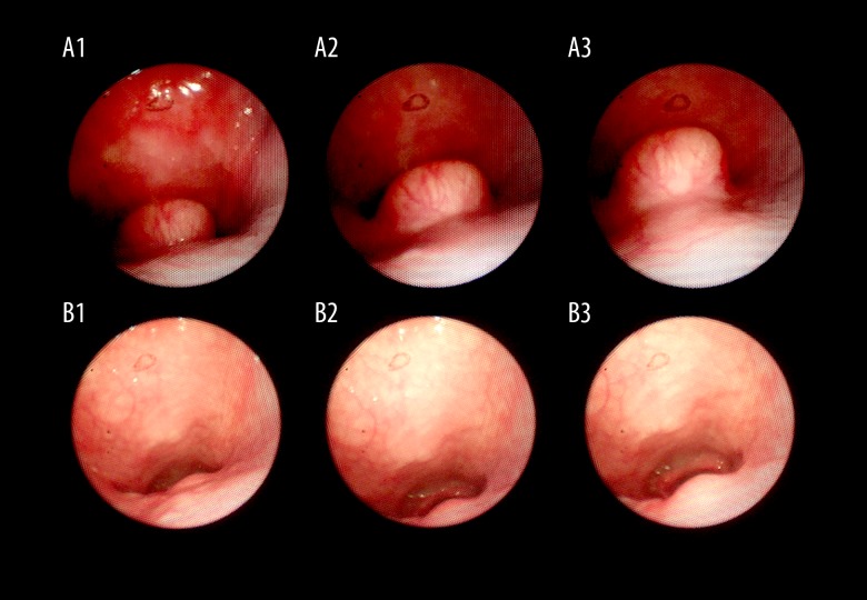
On initial oral and nasal physical examination, there were no significant clinical findings. However, on flexible fiberoptic nasopharyngoscopy imaging of the oropharyngeal passage, a tumor was seen on the midline of the posterior surface of the soft palate. The mass was approximately 3×2 cm in diameter (Figure 1A). On palpation of the tumor, there was no pain, and the mass was firm and ‘rubbery’ with well-defined margins. There was no significant lymph node enlargement in the head and neck.
Figure 1.
Pre-operative and postoperative flexible fiberoptic nasopharyngoscopy imaging of the oropharynx. (A) Pre-operative flexible fiberoptic nasopharyngoscopy imaging of the oropharyngeal passage shows obstruction by tumor. (B) Postoperative flexible nasopharyngoscopy evaluation. The oropharyngeal passage is open and the arytenoid cartilages can be seen.
The patient was evaluated using the Epworth sleepiness scale (ESS) to evaluate sleep quality and daytime sleepiness. The ESS is a short questionnaire of eight questions with a scale from 0–3 for each question, giving a possible total score out of 24. The patient’s ESS score was 4. Polysomnography (PSG), or a sleep study, was not performed.
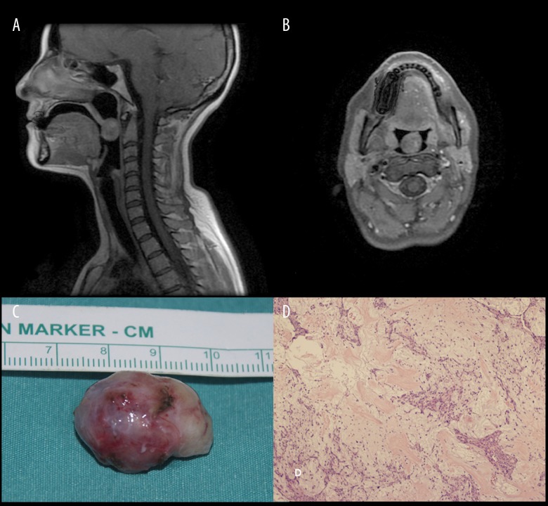
Pre-operative cranial sagittal and axial magnetic resonance imaging (MRI) of the oropharynx showed a round, well-defined mass, measuring 2.5×2 cm in diameter on the posterior surface of the soft palate that was obstructing the oropharyngeal passage (Figure 2A, 2B).
Figure 2.
Pre-operative magnetic resonance imaging (MRI) and postoperative macroscopic appearance and histopathology of the pleomorphic adenoma. Pre-operative cranial sagittal (A) and axial (B) magnetic resonance imaging (MRI) shows the oropharyngeal passage is occluded by the tumor. (C) The macroscopic appearance of the excised tumor. (D) Photomicrograph of the tumor histology shows a mixture of epithelial and myoepithelial cells in a myxoid stroma, consistent with a diagnosis of a benign pleomorphic adenoma arising from a minor salivary gland. Hematoxylin and eosin (H&E) stain. Objective magnification ×10.
Surgery was performed under general anesthesia, with orotracheal intubation. The oropharynx was exposed using a Davis-Crowe mouth gag and Russell-Davis tongue blade. The uvula was suspended, and part of the mass was observed on the posterior surface of the soft palate. A horizontal incision was made over the mass. A mucosal flap was removed using endoscopic visualization due to the limited view of the tumor margin. The tumor was removed using blunt dissection along the cleavage plane using 45° and 70° rigid endoscopes. Following en-bloc tumor resection, the mucosal flap was replaced and sutured with absorbable sutures.
The excised tumor was fixed in 10% neutral-buffered formalin. On macroscopic examination, the tumor was well-defined and measured 25×20×15 mm (Figure 2C). Sections were taken for histopathology. Light microscopic evaluation of the hematoxylin and eosin (H&E) stained tissue sections showed a biphasic population of epithelial and myoepithelial cells. Spindle-shaped and oval-shaped myoepithelial cells with hyperchromatic nuclei were located within myxoid stroma (Figure 2D). No cell atypia, no mitoses, no necrosis, and no malignant features were present histologically. The histological appearances were typical for a diagnosis of benign pleomorphic adenoma, which was completely excised. Following surgery, the patient was prescribed mild analgesia, consisting of paracetamol, 3×500 mg/day for seven days.
There were no postoperative complications, and the patient had no symptoms of velopharyngeal insufficiency (VPI), or inability to close the communication between the nasal cavity and the mouth, which can sometimes occur following surgery to the soft palate.
Postoperatively, the patient’s snoring was reduced, and her ESS score was reduced to 2 (out of 24). The patient remains symptom-free at six months following surgery, without tumor recurrence (Figure 1B), or speech disorder.
This case report was approved for publication by our institute and written informed consent was obtained from the patient.
Discussion
The most common benign tumor of the minor salivary glands is a pleomorphic adenoma, or ‘benign mixed tumor,’ which has an incidence ranging from 23–70% of all salivary neoplasms [2–4] The most common site for a pleomorphic adenoma of the minor salivary gland is the palate [5]. Pleomorphic adenoma can occur at any age, but the mean age is 47 years for women and 51 years for men, and the tumor is slightly more common in women [5].
The signs and symptoms of pleomorphic adenoma of the minor salivary gland vary according to their anatomical site of origin. The usual clinical history is that of a slow-growing tumor that has been present for a long time. The most frequent symptom is a non-ulcerative submucosal mass; other symptoms include obstruction of the nose, Eustachian tube dysfunction, altered vocalization, hearing loss, dyspnea, and hoarseness [6]. The usual approach to the treatment of pleomorphic adenoma is surgical excision [7]. Enucleation alone is not favored because of the possibility of local recurrence [7].
Review of the published literature has shown that previous case reports provide differing views of the effects of treatment of airway defects on sleep-related respiratory disorders. Nasal septum deviation or septal perforation repair surgery [8,9] has not been shown to change the pre-operative and postoperative apnea or hypopnea values [8,9]. However, postoperative reduction in apnea or hypopnea were shown following endoscopic sinus surgery in patients with nasal polyps, or after excision of parapharyngeal space tumors [10,11]. The location and size of the defect in the airway, and the nature of the underlying pathology may result in different responses to the effects of treatment on sleep disturbance. The obstructive effects of benign tumors in the oropharyngeal passage have been previously reported to be associated with sleep dysfunction, and effectively treated with surgery [12].
In this case report, due to the patient’s clinical history, pre-operative Epworth sleepiness scale (ESS) score of 4 (out of 24), and with consideration cost implications, we did not perform polysomnography (PSG), or a sleep study, before and after surgery. However, we believe that if the patient’s sleep-related symptoms had persisted following excision of the benign pleomorphic adenoma, located in a key part of the airway, PSG evaluation should have been performed. This view is supported by Casale and colleagues, who have suggested that if there is an underlying mechanical problem in patients with snoring, treatment of the underlying cause should be done first [13]. Also, a previous report in a patient with a giant pleomorphic adenoma arising from the right side of the pharynx, who was diagnosed with obstructive sleep apnea and treated with continuous positive airway pressure (CPAP) did not require CPAP therapy following surgery [14].
In this case report, a full ear, nose, and throat (ENT) examination were performed, including the use of a flexible fiberoptic nasopharyngoscope and laryngoscope, which were required to identify the tumor. Our strategy was to completely remove the tumor and the surrounding soft tissue under general anesthesia, sparing important tissues such as muscle and mucosa. From our experience of this case, for a well-encapsulated tumor, en-bloc excision with a mucosal flap created under endoscopic view is a recommended surgical option. In appropriate cases, minimally invasive surgery, rather than wide surgical resection may both reduce the risk of surgical complications and improve the postoperative quality of life for the patient, as in this case.
Conclusions
To our knowledge, this is the first case of a pleomorphic adenoma of the posterior surface of the soft palate, which was removed by endoscopic-assisted transoral surgery following pre-operative flexible fiberoptic nasopharyngoscopy imaging of the oropharyngeal passage. Review of the literature has also shown that there have been few previously reported cases of pleomorphic adenoma causing sleep disturbance.
Footnotes
Conflict of interest
None.
References:
- 1.Ansari MH. Salivary gland tumors in an Iranian population: A retrospective study of 130 cases. J Oral Maxillofac Surg. 2007;65:2187–94. doi: 10.1016/j.joms.2006.11.025. [DOI] [PubMed] [Google Scholar]
- 2.Qureshi MY, Khan TA, Dhurjati VN, et al. Pleomorphic adenoma in retromolar area: A very rare case report and review of literature. J Clin Diagn Res. 2016;10:ZD03–05. doi: 10.7860/JCDR/2016/16269.7067. [DOI] [PMC free article] [PubMed] [Google Scholar]
- 3.Ramesh M, Krishnan R, Paul G. Intraoral minor salivary gland tumours: A retrospective study from a dental and maxillofacial surgery centre in salem, Tamil Nadu. J Maxillofac Oral Surg. 2014;13:104–8. doi: 10.1007/s12663-013-0489-4. [DOI] [PMC free article] [PubMed] [Google Scholar]
- 4.Isacsson G, Shear M. Intraoral salivary gland tumors: A retrospective study of 201 cases. J Oral Pathol. 1983;12:57–62. doi: 10.1111/j.1600-0714.1983.tb00316.x. [DOI] [PubMed] [Google Scholar]
- 5.Takahashi H, Fujita S, Tsuda N, et al. Intraoral minor salivary gland tumors: A demographic and histologic study of 200 cases. Tohoku J Exp Med. 1990;161:111–28. doi: 10.1620/tjem.161.111. [DOI] [PubMed] [Google Scholar]
- 6.Maruyama A, Tsunoda A, Takahashi M, et al. Nasopharyngeal pleomorphic adenoma presenting as otitis media with effusion: Case report and literature review. Am J Otolaryngol. 2014;35:73–76. doi: 10.1016/j.amjoto.2013.08.019. [DOI] [PubMed] [Google Scholar]
- 7.Vlaykov A, Vicheva D. Nasal pleomorphic adenoma – a case report. Int J Sci Res. 2015;4:77–79. [Google Scholar]
- 8.Boynuegri S, Cayonu M, Tuna EU, et al. The effect of nasal septal perforation and its treatment on objective sleep and breathing parameters. Med Sci Monit. 2016;22:501–7. doi: 10.12659/MSM.897531. [DOI] [PMC free article] [PubMed] [Google Scholar]
- 9.Li HY, Lin Y, Chen NH, et al. Improvement in quality of life after nasal surgery alone for patients with obstructive sleep apnea and nasal obstruction. Arch Otolaryngol Head Neck Surg. 2008;134:429–33. doi: 10.1001/archotol.134.4.429. [DOI] [PubMed] [Google Scholar]
- 10.Uz U, Gunhan K, Yilmaz H, et al. The evaluation of pattern and quality of sleep in patients with chronic rhinosinusitis with nasal polyps. Auris Nasus Larynx. :2017. doi: 10.1016/j.anl.2017.01.015. [Epub ahead of print] [DOI] [PubMed] [Google Scholar]
- 11.Mulla O, Agada F, Dawson D, et al. Obstructive sleep apnoea and snoring – is examination necessary? Aust Fam Physician. 2011;40:886–88. [PubMed] [Google Scholar]
- 12.Wang AY, Wang JT, Levin B, et al. Parapharyngeal pleomorphic adenoma as a cause of severe obstructive sleep apnoea. ANZ J Surg. 2014;84:883–84. doi: 10.1111/ans.12330. [DOI] [PubMed] [Google Scholar]
- 13.Casale M, Salvinelli F, Mallio CA, et al. Upper airway study should always come before any sleep study in OSAS evaluation: A giant parapharyngeal lipoma behind OSAS. Eur Rev Med Pharmacol Sci. 2012;16(S4):106–9. [PubMed] [Google Scholar]
- 14.Adams AJ, Patterson AR, Brady G, et al. Resolution of obstructive sleep apnoea after resection of a pleomorphic salivary adenoma. Br J Oral Maxillofac Surg. 2008;46:53–54. doi: 10.1016/j.bjoms.2006.09.015. [DOI] [PubMed] [Google Scholar]


