Abstract

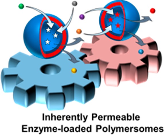
Enzyme loading of polymersomes requires permeability to enable them to interact with the external environment, typically requiring addition of complex functionality to enable porosity. Herein, we describe a synthetic route toward intrinsically permeable polymersomes loaded with functional proteins using initiator-free visible light-mediated polymerization-induced self-assembly (photo-PISA) under mild, aqueous conditions using a commercial monomer. Compartmentalization and retention of protein functionality was demonstrated using green fluorescent protein as a macromolecular chromophore. Catalytic enzyme-loaded vesicles using horseradish peroxidase and glucose oxidase were also prepared and the permeability of the membrane toward their small molecule substrates was revealed for the first time. Finally, the interaction of the compartmentalized enzymes between separate vesicles was validated by means of an enzymatic cascade reaction. These findings have a broad scope as the methodology could be applied for the encapsulation of a large range of macromolecules for advancements in the fields of nanotechnology, biomimicry, and nanomedicine.
Compartmentalization is essential for all forms of life. For instance, cells and organelles can interact with one another through enzyme cascades, and through transport of signaling molecules, energy and nutrients. Mimicry of these natural constructs using synthetic materials is of fundamental scientific interest and could also lead to advances in bionanotechnology and nanomedicine.1−3 Self-assembled bilayer structures, such as liposomes, used to encapsulate functional macromolecules, can be considered as minimal artificial cells or protocells.2,4 Amphiphilic block copolymer vesicles (also termed polymersomes) have been studied widely as such protocells, owing to their higher mechanical strength and easier functionalization when compared to liposomes. For example, Lecommondoux and van Hest et al. demonstrated that poly(styrene)-b-poly(3-(isocyano-l-alanyl-aminoethyl)thiophene)) (PS-b-PIAT) vesicles loaded with enzymes could be encapsulated inside a larger poly(butadiene)-b-poly(ethylene glycol) (PB-b-PEG) polymersome to create a multicompartmentalized polymersome-in-polymersome system, which structurally resembled a cell and its organelles.5 The encapsulated enzymes were able to interact via an enzymatic cascade reaction. Reactants and products were able to diffuse between the enzyme-containing compartments owing to the intrinsic permeability of these PS-b-PIAT polymersomes, comprising specialty monomers and polymerization techniques. While examples of enzyme-loaded polymersome nanoreactors are numerous,1−14 examples of membrane-forming polymers with intrinsic permeability are rarely reported. As such, the nonpermeable nature of the membrane must be overcome by the incorporation of membrane proteins13,15,16 or DNA nanopores17 into the polymersome membrane, postassembly radical photoreactions,12 or the use of stimuli-responsive membranes14,18−20 to impart permeability. Furthermore, the preparation of block copolymer vesicles often requires the use of organic solvents, which may be incompatible with the protein of interest. Additionally, conventional self-assembly procedures are typically performed at low concentrations, and require multiple synthetic and purification steps, which limits their scalability.
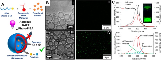
Herein, we report the intrinsic permeability of poly(ethylene glycol)-b-poly(2-hydroxypropyl methacrylate) (PEG-b-PHPMA) vesicles formed by a one-step aqueous, initiator-free, visible light-mediated polymerization-induced self-assembly (photo-PISA) route using commercial reagents (Figure 1A). This approach overcomes many of the challenges discussed for conventional protein-loaded block copolymer vesicles, such as their lack of intrinsic permeability. PISA shows numerous advantages over other self-assembly techniques, such as the use of purely aqueous conditions and high concentrations (in this case, 110 mg·mL–1).21−31 This single, rapid assembly methodology could feasibly be applied to the encapsulation of a range of functional proteins. Until very recently, bovine serum albumin (BSA), a robust nonfunctional protein, was the only protein to have been successfully encapsulated inside a PISA-derived vesicle.26,32 In more recent work, BSA-functionalized PISA-derived nanoparticles were prepared by direct polymerization of HPMA from the chain transfer agent (CTA)-functionalized protein.33 Very recently, Tan and Zhang et al. described enzyme-assisted photoinitiated PISA using glucose oxidase to degas the solution by reduction of dissolved oxygen.34 While this achieved enzyme-loaded vesicles by PISA, the permeable nature of the PHPMA membrane was not investigated and so the authors were unable to show that the enzyme remained active while encapsulated inside the vesicle.
Figure 1.
(A) Preparation of inherently permeable protein-loaded nanoreactors by aqueous PISA. (B) Cryo-TEM image and fluorescence micrograph of empty vesicles (I, II) and GFP-loaded vesicles (III, IV). (C; top) Fluorescence spectra of the 1st supernatant from the purification (red) and the untreated protein (black). The inset shows a digital photograph of the crude reaction mixture under a UV lamp. (C; bottom) Fluorescence spectra of the 1st supernatant (red), disassembled vesicle solution (green), and 2nd supernatant (blue) in a 5:1 methanol/water solvent mixture. In each case, the excitation spectra (λEm = 512 nm, dashed traces) and the emission spectra (λEx = 495 nm, solid traces) are shown.
Polymer Synthesis and Preparation of GFP-Loaded Vesicles
Aqueous photo-PISA without the need for an initiator was employed to prepare polymersomes comprising a PEG shell and a PHPMA membrane (Figure 1A). Such membranes are highly hydrated,22 which was hypothesized to allow size-selective transport of small molecules while retaining the encapsulated protein. From our previous work on photoinitiated PISA,35 a PEG113-b-PHPMA400 block copolymer formed at 10 wt % HPMA resulted in the reliable preparation of unilamellar vesicles when photoinitiated PISA was employed at 37 °C. This point on the phase diagram was used for the preparation of all protein-loaded vesicles discussed. Size exclusion chromatography (SEC) of each of the polymers confirmed that the diblock copolymer synthesis was not strongly affected by the presence of the encapsulated proteins in the reaction mixture (Figure S1). The empty vesicles were characterized by dry state stained transmission electron microscopy (TEM), cryogenic TEM (cryo-TEM) and dynamic light scattering (DLS) analysis (Figure S2). Cryo-TEM confirmed the vesicles’ unilamellar character (Figure 1B-I), and statistical analysis from 200 particles revealed an average membrane thickness of 25 ± 4 nm.
Encapsulation of a recombinant enhanced green fluorescent protein (GFP) was achieved by dissolution of the PEG113 macro CTA and HPMA monomer in an aqueous solution of GFP followed by irradiation at 400–410 nm at 37 °C to initiate the PISA. The GFP-loaded vesicles were purified from the unencapsulated protein by three centrifugation-resuspension cycles at relatively low centrifugal forces (16000 × g for 10 min) such that the unencapsulated protein remained in the supernatant after each cycle. The vesicles were fully characterized after purification (Figures S3 and 1B-III). The average membrane thickness of the vesicles was determined by cryo-TEM to be 52 ± 12 nm. This membrane thickness was significantly higher than that of the empty vesicles (25 ± 4 nm). This was attributed to the presence of residual salts present following the protein purification, which have an effect on the PISA assembly process, as will be discussed later when glucose oxidase (GOx)-loaded vesicles are explored. Fluorescence spectroscopy of the first supernatant from the purification revealed a loss in fluorescence after the PISA process relative to fresh GFP, which was attributed to photobleaching during the light-mediated polymerization;36 however, 56% of the protein’s fluorescence was retained (Figure 1C, top). Additionally, the purified vesicles’ fluorescence could be assessed in order to calculate the loading efficiency of the protein. A loading efficiency of 11 ± 2%, was calculated upon disassembly of the vesicles followed by fluorescence spectroscopy (Figure 1C, bottom). Figure 1B shows a fluorescence micrograph of the GFP-loaded vesicles, which demonstrated that the fluorescent protein was compartmentalized into discrete pockets inside the vesicles’ lumens. Control experiments wherein empty PISA vesicles containing no GFP were incubated with GFP and irradiated with light under the same conditions, followed by an identical purification procedure, revealed no fluorescent features (Figure S4), demonstrating that the fluorescence observed was from encapsulated protein and not from protein adhered to the particles’ surface.
Catalytic HRP-Loaded Nanoreactors
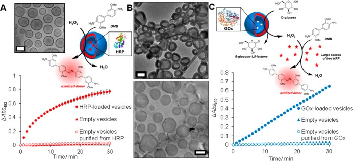
Horseradish peroxidase (HRP) was used as a model enzyme for encapsulation to prepare PISA-derived vesicles with a catalytic function. These HRP-loaded vesicles were prepared using a similar method to that used for the GFP-loaded vesicles and gave a broad, unimodal distribution of particle sizes, as characterized by DLS, dry state TEM, and cryo-TEM (Figures S5 and 2A). Again, the particles survived centrifugation and were also stable to purification by preparative SEC to ensure the complete removal of unencapsulated protein species. The activity of the HRP loaded inside the vesicle was assessed using a colorimetric assay, in which HRP was used to catalyze the oxidation of 3,3′-dimethoxybenzidine (DMB) to a colored dimer product (Figure 2A). It was found that the enzyme’s activity was retained despite its compartmentalization from the external medium. This demonstrated the permeability of the moderately hydrophobic PHPMA membrane-forming block toward DMB and hydrogen peroxide. To ensure complete removal of the free protein, the purification methodology was interrogated and several control experiments were performed. It was demonstrated that the proteins were not adhering to the particles’ surface by incubating the free protein with empty vesicles with light irradiation. Following an identical purification procedure, the activity of this control was shown to be negligible (Figure 2A). The preparative SEC traces of the HRP-loaded vesicles and of the free, unencapsulated HRP also revealed that the retention time of the very high molar mass vesicles was much lower than that of the free protein, such that the fractions collected for assessment of the vesicles’ activity did not contain any free enzyme in solution (Figure S6). These control experiments demonstrated that the observed activity in the HRP-loaded vesicles was solely a result of encapsulated protein and not that of protein adhered physically or chemically to the vesicle surface. Western blot analysis of the disassembled vesicles revealed a loading efficiency of 27.5%. This efficiency could also be increased to 54% by increasing the initial protein feed 5-fold. The encapsulated enzyme retained 57 ± 13% of its activity relative to the free enzyme (Figure S7). The loss in activity was attributed to the diffusive barrier of the membrane, which hampers the substrates in reaching the active site of the enzyme.
Figure 2.
(A, inset) Cryo-TEM image of HRP-loaded vesicles. (A, top) Schematic showing the HRP-catalyzed oxidation of DMB to its colored dimer product detected in the colorimetric assay. (A, bottom) Activity of the HRP-loaded vesicles against negative controls. (B) Cryo-TEM images of GOx-loaded vesicles using commercial GOx (top) and purified GOx (bottom). (C, top) Schematic showing the GOx-catalyzed oxidation of d-glucose to δ-glucono-1,5-lactone. Excess HRP in the external solution was then used to catalyze the oxidation of DMB using hydrogen peroxide product from the first step. (C, bottom) Activity of the GOx-loaded vesicles against negative controls. The error bars in A and C show the standard deviation from four repeats. All scale bars = 200 nm.
Preparation of GOx-Loaded Vesicles and Cascade Activity
To expand the scope of enzyme-loaded PISA vesicles, those loaded with GOx were prepared as this protein has been shown to be able to interact with HRP by means of a catalytic cascade reaction.8,14 GOx-loaded vesicles were prepared in a similar manner to HRP- and GFP-loaded vesicles and the purified particles were fully characterized (Figures S8 and 2B). Similar to the GFP-loaded vesicles, cryo-TEM of the GOx-loaded vesicles showed significantly thicker membranes than the empty vesicles or the HRP-loaded vesicles. This was attributed to the purity of the commercially available GOx, which is supplied as a lyophilized powder with salts and other stabilizing agents. Vesicles loaded with a purified, desalted GOx had an identical membrane thickness to the empty vesicles, within error, indicating that the protein itself did not have an effect on the PISA assembly process (Figure 2B). GOx catalyzes the oxidation of d-glucose to δ-glucono-1,5-lactone and produces hydrogen peroxide as a byproduct. Since this reaction gives no colorimetric output, a large excess of fresh, unencapsulated HRP was employed, along with DMB, in order to facilitate the oxidation of DMB to its colored dimer product using hydrogen peroxide as a rate limiting substrate (Figure 2C). In this way, the activity of the GOx could be measured. Similar to HRP-loaded vesicles, the GOx-loaded vesicles also showed catalytic activity, whereas the empty vesicles, and the empty vesicles purified from a GOx-containing solution after light irradiation, showed no appreciable activity (Figure 2C). Similar to HRP, Western blot analysis of the disassembled vesicles revealed a loading efficiency of 24%, and again, this could be increased to 41% by increasing the initial protein feed 5-fold. Relative to the free enzyme, the encapsulated enzyme retained 46 ± 1% of its activity, owing to the diffusive barrier imparted by the membrane (Figure S9).
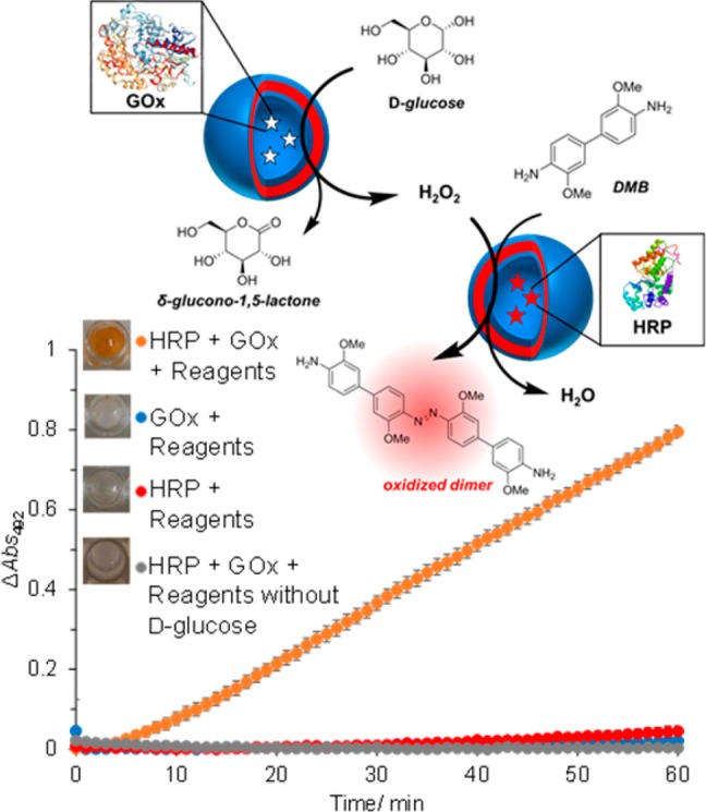
Once it had been shown that substrates for both GOx and HRP could permeate through the polymersome membrane, it was possible to assess if these two enzymes, separated in space by compartmentalization of their vesicle lumens, could interact with one another by utilization of a cascade reaction. HRP- and GOx-loaded vesicles were prepared in separate PISA formulations, purified as described previously and mixed in a 1:1 ratio in phosphate buffer. DMB was added as a reporter to give a colorimetric output, followed by d-glucose to initiate the cascade. Figure 3 shows that the cascade proceeded only when all components were present in solution. Controls where just one species was present, or when d-glucose was absent, showed no activity. This further demonstrated the permeability toward small molecules owing to the hydrated nature of PHPMA, allowing the crossing of substrates and products both in and out of the vesicle membrane. Relative to the free enzymes, the cascade proceeded with 46 ± 3% activity, owing to the hindered passage of substrates through the membrane (Figure S10).
Figure 3.

Schematic and activity of the cascade reaction between HRP- and GOx-loaded vesicles with negative controls. Error bars show the standard deviation from four repeats. Insets: End-point photographs of wells after 1.5 h.
We report for the first time the intrinsically permeable nature of PHPMA membranes toward a range of small molecule substrates. Functional proteins could be retained within compartmentalized domains and preserve their function, as revealed by fluorescence microscopy of GFP-loaded vesicles. Enzymes were also found to remain active, owing to the intrinsic permeability of the membrane, such that the compartmentalized enzymes could interact by way of a cascade reaction. This one-pot, mild approach is highly versatile and could be applied to a range of functional enzymes. As PHPMA is one of the most reported monomers for aqueous PISA found in the literature, it is expected that this phenomenon will be widely exploited for a range of biorelated applications.
Acknowledgments
This work was supported by EPSRC, BBSRC (BB/M017982/1), and ERC (638661 and 615142). We thank Dr. E. Fullam, UoW, for the GFP plasmid and Dr. M. Hasan, UoW, for assistance with protein extraction. Dr. S. Bakker and Mr. R. Keogh, UoW, are thanked for cryo-TEM assistance and Advanced BioImaging Research Technology Platform, BBSRC ALERT14 award BB/M01228X/1 is thanked for supporting cryo-TEM analysis.
Supporting Information Available
The Supporting Information is available free of charge on the ACS Publications website at DOI: 10.1021/acsmacrolett.7b00725.
Particle characterization, SEC traces including preparative SEC traces of the HRP-loaded vesicles, calculation of loading efficiencies and GFP control images (PDF).
Author Contributions
The manuscript was written through contributions of all authors. All authors have given approval to the final version of the manuscript.
The authors declare no competing financial interest.
Supplementary Material
References
- Gaitzsch J.; Huang X.; Voit B. Engineering Functional Polymer Capsules toward Smart Nanoreactors. Chem. Rev. 2016, 116, 1053–1093. 10.1021/acs.chemrev.5b00241. [DOI] [PubMed] [Google Scholar]
- Buddingh’ B. C.; van Hest J. C. M. Artificial Cells: Synthetic Compartments with Life-like Functionality and Adaptivity. Acc. Chem. Res. 2017, 50, 769–777. 10.1021/acs.accounts.6b00512. [DOI] [PMC free article] [PubMed] [Google Scholar]
- Palivan C. G.; Goers R.; Najer A.; Zhang X.; Car A.; Meier W. Bioinspired polymer vesicles and membranes for biological and medical applications. Chem. Soc. Rev. 2016, 45, 377–411. 10.1039/C5CS00569H. [DOI] [PubMed] [Google Scholar]
- Roodbeen R.; van Hest J. C. M. Synthetic cells and organelles: compartmentalization strategies. BioEssays 2009, 31, 1299–1308. 10.1002/bies.200900106. [DOI] [PubMed] [Google Scholar]
- Peters R. J. R. W.; Marguet M.; Marais S.; Fraaije M. W.; van Hest J. C. M.; Lecommandoux S. Synthetic cells and organelles: compartmentalization strategies. Angew. Chem., Int. Ed. 2014, 53, 146–150. 10.1002/anie.201308141. [DOI] [PubMed] [Google Scholar]
- Vriezema D. M.; Garcia P. M. L.; Oltra N. S.; Hatzakis N. S.; Kuiper S. M.; Nolte R. J. M.; Rowan A. E.; van Hest J. C. M. Positional assembly of enzymes in polymersome nanoreactors for cascade reactions. Angew. Chem., Int. Ed. 2007, 46, 7378–7382. 10.1002/anie.200701125. [DOI] [PubMed] [Google Scholar]
- Nallani M.; de Hoog H.-P. M.; Cornelissen J. J. L. M.; Palmans A. R. A.; van Hest J. C. M.; Nolte R. J. M. Polymersome Nanoreactors for Enzymatic Ring-Opening Polymerization. Biomacromolecules 2007, 8, 3723–3728. 10.1021/bm7005938. [DOI] [PubMed] [Google Scholar]
- van Dongen S. F. M.; Nallani M.; Cornelissen J.; Nolte R. J. M.; van Hest J. C. M. Three-Enzyme Cascade Reaction through Positional Assembly of Enzymes in a Polymersome Nanoreactor. Chem. - Eur. J. 2009, 15, 1107–1114. 10.1002/chem.200802114. [DOI] [PubMed] [Google Scholar]
- Schoonen L.; van Hest J. C. M. Compartmentalization Approaches in Soft Matter Science: From Nanoreactor Development to Organelle Mimic. Adv. Mater. 2016, 28, 1109–1128. 10.1002/adma.201502389. [DOI] [PubMed] [Google Scholar]
- Nijemeisland M.; Abdelmohsen L. K. E. A.; Huck W. T. S.; Wilson D. A.; van Hest J. C. M. A Compartmentalized Out-of-Equilibrium Enzymatic Reaction Network for Sustained Autonomous Movement. ACS Cent. Sci. 2016, 2, 843–849. 10.1021/acscentsci.6b00254. [DOI] [PMC free article] [PubMed] [Google Scholar]
- Abdelmohsen L. K. E. A.; Nijemeisland M.; Pawar G. M.; Janssen G.-J. A.; Nolte R. J. M.; van Hest J. C. M.; Wilson D. A. A Compartmentalized Out-of-Equilibrium Enzymatic Reaction Network for Sustained Autonomous Movement. ACS Nano 2016, 10, 2652–2660. 10.1021/acsnano.5b07689. [DOI] [PMC free article] [PubMed] [Google Scholar]
- Spulber M.; Najer A.; Winkelbach K.; Glaied O.; Waser M.; Pieles U.; Meier W.; Bruns N. Photoreaction of a Hydroxyalkyphenone with the Membrane of Polymersomes: A Versatile Method To Generate Semipermeable Nanoreactors. J. Am. Chem. Soc. 2013, 135, 9204–9212. 10.1021/ja404175x. [DOI] [PubMed] [Google Scholar]
- Langowska K.; Palivan C. G.; Meier W. Polymer nanoreactors shown to produce and release antibiotics locally. Chem. Commun. 2013, 49, 128–130. 10.1039/C2CC36345C. [DOI] [PubMed] [Google Scholar]
- Grafe D.; Gaitzsch J.; Appelhans D.; Voit B. Cross-linked polymersomes as nanoreactors for controlled and stabilized single and cascade enzymatic reactions. Nanoscale 2014, 6, 10752–10761. 10.1039/C4NR02155J. [DOI] [PubMed] [Google Scholar]
- Einfalt T.; Goers R.; Dinu I. A.; Najer A.; Spulber M.; Onaca-Fischer O.; Palivan C. G. Stimuli-Triggered Activity of Nanoreactors by Biomimetic Engineering Polymer Membranes. Nano Lett. 2015, 15, 7596–7603. 10.1021/acs.nanolett.5b03386. [DOI] [PubMed] [Google Scholar]
- Nardin C.; Thoeni S.; Widmer J.; Winterhalter M.; Meier W. Nanoreactors based on (polymerized) ABA-triblock copolymer vesicles. Chem. Commun. 2000, 1433–1434. 10.1039/b004280n. [DOI] [Google Scholar]
- Messager L.; Burns J. R.; Kim J.; Cecchin D.; Hindley J.; Pyne A. L. B.; Gaitzsch J.; Battaglia G.; Howorka S. Biomimetic Hybrid Nanocontainers with Selective Permeability. Angew. Chem., Int. Ed. 2016, 55, 11106–11109. 10.1002/anie.201604677. [DOI] [PMC free article] [PubMed] [Google Scholar]
- Gaitzsch J.; Appelhans D.; Wang L.; Battaglia G.; Voit B. Synthetic Bio-nanoreactor: Mechanical and Chemical Control of Polymersome Membrane Permeability. Angew. Chem., Int. Ed. 2012, 51, 4448–4451. 10.1002/anie.201108814. [DOI] [PubMed] [Google Scholar]
- Kim K. T.; Cornelissen J. J. L. M.; Nolte R. J. M.; van Hest J. C. M. A Polymersome Nanoreactor with Controllable Permeability Induced by Stimuli-Responsive Block Copolymers. Adv. Mater. 2009, 21, 2787–2791. 10.1002/adma.200900300. [DOI] [Google Scholar]
- Yao C.; Wang X.; Liu G.; Hu J.; Liu S. Distinct Morphological Transitions of Photoreactive and Thermoresponsive Vesicles for Controlled Release and Nanoreactor. Macromolecules 2016, 49, 8282–8295. 10.1021/acs.macromol.6b01374. [DOI] [Google Scholar]
- Warren N. J.; Armes S. P. Polymerization-Induced Self-Assembly of Block Copolymer Nano-objects via RAFT Aqueous Dispersion Polymerization. J. Am. Chem. Soc. 2014, 136, 10174–10185. 10.1021/ja502843f. [DOI] [PMC free article] [PubMed] [Google Scholar]
- Warren N. J.; Mykhaylyk O. O.; Ryan A. J.; Williams M.; Doussineau T.; Dugourd P.; Antoine R.; Portale G.; Armes S. P. Testing the Vesicular Morphology to Destruction: Birth and Death of Diblock Copolymer Vesicles Prepared via Polymerization-Induced Self-Assembly. J. Am. Chem. Soc. 2015, 137, 1929–1937. 10.1021/ja511423m. [DOI] [PMC free article] [PubMed] [Google Scholar]
- Canning S. L.; Smith G. N.; Armes S. P. A Critical Appraisal of RAFT-Mediated Polymerization-Induced Self-Assembly. Macromolecules 2016, 49, 1985–2001. 10.1021/acs.macromol.5b02602. [DOI] [PMC free article] [PubMed] [Google Scholar]
- Blanazs A.; Madsen J.; Battaglia G.; Ryan A. J.; Armes S. P. Mechanistic Insights for Block Copolymer Morphologies: How Do Worms Form Vesicles?. J. Am. Chem. Soc. 2011, 133, 16581–16587. 10.1021/ja206301a. [DOI] [PubMed] [Google Scholar]
- Blanazs A.; Ryan A. J.; Armes S. P. Predictive Phase Diagrams for RAFT Aqueous Dispersion Polymerization: Effect of Block Copolymer Composition, Molecular Weight, and Copolymer Concentration. Macromolecules 2012, 45, 5099–5107. 10.1021/ma301059r. [DOI] [Google Scholar]
- Tan J.; Sun H.; Yu M.; Sumerlin B. S.; Zhang L. Photo-PISA: Shedding Light on Polymerization-Induced Self-Assembly. ACS Macro Lett. 2015, 4, 1249–1253. 10.1021/acsmacrolett.5b00748. [DOI] [PubMed] [Google Scholar]
- Tan J.; Bai Y.; Zhang X.; Zhang L. Room temperature synthesis of poly(poly(ethylene glycol) methyl ether methacrylate)-based diblock copolymer nano-objects via Photoinitiated Polymerization-Induced Self-Assembly (Photo-PISA). Polym. Chem. 2016, 7, 2372–2380. 10.1039/C6PY00022C. [DOI] [Google Scholar]
- Tan J.; Zhang X.; Liu D.; Bai Y.; Huang C.; Li X.; Zhang L. Facile Preparation of CO2-Responsive Polymer Nano-Objects via Aqueous Photoinitiated Polymerization-Induced Self-Assembly (Photo-PISA). Macromol. Rapid Commun. 2017, 38, 1600508. 10.1002/marc.201600508. [DOI] [PubMed] [Google Scholar]
- Yeow J.; Boyer C. Photoinitiated Polymerization-Induced Self-Assembly (Photo-PISA): New Insights and Opportunities. Adv. Sci. 2017, 4, 1700137. 10.1002/advs.201700137. [DOI] [PMC free article] [PubMed] [Google Scholar]
- Yeow J.; Chapman R.; Xu J.; Boyer C. Oxygen tolerant photopolymerization for ultralow volumes. Polym. Chem. 2017, 8, 5012. 10.1039/C7PY00007C. [DOI] [Google Scholar]
- Pei Y.; Lowe A. B.; Roth P. J. Stimulus-Responsive Nanoparticles and Associated (Reversible) Polymorphism via Polymerization Induced Self-assembly (PISA). Macromol. Rapid Commun. 2017, 38, 1600528. 10.1002/marc.201600528. [DOI] [PubMed] [Google Scholar]
- Mable C. J.; Gibson R. R.; Prevost S.; McKenzie B. E.; Mykhaylyk O. O.; Armes S. P. Loading of Silica Nanoparticles in Block Copolymer Vesicles during Polymerization-Induced Self-Assembly: Encapsulation Efficiency and Thermally Triggered Release. J. Am. Chem. Soc. 2015, 137, 16098–16108. 10.1021/jacs.5b10415. [DOI] [PMC free article] [PubMed] [Google Scholar]
- Ma C.; Liu X.; Wu G.; Zhou P.; Zhou Y.; Wang L.; Huang X. Efficient Way to Generate Protein-Based Nanoparticles by in-Situ Photoinitiated Polymerization-Induced Self-Assembly. ACS Macro Lett. 2017, 6, 689–694. 10.1021/acsmacrolett.7b00422. [DOI] [PubMed] [Google Scholar]
- Tan J.; Liu D.; Bai Y.; Huang C.; Li X.; He J.; Xu Q.; Zhang L. Efficient Way to Generate Protein-Based Nanoparticles by in-Situ Photoinitiated Polymerization-Induced Self-Assembly. Macromolecules 2017, 50, 5798–5806. 10.1021/acs.macromol.7b01219. [DOI] [PubMed] [Google Scholar]
- Blackman L. D.; Doncom K. E. B.; Gibson M. I.; O’Reilly R. K. Comparison of photo- and thermally initiated polymerization-induced self-assembly: a lack of end group fidelity drives the formation of higher order morphologies. Polym. Chem. 2017, 8, 2860–2871. 10.1039/C7PY00407A. [DOI] [PMC free article] [PubMed] [Google Scholar]
- White J.; Stelzer E. Photobleaching GFP reveals protein dynamics inside live cells. Trends Cell Biol. 1999, 9, 61–65. 10.1016/S0962-8924(98)01433-0. [DOI] [PubMed] [Google Scholar]
Associated Data
This section collects any data citations, data availability statements, or supplementary materials included in this article.


