Abstract
This Special Collection will gather all studies highlighting recent advances in theoretical and experimental studies of arrhythmia, with a specific focus on research seeking to elucidate links between calcium homeostasis in cardiac cells and organ-scale disruption of heart rhythm.
Cardiac arrhythmia, caused by disruption of the coordinated electrical activity of heart, is among the leading causes of sudden cardiac death in United States.1 Intracellular calcium dynamics in cardiac cells have been recognized as an important contributor in life-threatening ventricular arrhythmia (ventricular tachycardia and ventricular fibrillation)2,3 as well as increasingly prevalent atrial arrhythmias (atrial fibrillation [AF] and flutter).4,5 We have assembled this special supplement which highlights recent advances in theoretical and experimental studies of arrhythmia, with a specific focus on research seeking to elucidate links between calcium homeostasis in cardiac cells and organ-scale disruption of heart rhythm.
Various cell types in heart, such as ventricular myocytes, Purkinje cells, and atrial cells, exhibit remarkably distinct calcium homeostasis which could describe their role in initiation and maintenance of arrhythmia. In this supplement, Limbu et al6 highlight the morphological and electrophysiological differences in cardiac Purkinje cells compared with ventricular myocytes which may explain their increased propensity to early afterdepolarization (EAD)-induced and delayed afterdepolarization (DAD)–induced arrhythmia. The authors use a detailed biophysical numerical model of murine cardiac cells to study the effects of distinct cytosolic calcium dynamics in Purkinje cells on their action potentials. The authors observed that cytosolic calcium diffusion waves in Purkinje cells were responsible for their peculiar action potential morphology, and altering the diffusion properties of these waves has a direct effect on the action potential duration. This numerical study is very important in understanding the link between distinct calcium homeostasis in cardiac Purkinje cells and their arrhythmogenic propensity in acquired and inherited channelopathies.7
The article by Weinberg8 is a computational study integrating electrical and calcium dynamics into a model of cardiac tissue to study the irregular rhythm known as alternans, a beat-to-beat alternation in the action potential, and/or intracellular calcium transient. The alternation of the action potential and the calcium transient can be in-phase or out-of-phase, both within the same cell or throughout the tissue. Out-of-phase alternation at the cellular level, electromechanical discordance, and at the tissue level, spatial discordance, promotes arrhythmias. Weinberg shows that sarcoplasmic reticulum calcium uptake and release regulate the heart rates at which the alternans occur and further that there is a complex interaction between regimes for electromechanical and spatial discordance. The study also demonstrates the advantages of the discrete-time coupled map modeling approach used to simulate coupled electrical and calcium signaling in a 1-dimensional cardiac tissue, for which more than 800 000 parameter combinations were tested.
Maleckar et al9 address the emerging area which sheds light on the critical role of cardiac structures at the subcellular scale and explores how such structures affect the calcium transient in both the normal and the diseased heart. The proteins governing calcium-induced calcium release (CICR) are arranged in a complex spatial pattern with respect to the T-tubule in the cardiac cell. These proteins include the L-type calcium channels as well as ryanodine receptors (RyR) and the SERCA (sarco/endoplasmic reticulum Ca2+-ATPase) pump, which are responsible for the release from and uptake of calcium back into the sarcoplasmic reticulum. Current imaging techniques are limited in their resolution, thus precluding a complete understanding of the intricate structural machinery of CICR. This review highlights the role of computational modeling in elucidating the functional implications of the intricate spatiotemporal dynamics of the various players in CICR in normal and pathophysiological conditions, such as heart failure (HF) and AF, and future directions are discussed.
The article by Edwards and Louch10 reviews critical species-dependent similarities and differences in the mechanisms regulating ionic currents, repolarization, and cardiac arrhythmias. The authors highlight molecular and functional differences in depolarizing and repolarizing currents, mechanisms underlying EADs, spontaneous RyR calcium release, and tissue-level properties. Rather than typical approaches of describing differences in distinct potassium currents, the authors take a subtly different approach of distinguishing between rapidly and slowly developing potassium currents, which is valuable when considering the action potentials of mouse and rat myocytes with distinctly longer action potentials observed in myocytes of larger mammals, including humans. The review highlights shortcomings in small rodent models to reproduce human ventricular electrophysiology in both healthy and disease states due to differences at the cellular level, in key ionic currents, and at the tissue level, due to size and regional differences. Edwards and Louch highlight the benefits of mathematical modeling as a valuable tool to bridge the translational gap between animal and human cardiac models.
Baumeister and Quinn11 review mechanistic links, mediated by altered intracellular calcium handling, between acute ischemia and ventricular arrhythmias. The authors carefully organize their review of relevant literature by spatial scales in the heart, ranging from the subcellular (ie, individual ion channels, metabolic factors, and the cell’s finely calibrated systems for handling the flow of calcium ions) and cell/tissue levels (eg, ischemia-induced changes in refractoriness and conduction velocity), all the way to the whole heart (ie, evolution of pro-arrhythmic triggers and substrate resulting from heterogeneous distribution of ischemic tissue). They also focus on recent salient literature in the area describing clever experimental techniques that can be used to carefully dissect the exact relationship between deranged calcium and the initiation of ectopic beats, such as the administration of localized catecholamine injections in Langendorff-perfused rabbit hearts.12
Pandit and Workman13 summarize the atrial electrophysiological remodeling occurring in the setting of ventricular HF. Heart failure increases the risk of AF, and the underlying causes remain poorly understood. In this review, cellular, ionic, and molecular mechanisms of atrial action potential and propagation changes are examined in different animal models of HF, in conjunction with relevant studies available from patients. The paper highlights salient points and gaps in knowledge that warrant further investigation, which may ultimately lead to better therapy for mitigating risk of AF in HF.
Karathanos et al14 present a timely review of light-based approaches currently being used to unravel complex mechanisms of arrhythmia initiation, perpetuation, and termination and, perhaps in the not-too-distant future, to transform the way care is delivered to patients having debilitating heart rhythm disorders. Broadly speaking, the authors’ focus is 2-fold: first, they consider cardiac applications of optogenetics,15 an emerging technology in which light sensitivity is inscribed in excitable tissue via gene or cell therapy and illumination is used to modulate electrophysiological behavior. They discuss the most recent important advances, ranging from the use of optogenetics to conduct noncontact, high-throughput cardiac electrophysiology assays to the use of multiscale computational simulations to explore potential translational and/or clinical applications such as light-based defibrillation. Second, they review recent developments in cardiac applications of photodynamic therapy, in which light-sensitized tissue is subjected to illumination to achieve highly targeted ablation. This technology has the potential to deliver a highly effective and cell-selective means of rendering excitable tissue nonconductive, which could be a very disruptive development in the field of clinical arrhythmia treatment.
Connolly and Bishop16 provide a detailed and nuanced overview of the many different computational strategies used to represent infarcted myocardium in simulations of cardiac electrophysiology. Given the recent surge of interest in the use of simulations conducted in patient-specific models reconstructed from clinical imaging data to derive useful insights for arrhythmia treatment, such as custom-tailored ablation planning, this is a timely and highly relevant review. The authors discuss the mathematical formulations and experimental evidence in support of 2 broadly defined categories of infarct modeling: “necrotic,” in which the infarct is represented by structurally intact but completely nonconductive tissue, and “passive,” where electrotonic coupling between viable myocytes around the scar and myofibroblasts within the infarct gives rise to distinct behavior. They comprehensively describe how dynamics of wavefront propagation, including initiation of reentry, are affected by the different methods in terms of action potential duration and effective refractory period. They conclude that there is currently not enough definitive evidence to decide which approach should be treated as the “gold standard” in the modeling field, but they also helpfully explain how results from modeling can be used to steer the development of common-sense experiments that could shed important new light on this important scientific question.
The article by Berenfeld17 examines the implications of electrical heterogeneity in the atria on the mechanisms of AF. Many experimental, computational, and clinical studies now suggest that rotors or spiral waves may play an important role in sustaining AF.18 However, whether such rotors are stable, or drift, remains a matter of debate. The junction between the pulmonary vein (PV) region and the posterior left atrium (LA) is known to harbor sources (rotors) that drive paroxysmal AF, and ablation and electrical isolation of PV ostia stop the recurrence of this predominant arrhythmia.19 Using a realistic tissue model of the PV-LA that incorporates the disparate ion channel densities between these regions, Berenfeld investigates and identifies the inward rectifier K+ channel, IK1, to be a key determinant of the rotor drift toward the region of lower excitability, ie, the PV. This finding may explain in part the key role played by the PV-LA region in maintaining sources that maintain paroxysmal AF.
In this issue, we have been able to include several important aspects of contemporary trajectories in the field of cardiac arrhythmia research, focusing in particular on calcium handling in various cardiac cell types and consequences of altered calcium dynamics in the context of heart rhythm disorders. Despite enduring research advances in this area, our understanding about the mechanistic relationship between cellular calcium (mis)handling and cardiac arrhythmia remains inadequate.
Biography
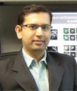
Lead Guest Editor DR MAKARAND DEO
Dr Makarand Deo is an Assistant Professor of Electronics Engineering at Norfolk State University, Norfolk, VA, USA. He completed his PhD at University of Calgary, Canada, and has previously worked at the University of Michigan and University of Toledo. He now works primarily in computational cardiac engineering and biosensing devices. Dr Deo has published numerous high-quality research articles in top engineering and medical journals and conferences. Dr Deo holds editorial appointments at Clinical Medicine Insights: Cardiology journal. Learn more about Dr Deo and his research by visiting his institutional Web page: https://www.nsu.edu/cset/engineering/research-webpages/ccblab.
Email: mdeo@nsu.edu

Guest Editors
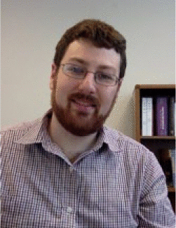
DR SETH H WEINBERG
Dr Seth H Weinberg is an Assistant Professor in Biomedical Engineering at Virginia Commonwealth University. He completed his PhD at the Johns Hopkins University and has previously worked at the College of William and Mary and Old Dominion University. He now works primarily in computational modeling of physiological systems. Dr Weinberg is the author or co-author of 29 published papers and has presented at 38 conferences and holds editorial appointments at Frontiers in Computational Physiology and Medicine. Learn more about Dr Weinberg by visiting his institutional website: shweinberglab.com.
Email: shweinberg@vcu.edu

Guest Editors
DR PATRICK M BOYLE
Dr Patrick M Boyle is an Assistant Research Professor of Biomedical Engineering at Johns Hopkins University. He completed his PhD at the University of Calgary. His research is focused on projects concerning the exploration of optogenetics as a novel means of manipulating cardiac electrophysiology, the use of image-based models to develop personalized treatment plans for complex atrial ablation procedures, and the role of the heart’s specialized conduction system in perpetuating arrhythmia. Dr Boyle is the author or co-author of 38 published papers and is recognized as an emerging leader in the field of computational cardiac electrophysiology. His research is supported by a Scientist Development Grant from the American Heart Association (16-SDG-30440006).
Email: pmjboyle@jhu.edu.

Footnotes
Funding:The author(s) received no financial support for the research, authorship, and/or publication of this article.
Declaration of Conflicting Interests:The author(s) declared no potential conflicts of interest with respect to the research, authorship, and/or publication of this article.
References
- 1. Benjamin EJ, Blaha MJ, Chiuve SE, et al. Heart disease and stroke statistics-2017 update: a report from the American Heart Association. Circulation. 2017;135:e146–e603. doi: 10.1161/CIR.0000000000000485. [DOI] [PMC free article] [PubMed] [Google Scholar]
- 2. Eisner D. Calcium in the heart: from physiology to disease. Exp Physiol. 2014;99:1273–1282. doi: 10.1113/expphysiol.2013.077305. [DOI] [PubMed] [Google Scholar]
- 3. Wagner S, Maier LS, Bers DM. Role of sodium and calcium dysregulation in tachyarrhythmias in sudden cardiac death. Circ Res. 2015;116:1956–1970. doi: 10.1161/CIRCRESAHA.116.304678. [DOI] [PMC free article] [PubMed] [Google Scholar]
- 4. Heijman J, Voigt N, Nattel S, Dobrev D. Cellular and molecular electrophysiology of atrial fibrillation initiation maintenance, and progression. Circ Res. 2014;114:1483–1499. doi: 10.1161/CIRCRESAHA.114.302226. [DOI] [PubMed] [Google Scholar]
- 5. Nattel S, Dobrev D. The multidimensional role of calcium in atrial fibrillation pathophysiology: mechanistic insights and therapeutic opportunities. Eur Heart J. 2012;33:1870–1877. doi: 10.1093/eurheartj/ehs079. [DOI] [PubMed] [Google Scholar]
- 6. Limbu B, Shah K, Weinberg SH, Deo M. Role of cytosolic calcium diffusion in murine cardiac purkinje cells. Clin Med Insights Cardiol. 2016;10:17–26. doi: 10.4137/CMC.S39705. [DOI] [PMC free article] [PubMed] [Google Scholar]
- 7. Haissaguerre M, Vigmond E, Stuyvers B, Hocini M, Bernus O. Ventricular arrhythmias and the His–Purkinje system. Nat Rev Cardiol. 2016;13:155–166. doi: 10.1038/nrcardio.2015.193. [DOI] [PubMed] [Google Scholar]
- 8. Weinberg SH. Impaired sarcoplasmic reticulum calcium uptake, promote electromechanically release, spatially discordant alternans: a computational study. Clin Med Insights Cardiol. 2016;10:1–15. doi: 10.4137/CMC.S39709. [DOI] [PMC free article] [PubMed] [Google Scholar]
- 9. Maleckar MM, Edwards AG, Louch WE, Lines GT. Studying dyadic structure-function relationships: a review of current modeling approaches and new insights into Ca2+ (mis)handling [published online ahead of print April 12, 2017]. Clin Med Insights Cardiol. doi: 10.1177/1179546817698602. [DOI] [PMC free article] [PubMed] [Google Scholar]
- 10. Edwards AG, Louch WE. Species-dependent mechanisms of cardiac arrhythmia: a cellular focus [published online ahead of print February 2, 2017]. Clin Med Insights Cardiol. doi: 10.1177/1179546816686061. [DOI] [PMC free article] [PubMed] [Google Scholar]
- 11. Baumeister P, Quinn TA. Altered calcium handling and ventricular arrhythmias in acute ischemia. Clin Med Insights Cardiol. 2016;10:61–69. doi: 10.4137/CMC.S39706. [DOI] [PMC free article] [PubMed] [Google Scholar]
- 12. Myles RC, Wang L, Kang C, Bers DM, Ripplinger CM. Local β-adrenergic stimulation overcomes source-sink mismatch to generate focal arrhythmia. Circ Res. 2012;110:1454–1464. doi: 10.1161/CIRCRESAHA.111.262345. [DOI] [PMC free article] [PubMed] [Google Scholar]
- 13. Pandit SV, Workman AJ. Atrial electrophysiological remodeling and fibrillation in heart failure [published online ahead of print October 31, 2016]. Clin Med Insights Cardiol. doi: 10.4137/CMC.S39713. [DOI] [PMC free article] [PubMed] [Google Scholar]
- 14. Karathanos TV, Boyle PM, Trayanova NA. Light-based approaches to cardiac arrhythmia research: from basic science to translational applications. Clin Med Insights Cardiol. 2016;10:47–60. doi: 10.4137/CMC.S39711. [DOI] [PMC free article] [PubMed] [Google Scholar]
- 15. Entcheva E. Cardiac optogenetics. Am J Physiol Heart Circ Physiol. 2013;304:H1179–H1191. doi: 10.1152/ajpheart.00432.2012. [DOI] [PMC free article] [PubMed] [Google Scholar]
- 16. Connolly AJ, Bishop MJ. Computational representations of myocardial infarct scars and implications for arrhythmogenesis. Clin Med Insights Cardiol. 2016;10:27–40. doi: 10.4137/CMC.S39708. [DOI] [PMC free article] [PubMed] [Google Scholar]
- 17. Berenfeld O. The major role of IK1 in mechanisms of rotor drift in the atria: a computational study [published online ahead of print December 20, 2016]. Clin Med Insights Cardiol. doi: 10.4137/CMC.S39773. [DOI] [PMC free article] [PubMed] [Google Scholar]
- 18. Pandit SV, Jalife J. Rotors and the dynamics of cardiac fibrillation. Circ Res. 2013;112:849–862. doi: 10.1161/CIRCRESAHA.111.300158. [DOI] [PMC free article] [PubMed] [Google Scholar]
- 19. Quintanilla JG, Pérez-Villacastín J, Pérez-Castellano N, et al. Mechanistic approaches to detect, target, and ablate the drivers of atrial fibrillation. Circ Arrhythm Electrophysiol. 2016;9:e002481. doi: 10.1161/CIRCEP.115.002481. [DOI] [PMC free article] [PubMed] [Google Scholar]


