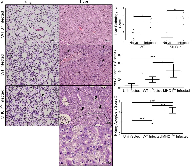
Fig 7A is incorrect. The label “CD8-/- Infected” should read “MHC I-/- Infected”. The authors have provided a corrected version here.
Fig 7. Histopathological comparison of MHC I-/- mice and WT mice infected with O. tsutsugamushi.
Foci of inflammation (arrows), including infiltration of macrophages and lymphocytes, were observed in infected mice (A, mag: 100x, 400x inset). Many apoptotic cells, possibly neutrophils, were observed in the liver of MHC I-/- mice. Increased necrosis and steatosis (arrows with circle end) were observed in the livers of MHC I-/- mice. Higher pathology scores indicating greater injury were observed in the livers of infected mice (B). There were significantly more apoptotic cells in the liver (C) and kidney (D) of MHC I-/- mice than their WT counterparts. All infected mice had increased apoptosis compared to uninfected mice. *, p<0.05; ***, p<0.001, n = 8; each tissue sample was blindly scored by four experienced investigators.
Reference
- 1.Xu G, Mendell NL, Liang Y, Shelite TR, Goez-Rivillas Y, Soong L, et al. (2017) CD8+ T cells provide immune protection against murine disseminated endotheliotropic Orientia tsutsugamushi infection. PLoS Negl Trop Dis 11(7): e0005763 https://doi.org/10.1371/journal.pntd.0005763 [DOI] [PMC free article] [PubMed] [Google Scholar]


