ABSTRACT
We describe a 59-year-old man who had nonunion of a right distal radius fracture after volar locking plate fixation. He underwent open reduction and internal fixation with a volar locking plate system for a dorsally displaced, unstable distal radius fracture at a previous hospital 5 months ago. Radiographs of the injured wrist showed nonunion of the distal radius with 1.5-mm ulnar minus variance. Radiographs of the unaffected wrist showed 3.5-mm ulnar plus variance. Intraoperative findings of surgical revision showed an unstable nonunion; thus, debridement of the nonunion, autogenous inlay bone grafting, and internal fixation using another type of volar locking plate system were performed. Healing of the re-operative site was confirmed radiographically 3 months postoperatively. We considered that volar locking plate fixation with excessive distraction of the fracture may lead to nonunion.
Key Words: Distal radius fracture, non-union, ulnar variance, unaffected wrist, volar locking plate
INTRODUCTION
Distal radius fractures are the most common type of upper limb fractures.1) However, nonunion of the distal radius is an extremely rare occurrence.2) Furthermore, there are few reports of nonunion with volar locking plate fixation.3) We describe a patient who had nonunion of a distal radius fracture after undergoing volar locking plate fixation. We considered that volar locking plate fixation with excessive distraction of the fracture may have led to the nonunion.
CASE PRESENTATION
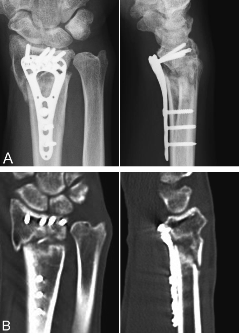
A 59-year-old man injured his right wrist in a motorcycle crash, and he presented to a regional hospital. He sustained a closed, dorsally displaced, unstable distal radius fracture (Figure 1). Two days later, open reduction and internal fixation with a volar locking plate system were performed at a previous hospital (Figure 2). Postoperatively, the wrist was immobilized with a short arm cast for 3 weeks. He was referred to our hospital because of prolonged right wrist pain for 5 months postoperatively. On initial examination, there was palpable crepitus on the volar aspect of the wrist with an active thumb or other fingers motion. The range of motion of his right wrist was 59% of that of the contralateral side, and his right grip strength was 8 kg. He was not a smoker, and he had no comorbidities such as diabetes, peripheral vascular disease, alcoholism, and obesity. There was no history of previous pain or injury to either side of the wrist. Radiographs of the right injured wrist showed nonunion of the distal radius with 1.5-mm ulnar minus variance and prominence of the volar plate at the watershed line (Figure 3A). A computed tomography scan showed segmental bone defects in the distal radius and sclerotic changes at the fracture edges (Figure 3B). Radiographs of the left unaffected wrist showed 3.5-mm ulnar plus variance (Figure 4).
Fig. 1.
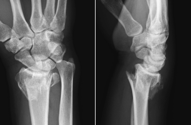
Posteroanterior and lateral radiographs of the affected wrist showing dorsally displaced distal radius fractures.
Fig. 2.
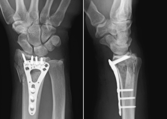
Radiographs showing open reduction and internal fixation with a volar locking plate system, which was performed at a previous hospital.
Fig. 3.
(A) Posteroanterior and lateral radiographs of the operated wrist showing a distal radius nonunion with 1.5-mm ulnar minus variance and prominence of the volar plate at the watershed line.
(B) Computed tomography scans showing segmental bone defects in the distal radius and sclerotic changes at the fracture edges.
Fig. 4.
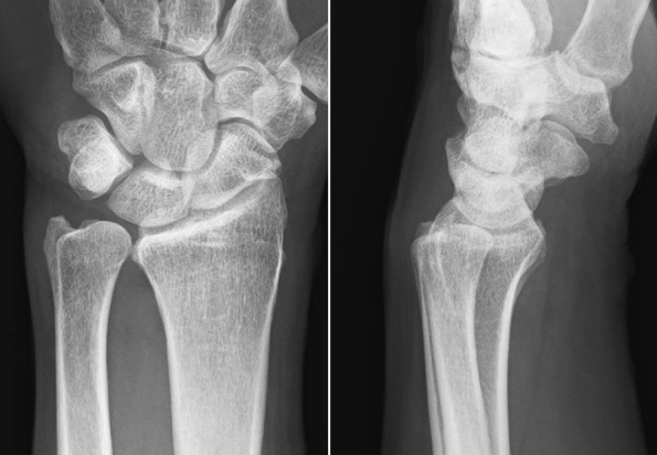
Posteroanterior and lateral radiographs of the unaffected wrist showing 3.5-mm ulnar plus variance.
Surgical treatment was performed for tendon attrition and nonunion of the distal radius. Intraoperative findings showed prominence of the distal edge of the plate and flexor tenosynovitis. The area of nonunion was identified after we removed the plate. Loosening of the distal locking screws and instability at the nonunion site, which was filled with fibrous scar tissue, were confirmed. First, tenosynovectomy was performed for flexor tenosynovitis, and then debridement of the nonunion, autogenous inlay iliac crest bone grafting, and internal fixation with another volar locking plate system were performed (Figure 5). Postoperatively, the wrist was immobilized with a short arm cast for 2 weeks. He started a gentle active range of motion exercise after the cast was removed. Healing of the nonunion was confirmed radiographically 3 months postoperatively (Figure 6). Three years postoperatively, he has experienced no pain and no problems with daily activities.
Fig. 5.
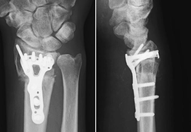
Posteroanterior and lateral radiographs showing debridement of the nonunion, autogenous inlay bone grafting, and internal fixation with another volar locking plate system, which were performed at our hospital.
Fig. 6.
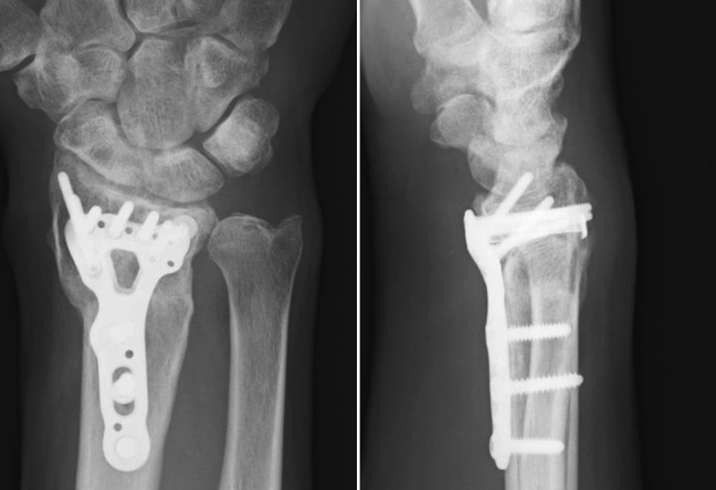
Posteroanterior and lateral radiographs showing healing of the nonunion at 3 months postoperatively.
DISCUSSION
Volar locking plate fixation of distal radius fractures has become an increasingly popular method because it offers assured and excellent fixation stability and maintains anatomic reduction. However, complications of volar locking plate fixation such as flexor and extensor tendon injury, and intra-articular screw placement have been reported.4) Yet, there are few reports of nonunion associated with volar locking plate fixation.3) Nonunion of the distal radius is an extremely rare occurrence.2) It has been reported that comorbid medical conditions such as diabetes, peripheral vascular disease, alcoholism, and morbid obesity may increase the risk of nonunion.5-7) Smoking may also play a role in the development of nonunion.5,7,8) Our patient was not a smoker, and he had no comorbidities.
There are several reports of nonunion of the distal radius. Nonunion of the distal radius is seen after internal fixation, external fixation, or non-operative treatment.9) Segalman and Clark6) suggested that excessive distraction of the fracture during the application of an external fixator is one of the causes of nonunion, because excessive distraction can create bony defects. In our case, although radiographs of the unaffected wrist showed 3.5-mm ulnar plus variance, radiographs of the operated wrist showed 1.5-mm ulnar minus variance. Hollevoet et al.10) reported that radiographic parameters, including the palmar tilt, radial inclination, and ulnar variance, have no significant differences between the right and left wrists. Therefore, volar locking plate fixation with excessive distraction of the fracture may lead to postoperative loss of correction and nonunion, although locked plates act as “internal external fixators” and provide very rigid fixation.11) On frontal view radiographs, the fracture line ran obliquely from the proximal radial side to the distal ulnar side in our patient. Therefore, evaluating the gap at the fracture site was difficult on postoperative lateral view radiographs because the X-ray beam was not perpendicular to the fracture line. There is a possibility that the fixing force of the locking screws was not enough, because only 4 locking screws were inserted into the distal radius fragment. However, in this fracture, although there was a bone fragment in the area of the distal radioulnar joint, dislocation and comminution of the bone were not large. Locking screws of sufficient length were inserted into the subchondral bone of the distal radius fragment, and cast immobilization was performed at 3 weeks postoperatively. This patient was a 59-year-old man with sufficient bone quality. If distraction of the fracture did not occur, we think that good bone healing would have been achieved. When performing treatment for a distal radius fracture, surgeons should refer to radiographs of the unaffected wrist.
Informed consent for publication of clinical data and photographs has been obtained from the patient.
REFERENCES
- 1).Angermann P, Lohmann M. Injuries to the hand and wrist: a study of 50,272 injuries. J Hand Surg Br, 1993; 18: 642-644. [DOI] [PubMed]
- 2).Bacom RW, Kurtzke JF. Colles’ fracture; a study of two thousand cases from the New York State Workmen’s Compensation Board. J Bone Joint Surg Am, 1953; 35-A: 643-658. [PubMed]
- 3).De Baere T, Lecouvet F, Barbier O. Breakage of a volar locking plate after delayed union of a distal radius fracture. Acta Orthop Belg, 2007; 73: 785-790. [PubMed]
- 4).Soong M, van Leerdam R, Guitton TG, Got C, Katarincic J, Ring D. Fracture of the distal radius: risk factors for complications after locked volar plate fixation. J Hand Surg Am, 2011; 36: 3-9. [DOI] [PubMed]
- 5).Ring D, Jupiter JB. Nonunion of the distal radius. Tech Hand Up Extrem Surg, 2002; 6: 6-9. [DOI] [PubMed]
- 6).Segalman KA, Clark GL. Un-united fractures of the distal radius: a report of 12 cases. J Hand Surg Am, 1998; 23: 914-919. [DOI] [PubMed]
- 7).Smith VA, Wright TW. Nonunion of the distal radius. J Hand Surg Br, 1999; 24: 601-603. [DOI] [PubMed]
- 8).Scolaro JA, Schenker ML, Yannascoli S, Baldwin K, Mehta S, Ahn J. Cigarette smoking increases complications following fracture. a systematic review. J Bone Joint Surg Am, 2014; 96: 674-681. [DOI] [PubMed]
- 9).Ring D. Nonunion of the distal radius. Hand Clin, 2005; 21: 443-447. [DOI] [PubMed]
- 10).Hollevoet N, Van Maele G, Van Seymortier P, Verdonk R. Comparison of palmar tilt, radial inclination and ulnar variance in left and right wrists. J Hand Surg Br, 2000; 25: 431-433. [DOI] [PubMed]
- 11).Egol KA, Kubiak EN, Fulkerson E, Kummer FJ, Koval KJ. Biomechanics of locked plates and screws. J Orthop Trauma, 2004; 18: 488-493. [DOI] [PubMed]


