Abstract
The sensation of touch is mediated by mechanosensory neurons that are embedded in skin and relay signals from the periphery to the central nervous system. During embryogenesis, axons elongate from these neurons to make contact with the developing skin. Concurrently, the epithelium of skin transforms from a homogeneous tissue into a heterogeneous organ that is made up of distinct layers and microdomains. Throughout this process, each neuronal terminal must form connections with an appropriate skin region to serve its function. This Review presents current knowledge of the development of the sensory microdomains in mammalian skin and the mechanosensory neurons that innervate them.
KEY WORDS: Touch, Skin, Hair follicle, Merkel cell, Placode, Axon guidance
Summary: This Review presents current knowledge of the development of the sensory microdomains in mammalian skin and the mechanosensory neurons that innervate them.
Introduction
Like all senses, the sense of touch allows us to gather information about the people and things in the world around us. What sets touch apart is its intimacy, as it requires direct contact with skin, the sensory organ of tactile sensation. Humans constantly rely on tactile feedback for mundane tasks such as fishing keys out of a cluttered pocket as well as essential behaviors such as manipulating food to feed ourselves and our offspring. Touch is also required for normal cognitive development in mammals. Indeed, tactile hyper- or hypo-sensitivity is a hallmark of neurodevelopmental disorders in children, including autism spectrum disorder (see Box 1; Blakemore et al., 2006; Cascio et al., 2008; Tomchek and Dunn, 2007). Despite the profound implications of early tactile input in cognitive function, the mechanisms that mediate mammalian touch receptor development remain mysterious.
Box 1. Autism spectrum disorder and tactile processing: hand in hand.
Autism spectrum disorder (ASD) affects approximately one in 132 people per year (Baxter et al., 2015). Patients with ASD exhibit impaired social interactions, repetitive behaviors and often display tactile hyper- or hypo-sensitivity (Tomchek and Dunn, 2007; Voos et al., 2013). Several genes, such as Fmr1 and Mecp2, have been linked to ASD and ASD-related syndromes based on studies of human patients and rodents (Amir et al., 1999; Badr et al., 1987; Rogers et al., 2003). When globally mutated in mice, these genes cause social anxiety behaviors, and in some cases also cause aberrant tactile sensitivity (Orefice et al., 2016). Alterations in tactile responsiveness were previously hypothesized to involve solely central nervous system impairment. Surprisingly, when Mecp2 or Gabrb3 are conditionally knocked out in the peripheral nervous system, mutant mice exhibit tactile and behavioral deficits (Orefice et al., 2016). These results suggest that mutations that underlie ASD impair both the central and peripheral nervous systems. This work also underscores the importance of robust tactile stimulation during development for normal cognitive function. The link between touch and cognitive development is thus an exciting current area of research.
The neurons that transform touch stimuli at the skin surface into electrical impulses – the currency of the nervous system – are known as mechanoreceptors. Two main types of mechanoreceptors innervate mammalian skin: low-threshold mechanoreceptors (LTMRs; see Glossary, Box 2) are tuned to respond to gentle touch, whereas nociceptors (see Glossary, Box 2) encode forces in the noxious (harmful) range. LTMRs can be further classified based on functional properties and molecular markers (Table 1; Fig. 1). Aδ-LTMRs, for example, detect hair follicle deflection (Rutlin et al., 2014), whereas some types of Aβ-LTMRs allow for high-acuity sensation (Bai et al., 2015; Li et al., 2011; Wellnitz et al., 2010), and C-LTMRs are proposed to convey information about social touch (Liljencrantz and Olausson, 2014; Liu et al., 2007; Olausson et al., 2010; Vrontou et al., 2013; Wessberg et al., 2003). These classes were originally designated based on classic electrophysiological and histological studies. Aβ- and Aδ-LTMR neurons are distinguished by their medium-to-large diameter, myelinated axons and medium-to-fast conduction velocities (Table 1; Gasser, 1941; Horch et al., 1977). Conversely, C-fibers possess small axonal diameters and exhibit slow conduction velocities. In total, seven subclasses of low-threshold mechanoreceptors, a substantial portion of the 17 subtypes of putative somatosensory neurons (see Glossary, Box 2; Usoskin et al., 2015), have been further distinguished by a combination of genetic, morphometric and physiological approaches. This variety has spurred research into how distinct classes of mechanoreceptors are specified during development.
Box 2. Glossary.
Afferent. An axon that carries sensory information from peripheral organs to the central nervous system.
Dermis. A deep layer of skin mainly composed of fibroblasts and extracellular matrix proteins.
Dorsal root ganglia. Clusters of somatosensory neuron cell bodies that flank the spine.
Epidermis. A superficial layer of skin that forms the barrier between the internal organs and the outside world.
Hair cells. Mechanosensory cells that relay information about head position and sound and water flow to sensory neurons.
Intervertebral foramina. The space between two vertebrae that houses dorsal root ganglia.
Keratinocyte. A keratin-producing epithelial cell that is the predominant cell type in the epidermis.
Lateral line. A system of sense organs that detect movement, pressure gradients and vibration in aquatic animals.
Low-threshold mechanoreceptors. Somatosensory neurons that transduce gentle touch stimuli.
Merkel cells. Epidermal cells that display features of mechanosensory receptor cells and form synapse-like connections with a particular type of tactile afferent.
Neural crest cells. Ectodermal cells that delaminate from the neural tube during development and form sensory ganglia, melanocytes, bone, cartilage, smooth muscle and more.
Nociceptors. Sensory neurons that transduce noxious (or potentially tissue damaging) thermal, mechanical or chemical stimuli to the spinal cord.
Placode. Thickened regions of specialized epithelial cells that give rise to sensory structures (feathers, hair, inner ear) and auxiliary tissues (lens, teeth).
Proprioceptors. Sensory neurons that transmit information about limb position and muscle stretch.
Pseudounipolar axon. Axon that extends from the cell body of a neuron and splits to form distal and proximal branches.
Skin appendage. Mini-organs or structures embedded in the skin that serve specialized functions. Examples include hair, sweat and sebaceous glands, nails, feathers and smooth muscles.
Somatosensory neurons. Peripheral neurons that transduce tactile, proprioceptive, thermal, pruritic and nociceptive stimuli into electrical signals that are relayed to the central nervous system.
Taste cells. Specialized sensory cells that detect salty, sour, sweet, bitter and umami tastes.
Touch domes. Specialized raised epithelial structures that are found surrounding guard hairs. These structures contain Merkel cells and the neurons that innervate them are sensitive to gentle pressure on the skin surface.
Table 1.
Classification of mouse low-threshold mechanoreceptors
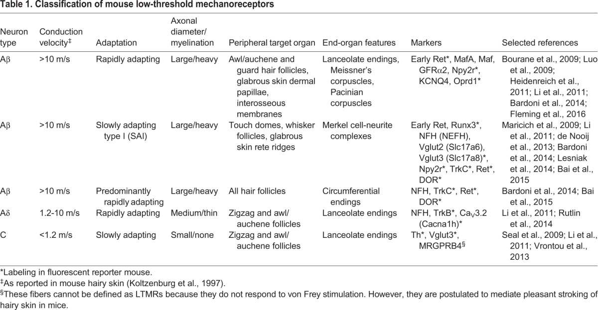
Fig. 1.
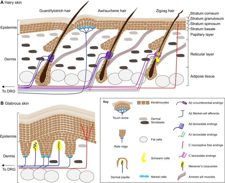
Mechanosensory end organs in skin. The touch receptors of hairy and glabrous skin are highly diverse. (A) Hairy skin is decorated with distinct types of hair follicles. Guard/tylotrich hairs are the most rare hair type, and also the longest. Awl/auchene hairs and zigzag hairs make up the bulk of the hairs in the mouse coat. Each hair type is associated with a unique complement of sensory neurons. Lanceolate and circumferential endings wrap around the bulge region of hair follicles, which stretches between the sebaceous gland and the connection site of arrector pili muscles. Note that all lanceolate endings intercalate with the protrusions of terminal Schwann cells, one of which is shown in the schematic (yellow). Other neurons innervate touch domes, which are discrete, raised zones of the skin adjacent to guard hairs. These neurons innervate epidermal Merkel cells (teal). (B) Instead of hairs, hallmarks of glabrous skin include invaginations of the epidermis called rete ridges. Dermal zones between rete ridges are called dermal papillae. Merkel cells and their associated afferents are found at the base of rete ridges, whereas Meissner's corpuscles protrude into dermal papillae.
Although they are diverse, mechanoreceptors and other somatosensory neurons share a basic body plan. The cell bodies of LTMRs are housed with those of other somatosensory neurons in dorsal root ganglia (DRG; see Glossary, Box 2). Each somatosensory neuron bears a pseudounipolar axon, or afferent (see Glossary, Box 2). Peripheral branches innervate skin or other target organs, whereas central branches innervate the spinal cord and/or hindbrain. Thus, afferents carry sensory signals from the periphery to the central nervous system.
At the periphery, distinct complements of LTMRs innervate hair-bearing skin compared with glabrous, or hairless, skin on the palms and soles of feet (Fig. 1; Dell and Munger, 1986; Halata and Munger, 1983; Li et al., 2011; Miller et al., 1958; Munger and Halata, 1983; Munger and Ide, 1988; Vallbo et al., 1999). In primates, glabrous skin contains four principal types of Aβ-LTMRs (Johnson et al., 2000; Miller et al., 1958). Complexes between Merkel cells (see Glossary, Box 1) and Aβ-LTMRs, which are arranged at the base of rete ridges in glabrous skin, detect sustained touch as well as object features such as edges and curvature (Goodwin et al., 1995; Iggo and Muir, 1969; Phillips et al., 1990). Also present are encapsulated endings such as Meissner corpuscles, which respond best to low-frequency vibration (Talbot et al., 1968), Pacinian corpuscles, which encode high-frequency vibrations (Talbot et al., 1968), and Ruffini endings, which detect skin stretch (Johansson and Vallbo, 1979; Johnson et al., 2000). The complement of neuronal endings differs in hairy skin, as it is specialized less for high-acuity sensation and more for sensation of movement along the body surface. In the mouse coat, for example, distinct combinations of C-, Aβ- and Aδ-LTMRs form collars around each type of hair follicle (Fig. 1; Li and Ginty, 2014; Wu et al., 2012). These collars are complex, consisting of both fence-shaped lanceolate endings, and neurons that wrap around these fences, called circumferential endings. Moreover, Merkel cell-neurite complexes form discrete clusters in touch domes (see Glossary, Box 2), which are specialized epidermal structures that surround guard hairs. These structures, which are visible to the naked eye, are raised owing to the presence of a thickened epidermis and columnar-shaped keratinocytes (see Glossary, Box 2) and increased vascularization (Fig. 1; Doucet et al., 2013; Halata et al., 2003; Iggo and Muir, 1969; Moll et al., 1996; Moll et al., 1993; Pinkus, 1902; Reinisch and Tschachler, 2005; Smith, 1977; Woo et al., 2010). The LTMRs that innervate hair follicles and touch domes are exquisitely sensitive to gentle touch stimulation.
Recent studies, many of which have established the hairy skin of the mouse as a tractable model system for studying LTMR afferent targeting, have shown that hair follicles and touch domes are discrete mechanosensory units that are innervated by unique complements of LTMRs. This finding, combined with concurrent advances in the field of skin development, set the stage to reveal how LTMRs establish and maintain appropriate innervation patterns. In this Review, we summarize the current state of knowledge about how skin and sensory neurons develop in unison to enable a sense of touch. We first provide an overview of the cellular and molecular mechanisms that drive specification of sensory neurons. Next, the process of skin development is outlined. We then describe current models for how touch receptors develop, with a particular focus on Merkel cell-neurite complexes and lanceolate endings. Finally, we highlight research in invertebrate model systems that provides clues into the evolutionarily conserved mechanisms of touch receptor development.
The early development and specification of mechanoreceptors
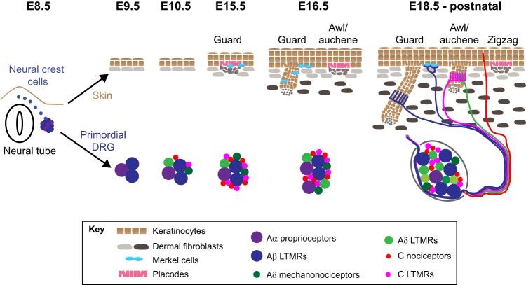
All somatosensory neurons, including LTMRs, arise from precursors called neural crest cells (see Glossary, Box 2) at early developmental stages (Weston, 1970). Neural crest cells are transient, multipotent cells that delaminate from the dorsal neural tube and migrate to the periphery in mice between embryonic day (E) 8.5 and E10 (Fig. 2; Serbedzija et al., 1990). A subset of neural crest cells follow a ventromedial path to form clusters on each side of the neural tube (Serbedzija et al., 1990; Thiery et al., 1982). As the embryo continues to grow from E9.5 to E13.5, these cells give rise to somatosensory neurons that coalesce into pairs of DRGs located in the intervertebral foramina (see Glossary, Box 2) at each spinal level (Fig. 2; Frank and Sanes, 1991; Marmigère and Ernfors, 2007).
Fig. 2.
Timelines of skin and sensory neuron development. The skin originates from the ectoderm (brown) and mesoderm (gray), which give rise to epidermis and dermis, respectively. Guard hairs are specified as epidermal placodes (pink) with dermal clusters or condensates of fibroblasts beneath them (gray); Merkel cells (teal) appear within and adjacent to developing guard hairs by E15.5. At roughly E16.5, a new crop of placodes appears that will become awl/auchene hairs. From E18.5 onward, additional waves of folliculogenesis generate zigzag hairs. All hair types elongate down into the dermis until P21, when folliculogenesis is complete. Development of dorsal root ganglia (DRG) occurs in parallel with skin development. At E8.5, neural crest cells delaminate from the neural tube and coalesce to form DRG. Large diameter mechanosensitive neurons are born in the first wave of specification at E9.5. Small diameter neurons are specified beginning one day later at E10.5. DRG neuron precursors continue to proliferate and generate large and small diameter neurons until E13.5. From E13.5 onward, somatosensory afferents elongate from DRG to innervate the skin. A subset of the tactile endings that form in the skin from E18.5 onward are illustrated.
Somatosensory neuron heterogeneity is first established by temporal waves of cell fate specification (Frank and Sanes, 1991; Ma et al., 1999). All somatosensory neurons require expression of either neurogenin 1 or 2 (Ngn1, 2; also known as Neurog1, 2), which encode basic helix-loop-helix transcription factors (Ma et al., 1999). Neurons specified early (E9.5 to E11.5) express Ngn2. These neurons generally include Aβ and Aδ afferents with large-diameter cell bodies (Fig. 2) and myelinated axons. They are fated to become either LTMRs or proprioceptors (see Glossary, Box 2), which sense joint position and muscle contraction (Banks et al., 2009; Goldstein et al., 1991; Lawson and Biscoe, 1979; Ma et al., 1999; Sherrington, 1906). A second wave of neuronal specification requires expression of Ngn1 and spans from E10.5 to E13.5. Neurons born during this period are unmyelinated C afferents with small-diameter cell bodies. They are fated to become nociceptors, a subset of which respond to thermal or itch stimuli (Box 2; Fig. 2; Bautista et al., 2014; Julius, 2013; Lawson and Biscoe, 1979; Ma et al., 1999; McKemy, 2013; Schepers and Ringkamp, 2010).
Transcription factors that specify mechanoreceptors include MafA (Bourane et al., 2009) and c-Maf, which is now known simply as Maf (Hu et al., 2012; Wende et al., 2012). Maf and MafA are both basic leucine zipper transcription factors that are expressed starting at roughly E11 and actively enhance specification of mechanosensory neurons, presumably by maintaining expression of the neurotrophin receptors Ret and Gfra2 (Bourane et al., 2009; Luo et al., 2009; Wende et al., 2012). Brn3a (Pou4f1) might also play a role in regulating touch receptor fate (Badea et al., 2012; Dykes et al., 2010). Brn3a encodes a POU-domain transcription factor that binds histone H3-acetylated chromatin in the runt-related transcription factor 3 (Runx3) locus and is thought to control Runx3 expression (Dykes et al., 2010). Runx3 promotes proprioceptor fate between E11.5 and E12 (Kramer et al., 2006). At E13.5, 59% of DRG neurons display rapidly inactivating mechanically gated currents, a hallmark of LTMRs (Lechner et al., 2009). A substantial proportion of these neurons are early-born, large-diameter neurons. By 2 weeks of postnatal age, mechanoreceptive neurons are not yet fully mature, but several types of A afferents can be distinguished in ex vivo electrophysiology preparations (Koltzenburg et al., 1997). Thus, mechanoreceptive neurons acquire sensitivity to touch very soon after specification, but require postnatal maturation to acquire their adult physiological properties.
The innervation of skin by mechanoreceptors
Following their specification, LTMRs and other somatosensory neurons simultaneously innervate the spinal cord and the periphery. During this process, the architecture and molecular environment of skin rapidly changes. The neural tube, which will become the spinal cord, transforms drastically as interneurons are born and specified (Lai et al., 2016). The process by which sensory axons target specific spinal cord subregions has been studied in detail and is the subject of several reviews (Gardiner, 2011; Masuda et al., 2009; O'Malley et al., 2014; Olson et al., 2016). Although the molecular and cellular mechanisms that mediate innervation of peripheral target tissues are less well-understood, recent parallel investigations of skin biology and somatosensory neurons have begun to reveal how skin microdomains and touch receptors develop in unison.
An overview of skin development
Mammalian skin begins as two layers of cells that transform into a multilayered structure by the time of birth (Fig. 2; Koster and Roop, 2007). The upper compartment consists of layers of keratinocytes that form the epidermis (see Glossary, Box 2), a protective barrier that prevents water from evaporating out of the body and protects internal organs from microbial insults and radiation (Hardman et al., 1998). The deeper layers of skin, by contrast, are composed of loosely packed fibroblasts and a collagen-rich extracellular matrix that together form the dermis (see Glossary, Box 2).
The epidermis derives from the ectoderm (Byrne et al., 1994; Fernandes et al., 2004; Jinno et al., 2010; Liu et al., 2013; Ohtola et al., 2008; Rendl et al., 2005; Rivera-Pérez and Hadjantonakis, 2014; Wong et al., 2006). The initial epidermal layer is the periderm, a protective layer that covers the skin surface and dies by apoptosis as the skin barrier develops in utero (Hardman et al., 1998; M'Boneko and Merker, 1988; Richardson et al., 2014). Between E9.5 and E18.5, the epidermis stratifies into distinct laminae: basal, spinous, granular and cornified (Fig. 2). These laminae are built sequentially, starting from the proliferative basal layer of cells and extending to enucleated keratinocytes in the surface cornified layer, which plays a key role in barrier function (Fig. 2; Byrne et al., 1994).
Beneath the epidermis, the dermis forms from multiple cellular sources, including neural crest and mesodermal cells (Fernandes et al., 2004; Jinno et al., 2010; Ohtola et al., 2008; Wong et al., 2006). This skin compartment also stratifies between E12.5 and E18.5, transforming into an upper papillary dermis and a lower reticular dermis (Driskell et al., 2013; Sorrell and Caplan, 2004). A deep layer called the hypodermis, primarily composed of fat cells, appears at postnatal stages (Driskell et al., 2013).
In summary, skin is highly dynamic when sensory innervation develops. This suggests that specialized mechanisms – perhaps mediated via secreted long-range signals and contact-dependent short-range cues – help to guide axons as they grow toward their cutaneous targets.
Mouse hair follicle development
Hair follicles, which are the most abundant tactile structures in mammalian skin, begin to develop in embryogenesis. In mice, for example, whisker follicles located on the lateral aspects of the face form at E12 and are innervated soon thereafter (Hasegawa et al., 2007; Munger and Rice, 1986; Schmidt-Ullrich and Paus, 2005). At E13.5, an early hair-follicle marker, keratin 17 (Krt17; also known as K17), dots the epithelium over most of the body's surface (Bianchi et al., 2005). These K17-positive patches form three types of hair follicles in successive waves. The first to develop are guard hairs, also known as tylotrich or primary hairs. These long hairs are sparse, composing roughly 1% of the adult coat hair (Fig. 1; Lechner and Lewin, 2013; Li et al., 2011). At E14.5, guard-hair keratinocyte precursors enlarge and elongate to form hair follicle placodes (see Glossary, Box 2), which are thickened patches of epithelial cells that proliferate and invaginate to form follicles (Fig. 2; Paus et al., 1999). Awl/auchene or secondary hair placodes, which represent roughly 20% of coat hairs (Lechner and Lewin, 2013; Li et al., 2011), arise in a subsequent wave of follicle specification between E15.5 and E16 (Paus et al., 1999). At this time, hairs also emerge on the tail. At later stages of development, ranging from E17 to postnatal ages, zigzag or tertiary hairs tile the body surface (Lechner and Lewin, 2013; Paus et al., 1999). Named after the bent appearance of their shafts, zigzag hairs make up the majority of the downy coat of mice. Although the precise function of each hair type is unknown, electrophysiological surveys of hair follicle LTMRs have shed light on the possible mechanosensory function of each follicle subtype (Adrian and Zotterman, 1926a,b; Bai et al., 2015; Brown and Iggo, 1967; Burgess et al., 1968; Iggo and Muir, 1969; Li and Ginty, 2014; Li et al., 2011; Rutlin et al., 2014; Zotterman, 1939).
The development of unique innervation patterns
Each murine hair type is associated with a different complement of LTMRs (Figs 1 and 2; for a review, see Abraira and Ginty, 2013). Guard hairs, for example, are innervated solely by myelinated Aβ-LTMR endings. The terminals of these neurons include lanceolate endings, which are fence-shaped structures that align with the length of the hair follicle, and circumferential endings that wrap around the lanceolate terminals in thick bundles (Bai et al., 2015; Halata, 1993; Ikeda et al., 2014; Li and Ginty, 2014; Li et al., 2011; Munger and Ide, 1988). In rodents, each guard hair is flanked by a touch dome containing Merkel cells innervated by slowly adapting Aβ-LTMRs. Other hair types also recruit distinct combinations of sensory innervation. Awl/auchene hairs are innervated by Aβ circumferential endings, as well as three molecularly distinct types of lanceolate endings that fall in to Aβ, Aδ and C classes. Finally, zigzag hairs are innervated by Aδ-LTMR lanceolate endings, Aβ-LTMR circumferential endings and C-fiber lanceolate endings. Thus, each hair follicle type is proposed to represent a distinctive mechanosensory unit (Bai et al., 2015; Li and Ginty, 2014; Li et al., 2011; Rutlin et al., 2014).
Two general models can account for how the unique innervation pattern of each hair type is achieved. First, given that hair follicles and DRG neurons are born in waves, the timing of the arrival of each neuronal subtype to the skin could correlate with the timing of hair follicle maturation. For example, zigzag hairs, the last hairs to arise on the skin surface, are innervated by a subset of afferents that excludes Aβ-LTMR lanceolate complexes. This could be due to the fact that zigzag hairs have not been specified at the time when Aβ-LTMR lanceolate endings innervate their targets. An alternative hypothesis is that each type of hair expresses a unique complement of molecular cues and that each LTMR class is programmed to express the cognate receptors. It is likely that a combination of the two strategies is employed to ensure that each hair follicle type receives an appropriate pattern of innervation. As discussed below, the search for cues that guide afferents to their target microdomains during development has begun to reveal how distinct mechanosensory organs form in skin. We first discuss lanceolate endings and Merkel cell-neurite complexes, as recent progress has uncovered key cell types and molecular mechanisms that drive their development. Current concepts about the development of other mechanoreceptors are then summarized.
The development of lanceolate endings: cutaneous cells and neurons get in touch
Aδ-LTMR lanceolate endings, which are also called down-hairs (D-hairs; Brown and Iggo, 1967; Brown et al., 1967; Rutlin et al., 2014), form hemi-collars on the caudal side of hair follicles (Figs 1 and 2; Li et al., 2011; Rutlin et al., 2014; Wu et al., 2012). The polarized organization of Aδ-LTMR endings allows these afferents to preferentially detect rostral hair movements (Rutlin et al., 2014). Aδ-LTMRs express TrkB (Ntrk2), a neurotrophin receptor that binds brain-derived neurotrophic factor (BDNF; Klein et al., 1991), whereas keratinocytes on the caudal side of hair follicles express BDNF and participate in the selective targeting of Aδ-LTMRs to these hair follicles (Rutlin et al., 2014). Indeed, the genetic deletion of keratinocyte-derived BDNF is sufficient to disrupt the organization of lanceolate endings, suggesting that BDNF acts as a target-derived cue that is responsible for promoting the guidance of afferent endings to their proper location (Rutlin et al., 2014). In this case, individual afferents are directionally selective but rostral preference is lost, underscoring the link between appropriate development of touch-receptor morphology and function (Rutlin et al., 2014). This example sets a precedent for specialized skin cells acting to promote accurate neural targeting. Given that the neurotrophin receptor Ret is required for the formation of Aβ lanceolate terminals (Bourane et al., 2009; Luo et al., 2009), it is possible that a subset of cells in guard and awl/auchene follicles express the Ret ligands glial cell line derived neurotrophic factor (GDNF), neurturin, artemin and persephin (Airaksinen and Saarma, 2002). Whether and how specific cell types orchestrate the patterning of circumferential endings and other LTMRs around hair follicles remain open questions.
Merkel-cell afferents: a coupling of rare neurons with special epithelial cells
Whereas hair follicle afferents signal passive touch, Merkel cells and the afferents that innervate them are essential for active, discriminative touch. First described by Friedrich Merkel in 1875 as ‘Tastzellen’ or ‘touch cells’, Merkel cells are epidermal-derived cells that cluster in high-tactile acuity areas of vertebrate skin (Merkel, 1875). Such areas include glabrous finger pads, lips, whisker follicles and touch domes (Halata et al., 2003; Lumpkin et al., 2003; Smith, 1977). The slowly adapting Aβ-LTMR afferents that innervate Merkel cells are specialized to mediate discriminative touch by encoding object features, such as shape and texture, as well as sustained pressure (Iggo and Muir, 1969; Johnson, 2001; Maricich et al., 2012; Wellnitz et al., 2010; Woodbury and Koerber, 2007).
The rarest of the four fundamental cell types that compose the epidermis, Merkel cells have long been shrouded in mystery. Merkel cells are unique because they are the only known neuron-like cells in vertebrate skin. Recent studies have confirmed the long-debated hypothesis that Merkel cells are mechanosensory cells required for tactile sensation (Ikeda et al., 2014; Maksimovic et al., 2014; Wallis et al., 2003; Woo et al., 2014). Like most other epithelial-derived sensory cells, Merkel cells form synapse-like contacts with primary afferents (Iggo and Muir, 1969; Merkel, 1875). Merkel cells also express presynaptic molecules (e.g. piccolo and synaptotagmin I) as well as an assortment of neuronal transcription factors (e.g. Atoh1, Gfi1, Isl1 and Lhx3; Ben-Arie et al., 2000; Haeberle et al., 2004). Intriguingly, Merkel cells share histochemical and morphological features with cells found in a rare and exceedingly aggressive cancer called Merkel cell carcinoma (see Box 3). Because of the unusual properties of Merkel cells, intense efforts have aimed to understand how these cells form and how they become integrated into functional mechanosensory organs.
Box 3. Merkel-cell carcinoma.
Merkel cell carcinoma (MCC) is a rare, aggressive type of skin cancer with a high rate of recurrence and an extremely low 5-year survival rate in cases that have metastasized to distant sites (13.5%; Agelli and Clegg, 2003; Albores-Saavedra et al., 2010; Harms et al., 2016). The cells that make up these tumors possess many morphological hallmarks of Merkel cells, including robust expression of cytokeratin 20 (also known as keratin 20) and dense-core secretory granules (Chan et al., 1997; Miettinen, 1995; Moll et al., 1992; Sibley et al., 1985; Tang and Toker, 1978). MCC is posited to arise in skin when an individual is infected with Merkel cell polyomavirus (MCPyV), though the precise mechanism of how the virus induces tumor progression is unknown (Carter et al., 2009; Feng et al., 2008; Kassem et al., 2008; Sharp et al., 2009). The cell type of origin of MCC is also an area of intense investigation (Tilling and Moll, 2012).
Merkel cell development
Merkel cell specification requires Atoh1, a helix-loop-helix transcription factor (Fig. 3). Atoh1 is an ortholog of atonal, a proneural gene required for the development of mechanosensory chordotonal organs in Drosophila (Jarman et al., 1993). Skin-specific ablation of Atoh1 totally abrogates Merkel cell specification (Morrison et al., 2009; Van Keymeulen et al., 2009). Downstream of Atoh1 activation, the transcription factor Sox2 is required to maintain Atoh1 expression (Perdigoto et al., 2014). Accordingly, the ablation of Sox2 results in a decrease in Merkel-cell number (Lesko et al., 2013). Conversely, the Polycomb complex subunits Ezh1 and Ezh2 negatively regulate Merkel cell formation by inhibiting Sox2 (Bardot et al., 2013; Perdigoto et al., 2016). Together, these findings highlight that Atoh1 and Sox2 work in concert to initiate and maintain Merkel cell fate (Fig. 3).
Fig. 3.
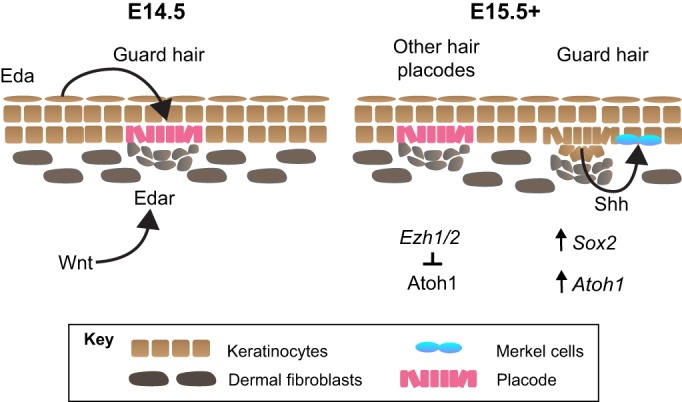
Factors involved in Merkel-cell formation. Guard, or tylotrich, hairs are specified at E14.5 in the trunk skin of mice. This process is driven by epidermal expression of Eda and dermal expression of Edar, which is induced by Wnt signaling in the dermis. The birth of nascent hair follicles is marked by the presence of epidermal placodes (pink) and the formation of dermal condensates (gray). One day later, Merkel cells (teal) appear with budding guard hairs. Their induction is postulated to result from Shh expression in the hair follicle. This leads to the expression of Atoh1 and Sox2, which encode transcription factors that work in concert to initiate and maintain Merkel cell specification. Ezh1/2 expression surrounding other hair follicle types represses Atoh1, preventing ectopic Merkel cell formation.
Merkel cells first appear in mice at E15.5 in the dorsal skin of the trunk (Ben-Arie et al., 2000; Morrison et al., 2009; Perdigoto et al., 2014), one day after the initial specification of guard hair follicles. Merkel cell specification depends on ectodysplasin-A (Eda), a tumor necrosis factor family member, and the Wnt and Sonic hedgehog (Shh) signaling pathways (Fig. 3). In Tabby mice, in which Eda signaling is disrupted, guard hair follicles and Merkel cells in touch domes fail to develop (Vielkind and Hardy, 1996; Vielkind et al., 1995). Dermal Wnt signaling activates Eda/Eda-receptor (Edar) signaling in hair follicle placodes, which in turn induces Shh expression in developing guard hair follicles (Xiao et al., 2016). This process is thought to spur expression of Atoh1 and Sox2 in nearby Merkel cell precursors (Xiao et al., 2016). Thus, a complex cascade of focal morphogen signaling presumably leads to the specific expression of Atoh1 in Merkel cell precursors (Fig. 3).
The formation of Merkel cell-neurite complexes
Aβ afferents begin to target Merkel cells during embryonic development (Nurse and Diamond, 1984; Vielkind et al., 1995). In Atoh1 knockout mice lacking Merkel cells, touch domes are innervated by Aβ afferents that are myelinated, have nodes of Ranvier and respond to low-threshold mechanical stimuli; however, these touch-dome afferents exhibit some striking phenotypic differences (Maksimovic et al., 2014; Maricich et al., 2009). For example, in the absence of Merkel cells, touch-dome afferents display a hyperbranched morphology (Maksimovic et al., 2014; Maricich et al., 2009; Morrison et al., 2009). They also show reduced firing rates during dynamic touch and truncation of the slowly adapting firing pattern seen in wild-type mice (Maksimovic et al., 2014). Additionally, the expression of several neurotrophin receptors, including TrkB and TrkC (Ntrk3), is decreased in large-diameter DRG cell bodies (Reed-Geaghan et al., 2016). Together, these studies suggest that Merkel cells express short-range cues that refine touch-dome afferents and influence their neuronal activity and gene expression patterns once they contact touch domes. These findings also suggest that Merkel cells are not the cellular source of long- or intermediate-range target-derived guidance cues for the afferents that contact them; instead, they mediate neuronal maturation and refinement.
Although the precise factors that are responsible for recruiting afferents to Merkel cell-enriched skin areas remain elusive, several lines of evidence point to neurotrophin signaling as playing a key role. Merkel cell afferents express TrkC, the receptor for the neurotrophin NT3 (Ntf3) (Cronk et al., 2002; Niu et al., 2014). Surprisingly, normal numbers of Merkel cell-neurite complexes are present at birth in the touch domes and glabrous skin of Ntf3 homozygous knockout animals (Airaksinen et al., 1996; Szeder et al., 2003); however, Merkel cell numbers decrease dramatically by 2-3 weeks of age (Airaksinen et al., 1996). This postnatal loss of Merkel cells also occurs in the whisker pad and is mediated by increased apoptosis (Halata et al., 2005). Merkel cell loss can be rescued by NT3 overexpression in the epidermis (Krimm et al., 2000, 2004). Thus, these findings suggest that NT3 is not required for target innervation of Merkel cell-enriched skin areas but rather plays an essential role in touch receptor maintenance. Similarly, Merkel cell-derived BDNF is not required for Aβ afferents to target touch domes; instead, Merkel cell-mediated secretion of BDNF at a specific time in embryogenesis (E16.5) influences neuronal firing patterns (Reed-Geaghan et al., 2016). It has also been shown that Merkel-cell afferents are reduced at postnatal day (P) 0 in Ret knockout mice, implicating GDNF family ligands in the embryonic development of these LTMRs (Bourane et al., 2009). These findings underscore the complex influence of neurotrophin signaling on afferent specification and maturation. Together, these studies also highlight that Merkel cells secrete BDNF and possibly other factors that influence neuronal development or activity. Uncovering the roles and downstream effects of specific neurotrophins, morphogens and transcription factors in the development of Merkel cells and LTMRs remain active areas of investigation in the field.
Mysterious mechanisms of encapsulated LTMR development
The formation of encapsulated endings such as Meissner corpuscles, Pacinian corpuscles and Ruffini endings has also been studied, although less is known about how these endings arise during development. During Meissner's corpuscle development, mechanoreceptive neurons innervate the glabrous skin dermal papillae, which are zones of the dermis that extend superficially into the epidermis, perpendicular to the skin surface (Cauna, 1956; Miller et al., 1958). At postnatal stages, these neurons become myelinated and their associated terminal Schwann cells transform into the lamellar cells that make up each corpuscle (Renehan and Munger, 1990; Zelená, 1994; Zelená et al., 1990). Although Meissner afferents fall into the early Ret-positive class of LTMRs, Ret is dispensable for their formation (Luo et al., 2009). By contrast, TrkB appears to be required for proper innervation of Meissner's corpuscles, as they are absent in TrkB mutant animals (González-Martínez et al., 2004; Luo et al., 2009; Perez-Pinera et al., 2008).
The mechanisms of development of Pacinian corpuscles and Ruffini endings are less well-understood, in part because these endings are rare in the mouse peripheral nervous system. Although Pacinian corpuscles are not found in mouse skin, Ret signaling is required for development of these corpuscles in mouse interosseous membranes (Luo et al., 2009; Zelená, 1981). Downstream of Ret, neuregulin 1 functions in the innervating axons of Pacinian corpuscles to trigger terminal Schwann cell maturation (Fleming et al., 2016). Ruffini endings are another touch receptor type that has rarely been located in mouse or primate skin (Honma et al., 2010; Paré et al., 2003, 2002; Rasmusson and Turnbull, 1986; Rice and Rasmusson, 2000; Ruffini, 1894). Notably, periodontal Ruffini endings are absent in TrkB mutant mice, highlighting conserved mechanisms of mechanoreceptor development across tissues (Matsuo et al., 2002).
Other sensory systems reveal principles of neuronal development
Many developmental mechanisms are modular programs that are used in different contexts throughout evolution (Fig. 4). A prime example is the essential role of Atoh orthologs in cell fate determination in various types of mechanosensory cells, including Merkel cells, Drosophila chordotonal organs and vertebrate inner ear hair cells (see Glossary, Box 2; Ben-Arie et al., 2000; Ben-Arie et al., 1996; Bermingham et al., 1999). As we discuss below, this and other similarities between Merkel cells and other sensory cell types can provide some insight into the development of mechanoreceptors.
Fig. 4.
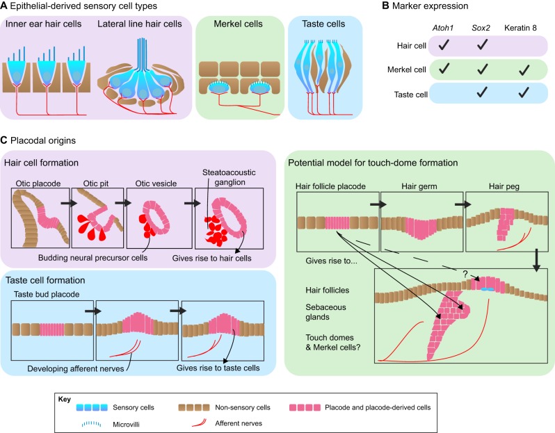
Mechanoreceptor development: insights from other sensory systems. Epidermal Merkel cells share many similarities with other sensory cell types, such as hair cells of the inner ear and taste cells. (A) All of these cell types possess apical protrusions of their cell membranes and are innervated by sensory afferents. (B) All of these cell types express similar complements of genes during their development. (C) Hair cells and taste cells develop from placodes. Based on this, we postulate that Merkel cells also arise from placodes; a potential model of placode-based Merkel cell formation is depicted.
Hair cells derive their name from bundles of microvilli-derived stereocilia protruding from their apical surfaces. When these tufts of stereocilia are deflected, hair cells translate sound and vestibular energy into electrical signals that trigger neurotransmitter release onto sensory afferents (Hudspeth and Jacobs, 1979). Hair cells are present in the inner ear and, in aquatic vertebrates, in the lateral line (see Glossary, Box 2). Merkel cells similarly possess apical microvilli (Fig. 4A; Sekerková et al., 2004; Sekerková et al., 2006). Like hair cells, Merkel cells are exquisitely mechanically sensitive (Ikeda et al., 2014; Maksimovic et al., 2014; Woo et al., 2014). Furthermore, both cell types derive from epithelial precursors and require Atoh1 and Sox2 for their specification (Fig. 4B; Bermingham et al., 1999; Dabdoub et al., 2008; Driver et al., 2013; Kiernan et al., 2005; Maricich et al., 2009; Morrison et al., 2009; Neves et al., 2013; Van Keymeulen et al., 2009).
Taste cells (see Glossary, Box 2) also bear similarities with Merkel cells. For example, both cell types arise from stratified epithelia but express keratin 8, a marker of simple epithelia (Fig. 4B). Both also express BDNF, which appears to influence the density and physiological hallmarks of their sensory afferents (Huang et al., 2015; Meng et al., 2015). Finally, like hair cells and Merkel cells, taste cells require Sox2 for proper development (Okubo et al., 2006).
Both hair cells and taste cells develop from placodes (Fig. 4C; Hall et al., 1999; Whitfield, 2015), which are thickened patches of specialized epithelial cells. Placodes also give rise to skin appendages (see Glossary, Box 2), such as hair follicles, sebaceous glands and feathers (Biggs and Mikkola, 2014; Piotrowski and Baker, 2014; Schlosser, 2005; Streit, 2004). The otic placode gives rise to the otic vesicle, which is the source of all inner ear cell types. Similarly, hair cells of zebrafish lateral line neuromasts develop from placodes that migrate along the body axis, depositing precursor cells along the way (Agarwala et al., 2015; Dambly-Chaudière et al., 2003; Metcalfe et al., 1985). Based on the similarities between Merkel cells, taste cells and hair cells, we hypothesize that Merkel cells are also placode-derived cells. The possibility that Merkel cells derive from placodes, in particular hair-follicle placodes, is suggested by several observations. First, Merkel cells initially appear both within and adjacent to the hair peg of guard hair follicles (Fig. 2; Ben-Arie et al., 2000; Morrison et al., 2009; Perdigoto et al., 2014; Vielkind et al., 1995). Second, in mature mice, several hair types contain Merkel cells within their follicles, including whiskers, hairs on the distal limb and a subset of guard hairs (Li et al., 2011; McIlwrath et al., 2007; Rice et al., 1997; Vielkind et al., 1995). Third, polycomb mutations cause Merkel cells to develop within all hair follicle subtypes (Perdigoto et al., 2016).
However, two recent findings throw a wrench in the simple model that Merkel cells derive from hair-follicle placodes. One line of evidence comes from analysis of ShhCre;Rosa26YFP mice, in which all hair follicle-derived cells are marked in a Cre-dependent manner (Levy et al., 2005). This analysis showed that, although Merkel cells arise concurrently with the appearance of developing guard hairs, only 11% of Merkel cells express YFP in ShhCre;Rosa26YFP mice, indicating that they are largely derived from a lineage that is distinct from the hair follicle (Xiao et al., 2016). Second, interfollicular Merkel cells are found in mice harboring an Fgf20 null mutation, which disrupts guard hair development (Xiao et al., 2016). These results indicate that Merkel cells in touch domes are not hair follicle derived. NCAM-expressing cells that localize to hair follicles have been proposed to serve as Merkel cell progenitors (Xiao et al., 2016). Alternatively, an as-yet-identified ‘touch-dome placode’ might give rise to interfollicular Merkel cells during development.
Another open question about Merkel cell progenitors is whether they are responsible for selectively recruiting touch-dome afferents. The lateral line system might provide some clues into this. The sensory neurons that innervate lateral line neuromasts target hair cells during development such that one subset of neurons encodes rostral hair-bundle movement and the other reports caudal hair-bundle movement. Thus, each set of neurons must make specific connections with appropriate hair cells. Live imaging has revealed that newly specified hair cells extend processes down into the deeper layers of skin to form physical contacts with developing neuronal endings (Dow et al., 2015). This result highlights an active role for hair cells themselves during the targeting process. Although Merkel cells are not required to recruit afferents to touch domes (Maricich et al., 2009), Merkel cell precursors or placode cells could actively recruit afferent terminals by similar contact-mediated mechanisms once the afferents have reached the vicinity. The live-imaging techniques and genetic mouse models required to address this question are on the horizon.
Evidence from invertebrate sensory structures also provides hints for how Merkel-cell afferents and other LTMRs might selectively target skin areas during development. With the use of powerful genetic tools, some signaling pathways that mediate somatosensory axon morphology have been dissected in Caenorabditis elegans. For example, it has been shown that menorin (MNR-1) and SAX-7 are required for the development of skin-targeted PVD neurons (Dong et al., 2013; Salzberg et al., 2013). PVD neurons extend processes along the length of the worm body wall that then branch and form a ‘menorah’ pattern (Dong et al., 2013; Salzberg et al., 2013). MNR-1 and SAX-7 influence neuronal morphology by forming a complex with the transmembrane receptor DMA-1. A series of expression analyses and epistasis experiments revealed that MNR-1 and SAX-7 are expressed by the hypodermis and that their combined expression is required for the establishment of the canonical menorah pattern. This evidence supports the hypothesis that there is a ‘combinatorial code’ of ligands and receptors that is required for the development of tactile afferents – a more complex model than a one-to-one pairing of neurotrophins and receptors. These findings also set the stage for investigation of vertebrate homologs of these proteins, which include Fam151 family members (Ruiz-Trillo et al., 2007; Salzberg et al., 2013), in the development of mammalian somatosensory neurons. This is an especially intriguing possibility for the development of morphologically similar lanceolate terminals, which also form fence- or menorah-like projections around hair follicles. Interestingly, lanceolate endings engage in close interactions with terminal Schwann cells, which are required for maintenance and regeneration of their fence-shaped terminals (Li and Ginty, 2014; Woo et al., 2012). The ligands and receptors involved in these interactions remain unknown. It will be interesting to examine whether Schwann cells mediate afferent targeting for Merkel-cell afferents, lanceolate endings or other mammalian touch receptors.
Development of the Drosophila melanogaster somatosensory system has been extensively studied and can also provide clues into vertebrate mechanoreceptor development. Indeed, a number of homologs of key molecular effectors of Drosophila somatosensory development have been implicated in mammals. For example, Frizzled mutations were first described in Drosophila, where they disrupt the planar polarity of sensory bristles and trichones, which are microscopic hair-like structures that decorate the wing (Fig. 5; Gubb and García-Bellido, 1982; Vinson and Adler, 1987). In mice, epidermal but not Merkel cell-derived frizzled 6 (Fzd6) regulates the planar organization of hair follicles and Merkel cells during development (Fig. 5; Chang and Nathans, 2013; Chang et al., 2016; Guo et al., 2004; Hua et al., 2014). In mature mice, hair shafts typically emerge from the skin in the rostral-to-caudal direction along the trunk of a mouse, and Merkel cell clusters form crescent shapes around the caudal mouth of guard-hair follicles. In Fzd6 mutant mice, both patterns are disrupted developmentally, which suggests that the same mechanisms that mediate polarity of hair follicles direct the clustering of Merkel cells on the caudal side of hair follicles (Chang and Nathans, 2013; Chang et al., 2016; Guo et al., 2004; Hua et al., 2014). Interestingly, members of the Wnt signaling family and planar cell polarity pathways (e.g. frizzled, Vangl and Celsr) are also involved in patterning the orientation of stereociliary bundles of inner ear hair cells (Fig. 5; Dabdoub et al., 2003; Montcouquiol et al., 2003; Wang et al., 2005; Wang et al., 2006). Given that Merkel cells and hair cells share many features, it is intriguing to speculate how planar cell polarity genes might play a role in Merkel cell organization and function.
Fig. 5.
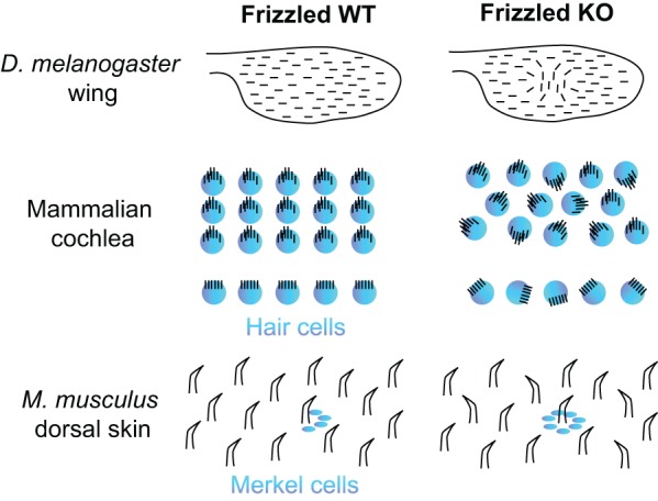
Planar cell polarity genes organize sensory structures across species. Frizzled genes in Drosophila and mice serve as a good example of how homologous planar cell polarity genes might influence tissue patterning. Bristles on the D. melanogaster wing are disrupted in clones with aberrant Frizzled signaling. Similarly, the apical microvilli in hair cells of the mammalian cochlea, which are usually highly organized, splay out in all directions in frizzled mutant cochleae. Strikingly, frizzled genes are also required for the patterning of hair follicles and Merkel cells in mouse (Mus musculus) dorsal skin; in frizzled mutants, hairs are oriented randomly rather than in a rostral-to-caudal direction, and Merkel cells form a ring around the entire follicle rather than forming a crescent on the caudal side. KO, knockout; WT, wild type.
Conclusions
Cutaneous mechanoreceptors are diverse, highly specialized and innervate distinct sensory structures. Thus, targeted innervation of the skin requires precision and accuracy. Although the cellular and molecular mechanisms responsible for recruiting afferents to specific areas of the skin are still largely elusive, recent studies of mouse hairy skin have begun to reveal basic principles of somatosensory development. These studies have highlighted that epithelial cells and placodes, which are crucial in other sensory systems for cell specification and innervation, should be investigated for their role in touch receptor development. This role is particularly interesting in the case of Merkel cell-enriched skin areas, such as touch domes, fingertips, lips and whisker follicles. In addition, given that human and non-human primates primarily use fingertips to actively explore tactile features of objects, future studies directed toward glabrous skin microdomain formation might shed light on the development and maintenance of touch receptors that are especially useful for active touch in humans.
The recent explosion of progress in the field of somatosensation has answered long-standing questions about mechanisms that govern development and function of touch receptors. In particular, this progress has highlighted that mammalian touch-receptor development research will benefit from studies of other model systems. Another avenue for the exploration and generation of new hypotheses will be to interrogate directly the link between defects in tactile sensitivity and abnormal cognitive development. The results of future studies have the power to reveal new methods for enhancing touch, for understanding the links between ASD and tactile processing, and for restoring tactile sensitivity lost after injury or through aging.
Acknowledgements
We thank Drs David Owens and Yanne Doucet for assistance with conception of this review, members of the Lumpkin laboratory for helpful discussions, and Drs Theanne Griffith, Yalda Moayedi and Cynthia Crawford for providing valuable comments on the manuscript.
Footnotes
Competing interests
The authors declare no competing or financial interests.
Funding
The authors are supported by funding from the National Institute of Neurological Disorders and Stroke (F31NS094023 to B.A.J., and R01NS073119 to E.A.L.), the National Institute of Arthritis and Musculoskeletal and Skin Diseases (R01AR051219 to E.A.L.) and the National Institute of General Medical Sciences (T32GM007367 to B.A.J.). Deposited in PMC for release after 12 months.
References
- Abraira V. E. and Ginty D. D. (2013). The sensory neurons of touch. Neuron 79, 618-639. 10.1016/j.neuron.2013.07.051 [DOI] [PMC free article] [PubMed] [Google Scholar]
- Adrian E. D. and Zotterman Y. (1926a). The impulses produced by sensory nerve endings: part 3. Impulses set up by Touch and Pressure. J. Physiol. 61, 465-483. 10.1113/jphysiol.1926.sp002308 [DOI] [PMC free article] [PubMed] [Google Scholar]
- Adrian E. D. and Zotterman Y. (1926b). The impulses produced by sensory nerve-endings: part II. The response of a Single End-Organ. J. Physiol. 61, 151-171. 10.1113/jphysiol.1926.sp002281 [DOI] [PMC free article] [PubMed] [Google Scholar]
- Agarwala S., Duquesne S., Liu K., Boehm A., Grimm L., Link S., König S., Eimer S., Ronneberger O. and Lecaudey V. (2015). Amotl2a interacts with the Hippo effector Yap1 and the Wnt/β-catenin effector Lef1 to control tissue size in zebrafish. Elife 4, e08201 10.7554/eLife.08201 [DOI] [PMC free article] [PubMed] [Google Scholar]
- Agelli M. and Clegg L. X. (2003). Epidemiology of primary Merkel cell carcinoma in the United States. J. Am. Acad. Dermatol. 49, 832-841. 10.1016/S0190-9622(03)02108-X [DOI] [PubMed] [Google Scholar]
- Airaksinen M. S. and Saarma M. (2002). The GDNF family: signalling, biological functions and therapeutic value. Nat. Rev. Neurosci. 3, 383-394. 10.1038/nrn812 [DOI] [PubMed] [Google Scholar]
- Airaksinen M. S., Koltzenburg M., Lewin G. R., Masu Y., Helbig C., Wolf E., Brem G., Toyka K. V., Thoenen H. and Meyer M. (1996). Specific subtypes of cutaneous mechanoreceptors require neurotrophin-3 following peripheral target innervation. Neuron 16, 287-295. 10.1016/S0896-6273(00)80047-1 [DOI] [PubMed] [Google Scholar]
- Albores-Saavedra J., Batich K., Chable-Montero F., Sagy N., Schwartz A. M. and Henson D. E. (2010). Merkel cell carcinoma demographics, morphology, and survival based on 3870 cases: a population based study. J. Cutan. Pathol. 37, 20-27. 10.1111/j.1600-0560.2009.01370.x [DOI] [PubMed] [Google Scholar]
- Amir R. E., Van den Veyver I. B., Wan M., Tran C. Q., Francke U. and Zoghbi H. Y. (1999). Rett syndrome is caused by mutations in X-linked MECP2, encoding methyl-CpG-binding protein 2. Nat. Genet. 23, 185-188. 10.1038/13810 [DOI] [PubMed] [Google Scholar]
- Badea T. C., Williams J., Smallwood P., Shi M., Motajo O. and Nathans J. (2012). Combinatorial expression of Brn3 transcription factors in somatosensory neurons: genetic and morphologic analysis. J. Neurosci. 32, 995-1007. 10.1523/JNEUROSCI.4755-11.2012 [DOI] [PMC free article] [PubMed] [Google Scholar]
- Badr G. G., Witt-Engerström I. and Hagberg B. (1987). Brain stem and spinal cord impairment in Rett syndrome: somatosensory and auditory evoked responses investigations. Brain Dev. 9, 517-522. 10.1016/S0387-7604(87)80076-1 [DOI] [PubMed] [Google Scholar]
- Bai L., Lehnert B. P., Liu J., Neubarth N. L., Dickendesher T. L., Nwe P. H., Cassidy C., Woodbury C. J. and Ginty D. D. (2015). Genetic identification of an expansive mechanoreceptor sensitive to skin stroking. Cell 163, 1783-1795. 10.1016/j.cell.2015.11.060 [DOI] [PMC free article] [PubMed] [Google Scholar]
- Banks R. W., Hulliger M., Saed H. H. and Stacey M. J. (2009). A comparative analysis of the encapsulated end-organs of mammalian skeletal muscles and of their sensory nerve endings. J. Anat. 214, 859-887. 10.1111/j.1469-7580.2009.01072.x [DOI] [PMC free article] [PubMed] [Google Scholar]
- Bardoni R., Tawfik V. L., Wang D., François A., Solorzano C., Shuster S. A., Choudhury P., Betelli C., Cassidy C., Smith K. et al. (2014). Delta opioid receptors presynaptically regulate cutaneous mechanosensory neuron input to the spinal cord dorsal horn. Neuron 81, 1312-1327. 10.1016/j.neuron.2014.01.044 [DOI] [PMC free article] [PubMed] [Google Scholar]
- Bardot E. S., Valdes V. J., Zhang J., Perdigoto C. N., Nicolis S., Hearn S. A., Silva J. M. and Ezhkova E. (2013). Polycomb subunits Ezh1 and Ezh2 regulate the Merkel cell differentiation program in skin stem cells. EMBO J. 32, 1990-2000. 10.1038/emboj.2013.110 [DOI] [PMC free article] [PubMed] [Google Scholar]
- Bautista D. M., Wilson S. R. and Hoon M. A. (2014). Why we scratch an itch: the molecules, cells and circuits of itch. Nat. Neurosci. 17, 175-182. 10.1038/nn.3619 [DOI] [PMC free article] [PubMed] [Google Scholar]
- Baxter A. J., Brugha T. S., Erskine H. E., Scheurer R. W., Vos T. and Scott J. G. (2015). The epidemiology and global burden of autism spectrum disorders. Psychol. Med. 45, 601-613. 10.1017/S003329171400172X [DOI] [PubMed] [Google Scholar]
- Ben-Arie N., McCall A. E., Berkman S., Eichele G., Bellen H. J. and Zoghbi H. Y. (1996). Evolutionary conservation of sequence and expression of the bHLH protein Atonal suggests a conserved role in neurogenesis. Hum. Mol. Genet. 5, 1207-1216. 10.1093/hmg/5.9.1207 [DOI] [PubMed] [Google Scholar]
- Ben-Arie N., Hassan B. A., Bermingham N. A., Malicki D. M., Armstrong D., Matzuk M., Bellen H. J. and Zoghbi H. Y. (2000). Functional conservation of atonal and Math1 in the CNS and PNS. Development 127, 1039-1048. [DOI] [PubMed] [Google Scholar]
- Bermingham N. A., Hassan B. A., Price S. D., Vollrath M. A., Ben-Arie N., Eatock R. A., Bellen H. J., Lysakowski A. and Zoghbi H. Y. (1999). Math1: an essential gene for the generation of inner ear hair cells. Science 284, 1837-1841. 10.1126/science.284.5421.1837 [DOI] [PubMed] [Google Scholar]
- Bianchi N., Depianto D., McGowan K., Gu C. and Coulombe P. A. (2005). Exploiting the keratin 17 gene promoter to visualize live cells in epithelial appendages of mice. Mol. Cell. Biol. 25, 7249-7259. 10.1128/MCB.25.16.7249-7259.2005 [DOI] [PMC free article] [PubMed] [Google Scholar]
- Biggs L. C. and Mikkola M. L. (2014). Early inductive events in ectodermal appendage morphogenesis. Semin. Cell Dev. Biol. 25-26, 11-21. 10.1016/j.semcdb.2014.01.007 [DOI] [PubMed] [Google Scholar]
- Blakemore S.-J., Tavassoli T., Calò S., Thomas R. M., Catmur C., Frith U. and Haggard P. (2006). Tactile sensitivity in Asperger syndrome. Brain Cogn. 61, 5-13. 10.1016/j.bandc.2005.12.013 [DOI] [PubMed] [Google Scholar]
- Bourane S., Garces A., Venteo S., Pattyn A., Hubert T., Fichard A., Puech S., Boukhaddaoui H., Baudet C., Takahashi S. et al. (2009). Low-threshold mechanoreceptor subtypes selectively express MafA and are specified by ret signaling. Neuron 64, 857-870. 10.1016/j.neuron.2009.12.004 [DOI] [PubMed] [Google Scholar]
- Brown A. G. and Iggo A. (1967). A quantitative study of cutaneous receptors and afferent fibres in the cat and rabbit. J. Physiol. 193, 707-733. 10.1113/jphysiol.1967.sp008390 [DOI] [PMC free article] [PubMed] [Google Scholar]
- Brown A. G., Iggo A. and Miller S. (1967). Myelinated afferent nerve fibers from the skin of the rabbit ear. Exp. Neurol. 18, 338-349. 10.1016/0014-4886(67)90053-2 [DOI] [PubMed] [Google Scholar]
- Burgess P. R., Petit D. and Warren R. M. (1968). Receptor types in cat hairy skin supplied by myelinated fibers. J. Neurophysiol. 31, 833-848. [DOI] [PubMed] [Google Scholar]
- Byrne C., Tainsky M. and Fuchs E. (1994). Programming gene expression in developing epidermis. Development 120, 2369-2383. [DOI] [PubMed] [Google Scholar]
- Carter J. J., Paulson K. G., Wipf G. C., Miranda D., Madeleine M. M., Johnson L. G., Lemos B. D., Lee S., Warcola A. H., Iyer J. G. et al. (2009). Association of Merkel cell polyomavirus-specific antibodies with Merkel cell carcinoma. J. Natl. Cancer Inst. 101, 1510-1522. 10.1093/jnci/djp332 [DOI] [PMC free article] [PubMed] [Google Scholar]
- Cascio C., McGlone F., Folger S., Tannan V., Baranek G., Pelphrey K. A. and Essick G. (2008). Tactile perception in adults with autism: a multidimensional psychophysical study. J. Autism Dev. Disord. 38, 127-137. 10.1007/s10803-007-0370-8 [DOI] [PMC free article] [PubMed] [Google Scholar]
- Cauna N. (1956). Nerve supply and nerve endings in Meissner's corpuscles. Am. J. Anat. 99, 315-350. 10.1002/aja.1000990206 [DOI] [PubMed] [Google Scholar]
- Chan J. K. C., Suster S., Wenig B. M., Tsang W. Y. W., Chan J. B. K. and Lau A. L. W. (1997). Cytokeratin 20 immunoreactivity distinguishes Merkel cell (primary cutaneous neuroendocrine) carcinomas and salivary gland small cell carcinomas from small cell carcinomas of various sites. Am. J. Surg. Pathol. 21, 226-234. 10.1097/00000478-199702000-00014 [DOI] [PubMed] [Google Scholar]
- Chang H. and Nathans J. (2013). Responses of hair follicle-associated structures to loss of planar cell polarity signaling. Proc. Natl. Acad. Sci. USA 110, E908-E917. 10.1073/pnas.1301430110 [DOI] [PMC free article] [PubMed] [Google Scholar]
- Chang H., Smallwood P. M., Williams J. and Nathans J. (2016). The spatio-temporal domains of Frizzled6 action in planar polarity control of hair follicle orientation. Dev. Biol. 409, 181-193. 10.1016/j.ydbio.2015.10.027 [DOI] [PMC free article] [PubMed] [Google Scholar]
- Cronk K. M., Wilkinson G. A., Grimes R., Wheeler E. F., Jhaveri S., Fundin B. T., Silos-Santiago I., Tessarollo L., Reichardt L. F. and Rice F. L. (2002). Diverse dependencies of developing Merkel innervation on the trkA and both full-length and truncated isoforms of trkC. Development 129, 3739-3750. [DOI] [PMC free article] [PubMed] [Google Scholar]
- Dabdoub A., Donohue M. J., Brennan A., Wolf V., Montcouquiol M., Sassoon D. A., Hseih J.-C., Rubin J. S., Salinas P. C. and Kelley M. W. (2003). Wnt signaling mediates reorientation of outer hair cell stereociliary bundles in the mammalian cochlea. Development 130, 2375-2384. 10.1242/dev.00448 [DOI] [PubMed] [Google Scholar]
- Dabdoub A., Puligilla C., Jones J. M., Fritzsch B., Cheah K. S. E., Pevny L. H. and Kelley M. W. (2008). Sox2 signaling in prosensory domain specification and subsequent hair cell differentiation in the developing cochlea. Proc. Natl. Acad. Sci. USA 105, 18396-18401. 10.1073/pnas.0808175105 [DOI] [PMC free article] [PubMed] [Google Scholar]
- Dambly-Chaudière C., Sapède D., Soubiran F., Decorde K., Gompel N. and Ghysen A. (2003). The lateral line of zebrafish: a model system for the analysis of morphogenesis and neural development in vertebrates. Biol. Cell 95, 579-587. 10.1016/j.biolcel.2003.10.005 [DOI] [PubMed] [Google Scholar]
- de Nooij J. C., Doobar S. and Jessell T. M. (2013). Etv1 inactivation reveals proprioceptor subclasses that reflect the level of NT3 expression in muscle targets. Neuron 77, 1055-1068. 10.1016/j.neuron.2013.01.015 [DOI] [PMC free article] [PubMed] [Google Scholar]
- Dell D. A. and Munger B. L. (1986). The early embryogenesis of papillary (sweat duct) ridges in primate glabrous skin: the dermatotopic map of cutaneous mechanoreceptors and dermatoglyphics. J. Comp. Neurol. 244, 511-532. 10.1002/cne.902440408 [DOI] [PubMed] [Google Scholar]
- Dong X., Liu O. W., Howell A. S. and Shen K. (2013). An extracellular adhesion molecule complex patterns dendritic branching and morphogenesis. Cell 155, 296-307. 10.1016/j.cell.2013.08.059 [DOI] [PMC free article] [PubMed] [Google Scholar]
- Doucet Y. S., Woo S.-H., Ruiz M. E. and Owens D. M. (2013). The touch dome defines an epidermal niche specialized for mechanosensory signaling. Cell Rep. 3, 1759-1765. 10.1016/j.celrep.2013.04.026 [DOI] [PMC free article] [PubMed] [Google Scholar]
- Dow E., Siletti K. and Hudspeth A. J. (2015). Cellular projections from sensory hair cells form polarity-specific scaffolds during synaptogenesis. Genes Dev. 29, 1087-1094. 10.1101/gad.259838.115 [DOI] [PMC free article] [PubMed] [Google Scholar]
- Driskell R. R., Lichtenberger B. M., Hoste E., Kretzschmar K., Simons B. D., Charalambous M., Ferron S. R., Herault Y., Pavlovic G., Ferguson-Smith A. C. et al. (2013). Distinct fibroblast lineages determine dermal architecture in skin development and repair. Nature 504, 277-281. 10.1038/nature12783 [DOI] [PMC free article] [PubMed] [Google Scholar]
- Driver E. C., Sillers L., Coate T. M., Rose M. F. and Kelley M. W. (2013). The Atoh1-lineage gives rise to hair cells and supporting cells within the mammalian cochlea. Dev. Biol. 376, 86-98. 10.1016/j.ydbio.2013.01.005 [DOI] [PMC free article] [PubMed] [Google Scholar]
- Dykes I. M., Lanier J., Eng S. R. and Turner E. E. (2010). Brn3a regulates neuronal subtype specification in the trigeminal ganglion by promoting Runx expression during sensory differentiation. Neural Dev. 5, 3 10.1186/1749-8104-5-3 [DOI] [PMC free article] [PubMed] [Google Scholar]
- Feng H., Shuda M., Chang Y. and Moore P. S. (2008). Clonal integration of a polyomavirus in human Merkel cell carcinoma. Science 319, 1096-1100. 10.1126/science.1152586 [DOI] [PMC free article] [PubMed] [Google Scholar]
- Fernandes K. J. L., McKenzie I. A., Mill P., Smith K. M., Akhavan M., Barnabé-Heider F., Biernaskie J., Junek A., Kobayashi N. R., Toma J. G. et al. (2004). A dermal niche for multipotent adult skin-derived precursor cells. Nat. Cell Biol. 6, 1082-1093. 10.1038/ncb1181 [DOI] [PubMed] [Google Scholar]
- Fleming M. S., Li J. J., Ramos D., Li T., Talmage D. A., Abe S.-I., Arber S. and Luo W. (2016). A RET-ER81-NRG1 signaling pathway drives the development of Pacinian corpuscles. J. Neurosci. 36, 10337-10355. 10.1523/JNEUROSCI.2160-16.2016 [DOI] [PMC free article] [PubMed] [Google Scholar]
- Frank E. and Sanes J. R. (1991). Lineage of neurons and glia in chick dorsal root ganglia: analysis in vivo with a recombinant retrovirus. Development 111, 895-908. [DOI] [PubMed] [Google Scholar]
- Gardiner N. J. (2011). Integrins and the extracellular matrix: key mediators of development and regeneration of the sensory nervous system. Dev. Neurobiol. 71, 1054-1072. 10.1002/dneu.20950 [DOI] [PubMed] [Google Scholar]
- Gasser H. S. (1941). The classification of nerve fibers. Ohio J. Sci. 41, 145-159. [Google Scholar]
- Goldstein M. E., House S. B. and Gainer H. (1991). NF-L and peripherin immunoreactivities define distinct classes of rat sensory ganglion cells. J. Neurosci. Res. 30, 92-104. 10.1002/jnr.490300111 [DOI] [PubMed] [Google Scholar]
- González-Martínez T., Germanà G. P., Monjil D. F., Silos-Santiago I., de Carlos F., Germanà G., Cobo J. and Vega J. A. (2004). Absence of Meissner corpuscles in the digital pads of mice lacking functional TrkB. Brain Res. 1002, 120-128. 10.1016/j.brainres.2004.01.003 [DOI] [PubMed] [Google Scholar]
- Goodwin A. W., Browning A. S. and Wheat H. E. (1995). Representation of curved surfaces in responses of mechanoreceptive afferent fibers innervating the monkey's fingerpad. J. Neurosci. 15, 798-810. [DOI] [PMC free article] [PubMed] [Google Scholar]
- Gubb D. and García-Bellido A. (1982). A genetic analysis of the determination of cuticular polarity during development in Drosophila melanogaster. J. Embryol. Exp. Morphol. 68, 37-57. [PubMed] [Google Scholar]
- Guo N., Hawkins C. and Nathans J. (2004). Frizzled6 controls hair patterning in mice. Proc. Natl. Acad. Sci. USA 101, 9277-9281. 10.1073/pnas.0402802101 [DOI] [PMC free article] [PubMed] [Google Scholar]
- Haeberle H., Fujiwara M., Chuang J., Medina M. M., Panditrao M. V., Bechstedt S., Howard J. and Lumpkin E. A. (2004). Molecular profiling reveals synaptic release machinery in Merkel cells. Proc. Natl. Acad. Sci. USA 101, 14503-14508. 10.1073/pnas.0406308101 [DOI] [PMC free article] [PubMed] [Google Scholar]
- Halata Z. (1993). Sensory innervation of the hairy skin (light- and electronmicroscopic study). J. Invest. Dermatol. 101, 75S-81S. 10.1111/1523-1747.ep12362877 [DOI] [PubMed] [Google Scholar]
- Halata Z. and Munger B. L. (1983). The sensory innervation of primate facial skin. II. Vermilion border and mucosa of lip. Brain Res. 286, 81-107. 10.1016/0165-0173(83)90022-X [DOI] [PubMed] [Google Scholar]
- Halata Z., Grim M. and Baumann K. I. (2003). [The Merkel cell: morphology, developmental origin, function]. Cas. Lek. Cesk. 142, 4-9. [PubMed] [Google Scholar]
- Halata Z., Kucera J., Kucera T. and Grim M. (2005). Apoptosis of Merkel cells in neurotrophin-3 null mice. Anat. Embryol. 209, 335-340. 10.1007/s00429-005-0455-0 [DOI] [PubMed] [Google Scholar]
- Hall J. M., Hooper J. E. and Finger T. E. (1999). Expression of sonic hedgehog, patched, and Gli1 in developing taste papillae of the mouse. J. Comp. Neurol. 406, 143-155. [DOI] [PubMed] [Google Scholar]
- Hardman M. J., Sisi P., Banbury D. N. and Byrne C. (1998). Patterned acquisition of skin barrier function during development. Development 125, 1541-1552. [DOI] [PubMed] [Google Scholar]
- Harms K. L., Healy M. A., Nghiem P., Sober A. J., Johnson T. M., Bichakjian C. K. and Wong S. L. (2016). Analysis of prognostic factors from 9387 merkel cell carcinoma cases forms the basis for the new 8th edition AJCC staging system. Ann. Surg. Oncol. 23, 3564-3571. 10.1245/s10434-016-5266-4 [DOI] [PMC free article] [PubMed] [Google Scholar]
- Hasegawa H., Abbott S., Han B.-X., Qi Y. and Wang F. (2007). Analyzing somatosensory axon projections with the sensory neuron-specific Advillin gene. J. Neurosci. 27, 14404-14414. 10.1523/JNEUROSCI.4908-07.2007 [DOI] [PMC free article] [PubMed] [Google Scholar]
- Heidenreich M., Lechner S. G., Vardanyan V., Wetzel C., Cremers C. W., De Leenheer E. M., Aránguez G., Moreno-Pelayo M. A., Jentsch T. J. and Lewin G. R. (2011). KCNQ4 K(+) channels tune mechanoreceptors for normal touch sensation in mouse and man. Nat. Neurosci. 15, 138-145. 10.1038/nn.2985 [DOI] [PubMed] [Google Scholar]
- Honma Y., Kawano M., Kohsaka S. and Ogawa M. (2010). Axonal projections of mechanoreceptive dorsal root ganglion neurons depend on Ret. Development 137, 2319-2328. 10.1242/dev.046995 [DOI] [PubMed] [Google Scholar]
- Horch K. W., Tuckett R. P. and Burgess P. R. (1977). A key to the classification of cutaneous mechanoreceptors. J. Investig. Dermatol. 69, 75-82. 10.1111/1523-1747.ep12497887 [DOI] [PubMed] [Google Scholar]
- Hu J., Huang T., Li T., Guo Z. and Cheng L. (2012). c-Maf is required for the development of dorsal horn laminae III/IV neurons and mechanoreceptive DRG axon projections. J. Neurosci. 32, 5362-5373. 10.1523/JNEUROSCI.6239-11.2012 [DOI] [PMC free article] [PubMed] [Google Scholar]
- Hua Z. L., Chang H., Wang Y., Smallwood P. M. and Nathans J. (2014). Partial interchangeability of Fz3 and Fz6 in tissue polarity signaling for epithelial orientation and axon growth and guidance. Development 141, 3944-3954. 10.1242/dev.110189 [DOI] [PMC free article] [PubMed] [Google Scholar]
- Huang T., Ma L. and Krimm R. F. (2015). Postnatal reduction of BDNF regulates the developmental remodeling of taste bud innervation. Dev. Biol. 405, 225-236. 10.1016/j.ydbio.2015.07.006 [DOI] [PMC free article] [PubMed] [Google Scholar]
- Hudspeth A. J. and Jacobs R. (1979). Stereocilia mediate transduction in vertebrate hair cells (auditory system/cilium/vestibular system). Proc. Natl. Acad. Sci. USA 76, 1506-1509. 10.1073/pnas.76.3.1506 [DOI] [PMC free article] [PubMed] [Google Scholar]
- Iggo A. and Muir A. R. (1969). The structure and function of a slowly adapting touch corpuscle in hairy skin. J. Physiol. 200, 763-796. 10.1113/jphysiol.1969.sp008721 [DOI] [PMC free article] [PubMed] [Google Scholar]
- Ikeda R., Cha M., Ling J., Jia Z., Coyle D. and Gu J. G. (2014). Merkel cells transduce and encode tactile stimuli to drive Aβ-afferent impulses. Cell 157, 664-675. 10.1016/j.cell.2014.02.026 [DOI] [PMC free article] [PubMed] [Google Scholar]
- Jarman A. P., Grau Y., Jan L. Y. and Jan Y. N. (1993). atonal is a proneural gene that directs chordotonal organ formation in the Drosophila peripheral nervous system. Cell 73, 1307-1321. 10.1016/0092-8674(93)90358-W [DOI] [PubMed] [Google Scholar]
- Jinno H., Morozova O., Jones K. L., Biernaskie J. A., Paris M., Hosokawa R., Rudnicki M. A., Chai Y., Rossi F., Marra M. A. et al. (2010). Convergent genesis of an adult neural crest-like dermal stem cell from distinct developmental origins. Stem Cells 28, 2027-2040. 10.1002/stem.525 [DOI] [PMC free article] [PubMed] [Google Scholar]
- Johansson R. S. and Vallbo A. B. (1979). Tactile sensibility in the human hand: relative and absolute densities of four types of mechanoreceptive units in glabrous skin. J. Physiol. 286, 283-300. 10.1113/jphysiol.1979.sp012619 [DOI] [PMC free article] [PubMed] [Google Scholar]
- Johnson K. O. (2001). The roles and functions of cutaneous mechanoreceptors. Curr. Opin. Neurobiol. 11, 455-461. 10.1016/S0959-4388(00)00234-8 [DOI] [PubMed] [Google Scholar]
- Johnson K. O., Yoshioka T. and Vega-Bermudez F. (2000). Tactile functions of mechanoreceptive afferents innervating the hand. J. Clin. Neurophysiol. 17, 539-558. 10.1097/00004691-200011000-00002 [DOI] [PubMed] [Google Scholar]
- Julius D. (2013). TRP channels and pain. Annu. Rev. Cell Dev. Biol. 29, 355-384. 10.1146/annurev-cellbio-101011-155833 [DOI] [PubMed] [Google Scholar]
- Kassem A., Schopflin A., Diaz C., Weyers W., Stickeler E., Werner M. and Zur Hausen A. (2008). Frequent detection of Merkel cell polyomavirus in human Merkel cell carcinomas and identification of a unique deletion in the VP1 gene. Cancer Res. 68, 5009-5013. 10.1158/0008-5472.CAN-08-0949 [DOI] [PubMed] [Google Scholar]
- Kiernan A. E., Pelling A. L., Leung K. K. H., Tang A. S. P., Bell D. M., Tease C., Lovell-Badge R., Steel K. P. and Cheah K. S. E. (2005). Sox2 is required for sensory organ development in the mammalian inner ear. Nature 434, 1031-1035. 10.1038/nature03487 [DOI] [PubMed] [Google Scholar]
- Klein R., Nanduri V., Jing S., Lamballe F., Tapley P., Bryant S., Cordon-Cardo C., Jones K. R., Reichardt L. F. and Barbacid M. (1991). The trkB tyrosine protein kinase is a receptor for brain-derived neurotrophic factor and neurotrophin-3. Cell 66, 395-403. 10.1016/0092-8674(91)90628-C [DOI] [PMC free article] [PubMed] [Google Scholar]
- Koltzenburg M., Stucky C. L. and Lewin G. R. (1997). Receptive properties of mouse sensory neurons innervating hairy skin. J. Neurophysiol. 78, 1841-1850. [DOI] [PubMed] [Google Scholar]
- Koster M. I. and Roop D. R. (2007). Mechanisms regulating epithelial stratification. Annu. Rev. Cell Dev. Biol. 23, 93-113. 10.1146/annurev.cellbio.23.090506.123357 [DOI] [PubMed] [Google Scholar]
- Kramer I., Sigrist M., de Nooij J. C., Taniuchi I., Jessell T. M. and Arber S. (2006). A role for Runx transcription factor signaling in dorsal root ganglion sensory neuron diversification. Neuron 49, 379-393. 10.1016/j.neuron.2006.01.008 [DOI] [PubMed] [Google Scholar]
- Krimm R. F., Davis B. M. and Albers K. M. (2000). Cutaneous overexpression of neurotrophin-3 (NT3) selectively restores sensory innervation in NT3 gene knockout mice. J. Neurobiol. 43, 40-49. [DOI] [PubMed] [Google Scholar]
- Krimm R. F., Davis B. M., Woodbury C. J. and Albers K. M. (2004). NT3 expressed in skin causes enhancement of SA1 sensory neurons that leads to postnatal enhancement of Merkel cells. J. Comp. Neurol. 471, 352-360. 10.1002/cne.20041 [DOI] [PubMed] [Google Scholar]
- Lai H. C., Seal R. P. and Johnson J. E. (2016). Making sense out of spinal cord somatosensory development. Development 143, 3434-3448. 10.1242/dev.139592 [DOI] [PMC free article] [PubMed] [Google Scholar]
- Lawson S. N. and Biscoe T. J. (1979). Development of mouse dorsal root ganglia: an autoradiographic and quantitative study. J. Neurocytol. 8, 265-274. 10.1007/BF01236122 [DOI] [PubMed] [Google Scholar]
- Lechner S. G. and Lewin G. R. (2013). Hairy sensation. Physiology 28, 142-150. 10.1152/physiol.00059.2012 [DOI] [PubMed] [Google Scholar]
- Lechner S. G., Frenzel H., Wang R. and Lewin G. R. (2009). Developmental waves of mechanosensitivity acquisition in sensory neuron subtypes during embryonic development. EMBO J. 28, 1479-1491. 10.1038/emboj.2009.73 [DOI] [PMC free article] [PubMed] [Google Scholar]
- Lesko M. H., Driskell R. R., Kretzschmar K., Goldie S. J. and Watt F. M. (2013). Sox2 modulates the function of two distinct cell lineages in mouse skin. Dev. Biol. 382, 15-26. 10.1016/j.ydbio.2013.08.004 [DOI] [PMC free article] [PubMed] [Google Scholar]
- Lesniak D. R., Marshall K. L., Wellnitz S. A., Jenkins B. A., Baba Y., Rasband M. N., Gerling G. J. and Lumpkin E. A. (2014). Computation identifies structural features that govern neuronal firing properties in slowly adapting touch receptors. Elife 3, 465-420 10.7554/eLife.01488 [DOI] [PMC free article] [PubMed] [Google Scholar]
- Levy V., Lindon C., Harfe B. D. and Morgan B. A. (2005). Distinct stem cell populations regenerate the follicle and interfollicular epidermis. Dev. Cell 9, 855-861. 10.1016/j.devcel.2005.11.003 [DOI] [PubMed] [Google Scholar]
- Li L. and Ginty D. D. (2014). The structure and organization of lanceolate mechanosensory complexes at mouse hair follicles. Elife 3, e01901 10.7554/eLife.01901 [DOI] [PMC free article] [PubMed] [Google Scholar]
- Li L., Rutlin M., Abraira V. E., Cassidy C., Kus L., Gong S., Jankowski M. P., Luo W., Heintz N., Koerber H. R. et al. (2011). The functional organization of cutaneous low-threshold mechanosensory neurons. Cell 147, 1615-1627. 10.1016/j.cell.2011.11.027 [DOI] [PMC free article] [PubMed] [Google Scholar]
- Liljencrantz J. and Olausson H. (2014). Tactile C fibers and their contributions to pleasant sensations and to tactile allodynia. Front. Behav. Neurosci. 8, 37 10.3389/fnbeh.2014.00037 [DOI] [PMC free article] [PubMed] [Google Scholar]
- Liu Q., Vrontou S., Rice F. L., Zylka M. J., Dong X. Z. and Anderson D. J. (2007). Molecular genetic visualization of a rare subset of unmyelinated sensory neurons that may detect gentle touch. Nat. Neurosci. 10, 946-948. 10.1038/nn1937 [DOI] [PubMed] [Google Scholar]
- Liu S., Zhang H. and Duan E. (2013). Epidermal development in mammals: key regulators, signals from beneath, and stem cells. IJMS 14, 10869-10895. 10.3390/ijms140610869 [DOI] [PMC free article] [PubMed] [Google Scholar]
- Lumpkin E. A., Collisson T., Parab P., Omer-Abdalla A., Haeberle H., Chen P., Doetzlhofer A., White P., Groves A., Segil N. et al. (2003). Math1-driven GFP expression in the developing nervous system of transgenic mice. Gene Expr. Patterns 3, 389-395. 10.1016/S1567-133X(03)00089-9 [DOI] [PubMed] [Google Scholar]
- Luo W., Enomoto H., Rice F. L., Milbrandt J. and Ginty D. D. (2009). Molecular identification of rapidly adapting mechanoreceptors and their developmental dependence on ret signaling. Neuron 64, 841-856. 10.1016/j.neuron.2009.11.003 [DOI] [PMC free article] [PubMed] [Google Scholar]
- Ma Q., Fode C., Guillemot F. and Anderson D. J. (1999). Neurogenin1 and neurogenin2 control two distinct waves of neurogenesis in developing dorsal root ganglia. Genes Dev. 13, 1717-1728. 10.1101/gad.13.13.1717 [DOI] [PMC free article] [PubMed] [Google Scholar]
- Maksimovic S., Nakatani M., Baba Y., Nelson A. M., Marshall K. L., Wellnitz S. A., Firozi P., Woo S.-H., Ranade S., Patapoutian A. et al. (2014). Epidermal Merkel cells are mechanosensory cells that tune mammalian touch receptors. Nature 509, 617-621. 10.1038/nature13250 [DOI] [PMC free article] [PubMed] [Google Scholar]
- Maricich S. M., Wellnitz S. A., Nelson A. M., Lesniak D. R., Gerling G. J., Lumpkin E. A. and Zoghbi H. Y. (2009). Merkel cells are essential for light-touch responses. Science 324, 1580-1582. 10.1126/science.1172890 [DOI] [PMC free article] [PubMed] [Google Scholar]
- Maricich S. M., Morrison K. M., Mathes E. L. and Brewer B. M. (2012). Rodents rely on merkel cells for texture discrimination tasks. J. Neurosci. 32, 3296-3300. 10.1523/JNEUROSCI.5307-11.2012 [DOI] [PMC free article] [PubMed] [Google Scholar]
- Marmigère F. and Ernfors P. (2007). Specification and connectivity of neuronal subtypes in the sensory lineage. Nat. Rev. Neurosci. 8, 114-127. 10.1038/nrn2057 [DOI] [PubMed] [Google Scholar]
- Masuda T., Yaginuma H., Sakuma C. and Ono K. (2009). Netrin-1 signaling for sensory axons: involvement in sensory axonal development and regeneration. Cell Adh. Migr. 3, 171-173. 10.4161/cam.3.2.7837 [DOI] [PMC free article] [PubMed] [Google Scholar]
- Matsuo S., Ichikawa H., Silos-Santiago I., Kiyomiya K.-I., Kurebe M., Arends J. J. A. and Jacquin M. F. (2002). Ruffini endings are absent from the periodontal ligament of trkB knockout mice. Somatosens. Mot. Res. 19, 213-217. 10.1080/0899022021000009134 [DOI] [PubMed] [Google Scholar]
- M'Boneko V. and Merker H.-J. (1988). Development and morphology of the periderm of mouse embryos (days 9-12 of gestation). Acta Anat. 133, 325-336. 10.1159/000146662 [DOI] [PubMed] [Google Scholar]
- McIlwrath S. L., Lawson J. J., Anderson C. E., Albers K. M. and Koerber H. R. (2007). Overexpression of neurotrophin-3 enhances the mechanical response properties of slowly adapting type 1 afferents and myelinated nociceptors. Eur. J. Neurosci. 26, 1801-1812. 10.1111/j.1460-9568.2007.05821.x [DOI] [PubMed] [Google Scholar]
- McKemy D. D. (2013). The molecular and cellular basis of cold sensation. ACS Chem. Neurosci. 4, 238-247. 10.1021/cn300193h [DOI] [PMC free article] [PubMed] [Google Scholar]
- Meng L., Ohman-Gault L., Ma L. and Krimm R. F. (2015). Taste bud-derived BDNF is required to maintain normal amounts of innervation to adult taste buds. eNeuro 2, ENEURO.0097-15.2015 10.1523/ENEURO.0097-15.2015 [DOI] [PMC free article] [PubMed] [Google Scholar]
- Merkel F. (1875). Tastzellen und Tastkörperchen bei den Haustieren und beim Menschen. Arch. Mikrosk. Anat. 11, 636-652. 10.1007/BF02933819 [DOI] [Google Scholar]
- Metcalfe W. K., Kimmel C. B. and Schabtach E. (1985). Anatomy of the posterior lateral line system in young larvae of the zebrafish. J. Comp. Neurol. 233, 377-389. 10.1002/cne.902330307 [DOI] [PubMed] [Google Scholar]
- Miettinen M. (1995). Keratin 20: immunohistochemical marker for gastrointestinal, urothelial, and Merkel cell carcinomas. Mod. Pathol. 8, 384-388. [PubMed] [Google Scholar]
- Miller M. R., Ralston H. J. III and Kasahara M. (1958). The pattern of cutaneous innervation of the human hand. Am. J. Anat. 102, 183-217. 10.1002/aja.1001020203 [DOI] [PubMed] [Google Scholar]
- Moll R., Lowe A., Laufer J. and Franke W. W. (1992). Cytokeratin 20 in human carcinomas. A new histodiagnostic marker detected by monoclonal antibodies. Am. J. Pathol. 140, 427-447. [PMC free article] [PubMed] [Google Scholar]
- Moll I., Troyanovsky S. M. and Moll R. (1993). Special program of differentiation expressed in keratinocytes of human haarscheiben: an analysis of individual cytokeratin polypeptides. J. Invest. Dermatol. 100, 69-76. 10.1111/1523-1747.ep12354535 [DOI] [PubMed] [Google Scholar]
- Moll I., Paus R. and Moll R. (1996). Merkel cells in mouse skin: intermediate filament pattern, localization, and hair cycle-dependent density. J. Investig. Dermatol. 106, 281-286. 10.1111/1523-1747.ep12340714 [DOI] [PubMed] [Google Scholar]
- Montcouquiol M., Rachel R. A., Lanford P. J., Copeland N. G., Jenkins N. A. and Kelley M. W. (2003). Identification of Vangl2 and Scrb1 as planar polarity genes in mammals. Nature 423, 173-177. 10.1038/nature01618 [DOI] [PubMed] [Google Scholar]
- Morrison K. M., Miesegaes G. R., Lumpkin E. A. and Maricich S. M. (2009). Mammalian Merkel cells are descended from the epidermal lineage. Dev. Biol. 336, 76-83. 10.1016/j.ydbio.2009.09.032 [DOI] [PMC free article] [PubMed] [Google Scholar]
- Munger B. L. and Halata Z. (1983). The sensory innervation of primate facial skin. I. Hairy skin. Brain Res. 286, 45-80. 10.1016/0165-0173(83)90021-8 [DOI] [PubMed] [Google Scholar]
- Munger B. L. and Ide C. (1988). The structure and function of cutaneous sensory receptors. Arch. Histol. Cytol. 51, 1-34. 10.1679/aohc.51.1 [DOI] [PubMed] [Google Scholar]
- Munger B. L. and Rice F. L. (1986). Successive waves of differentiation of cutaneous afferents in rat mystacial skin. J. Comp. Neurol. 252, 404-414. 10.1002/cne.902520309 [DOI] [PubMed] [Google Scholar]
- Neves J., Vachkov I. and Giraldez F. (2013). Sox2 regulation of hair cell development: incoherence makes sense. Hear. Res. 297, 20-29. 10.1016/j.heares.2012.11.003 [DOI] [PubMed] [Google Scholar]
- Niu J., Vysochan A. and Luo W. (2014). Dual innervation of neonatal Merkel cells in mouse touch domes. PLoS ONE 9, e92027 10.1371/journal.pone.0092027 [DOI] [PMC free article] [PubMed] [Google Scholar]
- Nurse C. A. and Diamond J. (1984). A fluorescent microscopic study of the development of rat touch domes and their Merkel cells. Neuroscience 11, 509-520. 10.1016/0306-4522(84)90041-1 [DOI] [PubMed] [Google Scholar]
- Ohtola J., Myers J., Akhtar-Zaidi B., Zuzindlak D., Sandesara P., Yeh K., Mackem S. and Atit R. (2008). beta-Catenin has sequential roles in the survival and specification of ventral dermis. Development 135, 2321-2329. 10.1242/dev.021170 [DOI] [PMC free article] [PubMed] [Google Scholar]
- Okubo T., Pevny L. H. and Hogan B. L. M. (2006). Sox2 is required for development of taste bud sensory cells. Genes Dev. 20, 2654-2659. 10.1101/gad.1457106 [DOI] [PMC free article] [PubMed] [Google Scholar]
- Olausson H., Wessberg J., Morrison I., McGlone F. and Vallbo A. (2010). The neurophysiology of unmyelinated tactile afferents. Neurosci. Biobehav. Rev. 34, 185-191. 10.1016/j.neubiorev.2008.09.011 [DOI] [PubMed] [Google Scholar]
- Olson W., Dong P., Fleming M. and Luo W. (2016). The specification and wiring of mammalian cutaneous low-threshold mechanoreceptors. Wiley Interdiscip. Rev. Dev. Biol. 5, 389-404. 10.1002/wdev.229 [DOI] [PMC free article] [PubMed] [Google Scholar]
- O'Malley A. M., Shanley D. K., Kelly A. T. and Barry D. S. (2014). Towards an understanding of semaphorin signalling in the spinal cord. Gene 553, 69-74. 10.1016/j.gene.2014.10.005 [DOI] [PubMed] [Google Scholar]
- Orefice L. L., Zimmerman A. L., Chirila A. M., Sleboda S. J., Head J. P. and Ginty D. D. (2016). Peripheral mechanosensory neuron dysfunction underlies tactile and behavioral deficits in mouse models of ASDs. Cell, 166, 299-313. 10.1016/j.cell.2016.05.033 [DOI] [PMC free article] [PubMed] [Google Scholar]
- Paré M., Smith A. M. and Rice F. L. (2002). Distribution and terminal arborizations of cutaneous mechanoreceptors in the glabrous finger pads of the monkey. J. Comp. Neurol. 445, 347-359. 10.1002/cne.10196 [DOI] [PubMed] [Google Scholar]
- Paré M., Behets C. and Cornu O. (2003). Paucity of presumptive ruffini corpuscles in the index finger pad of humans. J. Comp. Neurol. 456, 260-266. 10.1002/cne.10519 [DOI] [PubMed] [Google Scholar]
- Paus R., Müller-Röver S., van der Veen C., Maurer M., Eichmüller S., Ling G., Hofmann U., Foitzik K., Mecklenburg L. and Handjiski B. (1999). A comprehensive guide for the recognition and classification of distinct stages of hair follicle morphogenesis. J. Investig. Dermatol. 113, 523-532. 10.1046/j.1523-1747.1999.00740.x [DOI] [PubMed] [Google Scholar]
- Perdigoto C. N., Bardot E. S., Valdes V. J., Santoriello F. J. and Ezhkova E. (2014). Embryonic maturation of epidermal Merkel cells is controlled by a redundant transcription factor network. Development 141, 4690-4696. 10.1242/dev.112169 [DOI] [PMC free article] [PubMed] [Google Scholar]
- Perdigoto C. N., Dauber K. L., Bar C., Tsai P.-C., Valdes V. J., Cohen I., Santoriello F. J., Zhao D., Zheng D., Hsu Y.-C. et al. (2016). Polycomb-mediated repression and sonic hedgehog signaling interact to regulate merkel cell specification during skin development. PLoS Genet. 12, e1006151-e1006127 10.1371/journal.pgen.1006151 [DOI] [PMC free article] [PubMed] [Google Scholar]
- Perez-Pinera P., García-Suarez O., Germanà A., Díaz-Esnal B., de Carlos F., Silos-Santiago I., del Valle M. E., Cobo J. and Vega J. A. (2008). Characterization of sensory deficits in TrkB knockout mice. Neurosci. Lett. 433, 43-47. 10.1016/j.neulet.2007.12.035 [DOI] [PubMed] [Google Scholar]
- Phillips J. R., Johansson R. S. and Johnson K. O. (1990). Representation of braille characters in human nerve fibres. Exp. Brain Res. 81, 589-592. 10.1007/BF02423508 [DOI] [PubMed] [Google Scholar]
- Pinkus F. (1902). Uber einen bisher unbekannten Nebenapparat am Haarsystem des Menschen: Haarscheiben. Dermatol. Z. 9, 465-469. 10.1159/000241971 [DOI] [Google Scholar]
- Piotrowski T. and Baker C. V. H. (2014). The development of lateral line placodes: taking a broader view. Dev. Biol. 389, 68-81. 10.1016/j.ydbio.2014.02.016 [DOI] [PubMed] [Google Scholar]
- Rasmusson D. D. and Turnbull B. G. (1986). Sensory innervation of the raccoon forepaw: 2. Response properties and classification of slowly adapting fibers. Somatosens Res. 4, 63-75. 10.3109/07367228609144598 [DOI] [PubMed] [Google Scholar]
- Reed-Geaghan E. G., Wright M. C., See L. A., Adelman P. C., Lee K. H., Koerber H. R. and Maricich S. M. (2016). Merkel cell-driven BDNF signaling specifies SAI neuron molecular and electrophysiological phenotypes. J. Neurosci. 36, 4362-4376. 10.1523/JNEUROSCI.3781-15.2016 [DOI] [PMC free article] [PubMed] [Google Scholar]
- Reinisch C. M. and Tschachler E. (2005). The touch dome in human skin is supplied by different types of nerve fibers. Ann. Neurol. 58, 88-95. 10.1002/ana.20527 [DOI] [PubMed] [Google Scholar]
- Rendl M., Lewis L. and Fuchs E. (2005). Molecular dissection of mesenchymal-epithelial interactions in the hair follicle. PLoS Biol. 3, e331 10.1371/journal.pbio.0030331 [DOI] [PMC free article] [PubMed] [Google Scholar]
- Renehan W. E. and Munger B. L. (1990). The development of Meissner corpuscles in primate digital skin. Brain Res. Dev. Brain Res. 51, 35-44. 10.1016/0165-3806(90)90256-X [DOI] [PubMed] [Google Scholar]
- Rice F. L. and Rasmusson D. D. (2000). Innervation of the digit on the forepaw of the raccoon. J. Comp. Neurol. 417, 467-490. [DOI] [PubMed] [Google Scholar]
- Rice F. L., Fundin B. T., Arvidsson J., Aldskogius H. and Johansson O. (1997). Comprehensive immunofluorescence and lectin binding analysis of vibrissal follicle sinus complex innervation in the mystacial pad of the rat. J. Comp. Neurol. 385, 149-184. [DOI] [PubMed] [Google Scholar]
- Richardson R. J., Hammond N. L., Coulombe P. A., Saloranta C., Nousiainen H. O., Salonen R., Berry A., Hanley N., Headon D., Karikoski R. et al. (2014). Periderm prevents pathological epithelial adhesions during embryogenesis. J. Clin. Invest. 124, 3891-3900. 10.1172/JCI71946 [DOI] [PMC free article] [PubMed] [Google Scholar]
- Rivera-Pérez J. A. and Hadjantonakis A.-K. (2014). The dynamics of morphogenesis in the early mouse embryo. Cold Spring Harb. Perspect. Biol. 7, a015867 10.1101/cshperspect.a015867 [DOI] [PMC free article] [PubMed] [Google Scholar]
- Rogers S. J., Hepburn S. and Wehner E. (2003). Parent reports of sensory symptoms in toddlers with autism and those with other developmental disorders. J. Autism Dev. Disord. 33, 631-642. 10.1023/B:JADD.0000006000.38991.a7 [DOI] [PubMed] [Google Scholar]
- Ruffini A. (1894). Sur un nouvel organe nerveux terminal et sur la présence des corpuscules Golgi-Mazzoni dans le conjonctif sous-cutané de la pulpe des doigts de l'homme. Arch. Ital. Biol. 21, 249-265. [Google Scholar]
- Ruiz-Trillo I., Burger G., Holland P. W. H., King N., Lang B. F., Roger A. J. and Gray M. W. (2007). The origins of multicellularity: a multi-taxon genome initiative. Trends Genet. 23, 113-118. 10.1016/j.tig.2007.01.005 [DOI] [PubMed] [Google Scholar]
- Rutlin M., Ho C.-Y., Abraira V. E., Cassidy C., Bai L., Woodbury C. J. and Ginty D. D. (2014). The cellular and molecular basis of direction selectivity of Adelta-LTMRs. Cell 159, 1640-1651. 10.1016/j.cell.2014.11.038 [DOI] [PMC free article] [PubMed] [Google Scholar]
- Salzberg Y., Díaz-Balzac C. A., Ramirez-Suarez N. J., Attreed M., Tecle E., Desbois M., Kaprielian Z. and Bülow H. E. (2013). Skin-derived cues control arborization of sensory dendrites in Caenorhabditis elegans. Cell 155, 308-320. 10.1016/j.cell.2013.08.058 [DOI] [PMC free article] [PubMed] [Google Scholar]
- Schepers R. J. and Ringkamp M. (2010). Thermoreceptors and thermosensitive afferents. Neurosci. Biobehav. Rev. 34, 177-184. 10.1016/j.neubiorev.2009.10.003 [DOI] [PubMed] [Google Scholar]
- Schlosser G. (2005). Evolutionary origins of vertebrate placodes: insights from developmental studies and from comparisons with other deuterostomes. J. Exp. Zool. B Mol. Dev. Evol. 304B, 347-399. 10.1002/jez.b.21055 [DOI] [PubMed] [Google Scholar]
- Schmidt-Ullrich R. and Paus R. (2005). Molecular principles of hair follicle induction and morphogenesis. BioEssays 27, 247-261. 10.1002/bies.20184 [DOI] [PubMed] [Google Scholar]
- Seal R. P., Wang X., Guan Y., Raja S. N., Woodbury C. J., Basbaum A. I. and Edwards R. H. (2009). Injury-induced mechanical hypersensitivity requires C-low threshold mechanoreceptors. Nature 462, 651-655. 10.1038/nature08505 [DOI] [PMC free article] [PubMed] [Google Scholar]
- Sekerková G., Zheng L., Loomis P. A., Changyaleket B., Whitlon D. S., Mugnaini E. and Bartles J. R. (2004). Espins are multifunctional actin cytoskeletal regulatory proteins in the microvilli of chemosensory and mechanosensory cells. J. Neurosci. 24, 5445-5456. 10.1523/JNEUROSCI.1279-04.2004 [DOI] [PMC free article] [PubMed] [Google Scholar]
- Sekerková G., Zheng L., Loomis P. A., Mugnaini E. and Bartles J. R. (2006). Espins and the actin cytoskeleton of hair cell stereocilia and sensory cell microvilli. Cell. Mol. Life Sci. 63, 2329-2341. 10.1007/s00018-006-6148-x [DOI] [PMC free article] [PubMed] [Google Scholar]
- Serbedzija G. N., Fraser S. E. and Bronner-Fraser M. (1990). Pathways of trunk neural crest cell migration in the mouse embryo as revealed by vital dye labelling. Development 108, 605-612. [DOI] [PubMed] [Google Scholar]
- Sharp C. P., Norja P., Anthony I., Bell J. E. and Simmonds P. (2009). Reactivation and mutation of newly discovered WU, KI, and Merkel cell carcinoma polyomaviruses in immunosuppressed individuals. J. Infect. Dis. 199, 398-404. 10.1086/596062 [DOI] [PubMed] [Google Scholar]
- Sherrington C. S. (1906). The Integrative Action of the Nervous System. New York, USA: Scribner.
- Sibley R. K., Dehner L. P. and Rosai J. (1985). Primary neuroendocrine (Merkel cell?) carcinoma of the skin. I. A clinicopathologic and ultrastructural study of 43 cases. Am. J. Surg. Pathol. 9, 95-108. 10.1097/00000478-198502000-00004 [DOI] [PubMed] [Google Scholar]
- Smith K. R., Jr (1977). The Haarscheibe. J. Invest. Dermatol. 69, 68-74. 10.1111/1523-1747.ep12497883 [DOI] [PubMed] [Google Scholar]
- Sorrell J. M. and Caplan A. I. (2004). Fibroblast heterogeneity: more than skin deep. J. Cell Sci. 117, 667-675. 10.1242/jcs.01005 [DOI] [PubMed] [Google Scholar]
- Streit A. (2004). Early development of the cranial sensory nervous system: from a common field to individual placodes. Dev. Biol. 276, 1-15. 10.1016/j.ydbio.2004.08.037 [DOI] [PubMed] [Google Scholar]
- Szeder V., Grim M., Kucera J. and Sieber-Blum M. (2003). Neurotrophin-3 signaling in mammalian Merkel cell development. Dev. Dyn. 228, 623-629. 10.1002/dvdy.10403 [DOI] [PubMed] [Google Scholar]
- Talbot W. H., Darian-Smith I., Kornhuber H. H. and Mountcastle V. B. (1968). The sense of flutter-vibration: comparison of the human capacity with response patterns of mechanoreceptive afferents from the monkey hand. J. Neurophysiol. 31, 301-334. [DOI] [PubMed] [Google Scholar]
- Tang C.-K. and Toker C. (1978). Trabecular carcinoma of the skin: an ultrastructural study. Cancer 42, 2311-2321. [DOI] [PubMed] [Google Scholar]
- Thiery J. P., Duband J. L. and Delouvée A. (1982). Pathways and mechanisms of avian trunk neural crest cell migration and localization. Dev. Biol. 93, 324-343. 10.1016/0012-1606(82)90121-X [DOI] [PubMed] [Google Scholar]
- Tilling T. and Moll I. (2012). Which are the cells of origin in merkel cell carcinoma? J. Skin Cancer 2012, 680410 10.1155/2012/680410 [DOI] [PMC free article] [PubMed] [Google Scholar]
- Tomchek S. D. and Dunn W. (2007). Sensory processing in children with and without autism: a comparative study using the short sensory profile. Am. J. Occup. Ther. 61, 190-200. 10.5014/ajot.61.2.190 [DOI] [PubMed] [Google Scholar]
- Usoskin D., Furlan A., Islam S., Abdo H., Lönnerberg P., Lou D., Hjerling-Leffler J., Haeggström J., Kharchenko O., Kharchenko P. V. et al. (2015). Unbiased classification of sensory neuron types by large-scale single-cell RNA sequencing. Nat. Neurosci. 18, 145-153. 10.1038/nn.3881 [DOI] [PubMed] [Google Scholar]
- Vallbo A. B., Olausson H. and Wessberg J. (1999). Unmyelinated afferents constitute a second system coding tactile stimuli of the human hairy skin. J. Neurophysiol. 81, 2753-2763. [DOI] [PubMed] [Google Scholar]
- Van Keymeulen A., Mascre G., Youseff K. K., Harel I., Michaux C., De Geest N., Szpalski C., Achouri Y., Bloch W., Hassan B. A. et al. (2009). Epidermal progenitors give rise to Merkel cells during embryonic development and adult homeostasis. J. Cell Biol. 187, 91-100. 10.1083/jcb.200907080 [DOI] [PMC free article] [PubMed] [Google Scholar]
- Vielkind U. and Hardy M. H. (1996). Changing patterns of cell adhesion molecules during mouse pelage hair follicle development. 2. Follicle morphogenesis in the hair mutants, Tabby and downy. Acta Anat. 157, 183-194. 10.1159/000147880 [DOI] [PubMed] [Google Scholar]
- Vielkind U., Sebzda M. K., Gibson I. R. and Hardy M. H. (1995). Dynamics of Merkel cell patterns in developing hair follicles in the dorsal skin of mice, demonstrated by a monoclonal antibody to mouse keratin 8. Acta Anat. 152, 93-109. 10.1159/000147688 [DOI] [PubMed] [Google Scholar]
- Vinson C. R. and Adler P. N. (1987). Directional non-cell autonomy and the transmission of polarity information by the frizzled gene of Drosophila. Nature 329, 549-551. 10.1038/329549a0 [DOI] [PubMed] [Google Scholar]
- Voos A. C., Pelphrey K. A. and Kaiser M. D. (2013). Autistic traits are associated with diminished neural response to affective touch. Soc. Cogn. Affect Neurosci. 8, 378-386. 10.1093/scan/nss009 [DOI] [PMC free article] [PubMed] [Google Scholar]
- Vrontou S., Wong A. M., Rau K. K., Koerber H. R. and Anderson D. J. (2013). Genetic identification of C fibres that detect massage-like stroking of hairy skin in vivo. Nature 493, 669-673. 10.1038/nature11810 [DOI] [PMC free article] [PubMed] [Google Scholar]
- Wallis D., Hamblen M., Zhou Y., Venken K. J. T., Schumacher A., Grimes H. L., Zoghbi H. Y., Orkin S. H. and Bellen H. J. (2003). The zinc finger transcription factor Gfi1, implicated in lymphomagenesis, is required for inner ear hair cell differentiation and survival. Development 130, 221-232. 10.1242/dev.00190 [DOI] [PubMed] [Google Scholar]
- Wang J., Mark S., Zhang X., Qian D., Yoo S.-J., Radde-Gallwitz K., Zhang Y., Lin X., Collazo A., Wynshaw-Boris A. et al. (2005). Regulation of polarized extension and planar cell polarity in the cochlea by the vertebrate PCP pathway. Nat. Genet. 37, 980-985. 10.1038/ng1622 [DOI] [PMC free article] [PubMed] [Google Scholar]
- Wang Y., Guo N. and Nathans J. (2006). The role of Frizzled3 and Frizzled6 in neural tube closure and in the planar polarity of inner-ear sensory hair cells. J. Neurosci. 26, 2147-2156. 10.1523/JNEUROSCI.4698-05.2005 [DOI] [PMC free article] [PubMed] [Google Scholar]
- Wellnitz S. A., Lesniak D. R., Gerling G. J. and Lumpkin E. A. (2010). The regularity of sustained firing reveals two populations of slowly adapting touch receptors in mouse hairy skin. J. Neurophysiol. 103, 3378-3388. 10.1152/jn.00810.2009 [DOI] [PMC free article] [PubMed] [Google Scholar]
- Wende H., Lechner S. G., Cheret C., Bourane S., Kolanczyk M. E., Pattyn A., Reuter K., Munier F. L., Carroll P., Lewin G. R. et al. (2012). The transcription factor c-Maf controls touch receptor development and function. Science 335, 1373-1376. 10.1126/science.1214314 [DOI] [PubMed] [Google Scholar]
- Wessberg J., Olausson H., Fernström K. W. and Vallbo A. B. (2003). Receptive field properties of unmyelinated tactile afferents in the human skin. J. Neurophysiol. 89, 1567-1575. 10.1152/jn.00256.2002 [DOI] [PubMed] [Google Scholar]
- Weston J. A. (1970). The migration and differentiation of neural crest cells. Adv. Morphog. 8, 41-114. 10.1016/B978-0-12-028608-9.50006-5 [DOI] [PubMed] [Google Scholar]
- Whitfield T. T. (2015). Development of the inner ear. Curr. Opin. Genet. Dev. 32, 112-118. 10.1016/j.gde.2015.02.006 [DOI] [PubMed] [Google Scholar]
- Wong C. E., Paratore C., Dours-Zimmermann M. T., Rochat A., Pietri T., Suter U., Zimmermann D. R., Dufour S., Thiery J. P., Meijer D. et al. (2006). Neural crest-derived cells with stem cell features can be traced back to multiple lineages in the adult skin. J. Cell Biol. 175, 1005-1015. 10.1083/jcb.200606062 [DOI] [PMC free article] [PubMed] [Google Scholar]
- Woo S.-H., Stumpfova M., Jensen U. B., Lumpkin E. A. and Owens D. M. (2010). Identification of epidermal progenitors for the Merkel cell lineage. Development 137, 3965-3971. 10.1242/dev.055970 [DOI] [PMC free article] [PubMed] [Google Scholar]
- Woo S.-H., Baba Y., Franco A. M., Lumpkin E. A. and Owens D. M. (2012). Excitatory glutamate is essential for development and maintenance of the piloneural mechanoreceptor. Development 139, 740-748. 10.1242/dev.070847 [DOI] [PMC free article] [PubMed] [Google Scholar]
- Woo S.-H., Ranade S., Weyer A. D., Dubin A. E., Baba Y., Qiu Z., Petrus M., Miyamoto T., Reddy K., Lumpkin E. A. et al. (2014). Piezo2 is required for Merkel-cell mechanotransduction. Nature 509, 622-626. 10.1038/nature13251 [DOI] [PMC free article] [PubMed] [Google Scholar]
- Woodbury C. J. and Koerber H. R. (2007). Central and peripheral anatomy of slowly adapting type I low-threshold mechanoreceptors innervating trunk skin of neonatal mice. J. Comp. Neurol. 505, 547-561. 10.1002/cne.21517 [DOI] [PubMed] [Google Scholar]
- Wu H., Williams J. and Nathans J. (2012). Morphologic diversity of cutaneous sensory afferents revealed by genetically directed sparse labeling. Elife 1, e00181 10.7554/eLife.00181 [DOI] [PMC free article] [PubMed] [Google Scholar]
- Xiao Y., Thoresen D. T., Miao L., Williams J. S., Wang C., Atit R. P., Wong S. Y. and Brownell I. (2016). A cascade of Wnt, Eda, and Shh signaling is essential for touch dome Merkel cell development. PLoS Genet. 12, e1006150 10.1371/journal.pgen.1006150 [DOI] [PMC free article] [PubMed] [Google Scholar]
- Zelená J. (1981). Multiple innervation of rat pacinian corpuscles regenerated after neonatal axotomy. Neuroscience 6, 1675-1686. 10.1016/0306-4522(81)90234-7 [DOI] [PubMed] [Google Scholar]
- Zelena J. (1994). Nerves and Mechanoreceptors: The Role of Innervation in the Development and Maintenance of Mammalian Mechanoreceptors, 1st edn. London; New York: Chapman & Hall. [Google Scholar]
- Zelená J., Jirmanová I., Nitatori T. and Ide C. (1990). Effacement and regeneration of tactile lamellar corpuscles of rat after postnatal nerve crush. Neuroscience 39, 513-522. 10.1016/0306-4522(90)90287-E [DOI] [PubMed] [Google Scholar]
- Zotterman Y. (1939). Touch, pain and tickling: an electro-physiological investigation on cutaneous sensory nerves. J. Physiol. 95, 1-28. 10.1113/jphysiol.1939.sp003707 [DOI] [PMC free article] [PubMed] [Google Scholar]


