Fig. 1.
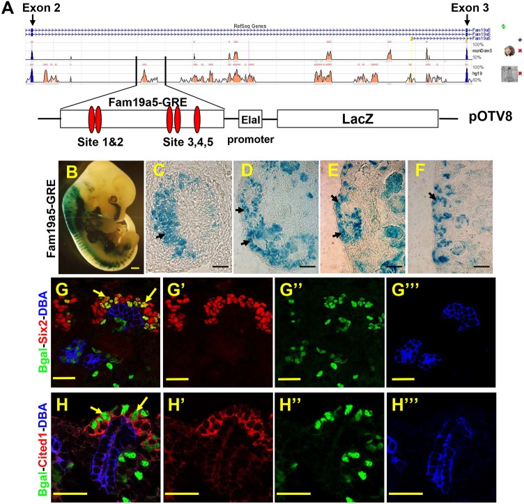
A 2.9-kb fragment in intron 2 of Fam19a5 activates transcription in the renewing nephron progenitor cell population. (A) Schematic of intron 2 of mouse Fam19a5. Peaks represent areas of homology between mouse, possum or human. The boxed area indicates the position of the fragment used to engineer the reporter. The 2.9-kb fragment was cloned into the pOTV8 plasmid containing a minimal promoter and a β-galactosidase cDNA. The relative positions of the five consensus Lef/Tcf-binding sites are indicated by red ovals. (B-F) Whole embryos (E12.5; B) and kidney sections from Fam19a5-GRE-stained 11.5 (C), E12.5 (D), E14.5 (E) and P1 (F) kidneys. For C-F, the cortical zone is to the left. (G-H‴) Sections of P1 Fam19a5-GRE kidneys stained with antibodies to Six2 (red in G,G′) or Cited1 (red in H,H′), β-gal (Bgal, green), the collecting duct marker DBA (blue in G,G‴,H,H‴). G′-G‴ and H′-H‴ are single-channel images; G and H are merged images. Arrows indicate the nephron progenitor cells. Scale bars: 500 μm (B); 50 μm (C-F); 30 μm (G-H‴).

