Description
A 75-year-old woman with a history of hypertension was admitted to our hospital for video-assisted thoracic surgery of the right caudal lung lobe because of a pT4N0M0 adenocarcinoma. After uneventful induction of anaesthesia, a difficult intubation was encountered due to a short stature and limited mobility of the neck. After multiple attempts a 35 French double-lumen tube was placed over a gum-elastic bougie with help of a video laryngoscope. To confirm correct placement a bronchoscopy was performed; however, the carina could not be visualised and the tube was repositioned several times.
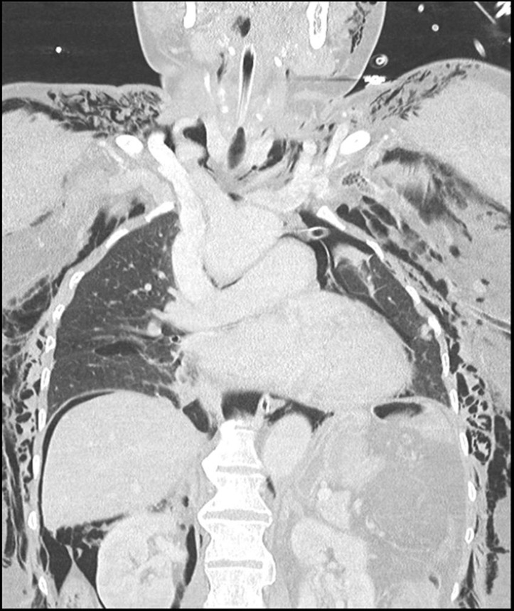
Suddenly a swelling of the abdomen and subcutaneous emphysema in the neck were noticed, ventilation pressures increased, and the patient developed bradycardia. A tension pneumothorax was suspected and bilateral needle thoracentesis was performed. The double-lumen tube was replaced by a size 6.5 single-lumen tube. Bilateral thorax drains were placed and the patient was admitted to the intensive care. Fibreoptic bronchoscopy revealed no tear of the trachea and an oesophagoscopy was unremarkable. A CT of the thorax and abdomen showed correct position of the thorax drains, massive pneumoperitoneum (figure 1), and diffuse subcutaneous and mediastinal emphysema (figure 2).
Figure 1.
Axial cross-section of the abdomen showing free air in the peritoneum and air in the retroperitoneal space on the right. Pay special notice to the ligamentum falciforme clearly visible in this coupe.
Figure 2.
Coronal cross-section depicting free air above the liver, pneumothorax on the left side with a drain in situ and pneumomediastinum. Also there is ample subcutaneous emphysema all around the thorax, the neck and even around the jaw on the left.
Ventilation became increasingly difficult on the intensive care with rapidly increasing driving pressures and concomitant haemodynamic instability. Clinical examination revealed a marked increase in abdominal distension compared with when the patient was admitted. We hypothesised that the pneumoperitoneum impeded our ability to properly ventilate. Nearly all case reports about combined pneumothorax and pneumoperitoneum are associated with a traumatic injury mechanism where surgery will be performed anyway to exclude intestinal perforation, thus resolving the pneumoperitoneum.
Since we did not suspect intestinal perforation, we decided to deflate the abdomen using an intramuscular needle (online supplementary video 1). After deflating the abdomen driving pressures dropped spectacularly and haemodynamic support could be withdrawn. Several days later the patient was successfully extubated. A culprit lesion was never found.
bcr-2017-223275supp001.mp4 (403.9KB, mp4)
Tension pneumoperitoneum is a rare sequela of a tension pneumothorax. Numerous reports have been written about pneumothorax-associated pneumoperitoneum in combination with a diaphragm defect allowing air to directly enter the peritoneum. A second proposed mechanism is that as a result of high intrathoracic pressure, air escaping from ruptured alveoli, or in this case perhaps a trachea lesion, leaks into the mediastinum through the diaphragm openings into the retroperitoneum and peritoneum.1 Even though a culprit lesion was never found, this mechanism explains fully the development of pneumoperitoneum in our patient and the rapid improvement in clinical condition after abdominal deflation.
Learning points.
Tracheobronchial rupture is a rare but life-threatening complication of endotracheal intubation possibly associated with female sex and short stature.2
Respiratory failure combined with haemodynamic instability was treated by deflating the pneumoperitoneum.
Footnotes
Contributors: MMdO wrote the manuscript and collected the images under supervision of LEMH. MV coauthored the manuscript. DN reviewed all the CT imaging and helped select the most illustrative images. LEMH acquired informed consent and is the corresponding author.
Competing interests: None declared.
Patient consent: Obtained.
Provenance and peer review: Not commissioned; externally peer reviewed.
References
- 1.Zotos PG, Kontogiannis AG, Dimakopoulos AD, et al. Tension pneumoperitoneum in association with tension pneumothorax. Am J Respir Crit Care Med 2012;186:1306 doi:10.1164/rccm.201205-0940IM [DOI] [PubMed] [Google Scholar]
- 2.Miñambres E, González-Castro A, Burón J, et al. Management of postintubation tracheobronchial rupture: our experience and a review of the literature. Eur J Emerg Med 2007;14:177–9. doi:10.1097/MEJ.0b013e3280bef8f0 [DOI] [PubMed] [Google Scholar]
Associated Data
This section collects any data citations, data availability statements, or supplementary materials included in this article.
Supplementary Materials
bcr-2017-223275supp001.mp4 (403.9KB, mp4)


