Abstract
Potassium channel tetramerization domain containing 15 (Kctd15) was previously found to have a role in early neural crest (NC) patterning, specifically delimiting the region where NC markers are expressed via repression of transcription factor AP-2a and inhibition of Wnt signaling. We used transcription activator-like effector nucleases (TALENs) to generate null mutations in zebrafish kctd15a and kctd15b paralogs to study the in vivo role of Kctd15. We found that while deletions producing frame-shift mutations in each paralog showed no apparent phenotype, kctd15a/b double mutant zebrafish are smaller in size and show several phenotypes including some affecting the NC, such as expansion of the early NC domain, increased pigmentation, and craniofacial defects. Both melanophore and xanthophore pigment cell numbers and early markers are up-regulated in the double mutants. While we find no embryonic craniofacial defects, adult mutants have a deformed maxillary segment and missing barbels. By confocal imaging of mutant larval brains we found that the torus lateralis (TLa), a region implicated in gustatory networks in other fish, is absent. Ablation of this brain tissue in wild type larvae mimics some aspects of the mutant growth phenotype. Thus kctd15 mutants show deficits in the development of both neural crest derivatives, and specific regions within the central nervous system, leading to a strong reduction in normal growth rates.
Introduction
The family of potassium channel tetramerization domain (KCTD) proteins has diverse biological functions, including protein degradation, DNA replication, regulating the hedgehog pathway, and transcriptional repression [1]. While these proteins all share a BTB/POZ (BR-C, ttk and bab/Pox virus and Zinc finger) protein-protein interaction domain near the N-terminus, they are structurally very different outside of this region [1–4]. Mutations or variants in several KCTD members have been implicated in various human diseases, including cancer, neurological diseases and metabolic disorders (reviewed in [5]), providing medical significance for the further study of this family of proteins.
In zebrafish, we previously reported that Kctd15 has a function during embryogenesis in the neural crest (NC) [6, 7]. The NC is a population of cells unique to vertebrates whose derivatives migrate and differentiate into a variety of cell types throughout the body, including craniofacial bones, pigment cells and much of the peripheral nervous system [8, 9]. Kctd15 is expressed first in the neural plate border region adjacent to the NC, and has a role in defining the NC region by repressing transcription factor AP-2 activity and inhibiting Wnt signaling [6, 7].
Kctd15 function has also been examined in other organisms. In the fruit fly Drosophila, Kctd15 is involved in both male aggression [10] and feeding frequency, as loss of the Kctd15 ortholog Twz in octopaminergic neurons resulted in flies consuming more food [11]. Genome wide association studies (GWAS) in humans have found significant linkage between KCTD15 and obesity [12–15]. Additionally, kctd15 gene expression levels in the hypothalamus of chickens and mice are related to diet, further supporting a role of Kctd15 in obesity [16, 17].
Here, we report the generation and characterization of zebrafish Kctd15 mutants. Zebrafish have two kctd15 paralogs (kctd15a and kctd15b) that have overlapping expression domains in the early NC, but then diverge as embryogenesis progresses [6, 18]. We find that homozygous mutants in either paralog alone show no phenotype, but kctd15a/b double mutants, while viable and fertile, are smaller in size and exhibit abnormalities in several NC-derived tissues including pigment cells and craniofacial bones. These mutants are lacking a fully formed maxilla bone in the upper jaw of adult fish, as well as the sensory barbels normally present on this craniofacial segment. Additionally, these mutants are missing the torus lateralis (TLa), a midbrain region which is implicated in gustatory networks in other fish species [19–22]. Ablation of this region resulted in a significant growth delay in mutant larvae compared to controls, though kctd15 mutant larvae were still smaller. Together, these results suggest that it is a combination of the deformed maxillary bone and sensory problems that lead to the smaller size of the mutants.
Results
Generation of zebrafish kctd15 mutants
Zebrafish have two kctd15 paralogs, kctd15a and kctd15b, which are 91% identical and 98% similar at the amino acid level, with complete identity in the protein-protein interacting BTB domain. We used transcription activator-like effector nucleases (TALENs; [23]) to generate null mutations in each kctd15 paralog for further characterization and functional studies (S1 Fig). Several mutations were recovered, with ~30% fish carrying at least one mutation. For further characterization, we chose kctd15aΔ23, carrying a 23 base-pair (bp) deletion upstream of the BTB-encoding sequence in exon 2, and kctd15bΔ19 carrying a 19 bp deletion at the beginning of the BTB-containing region in the third exon. Both deletions lead to frame-shifts and introduced premature stop codons (S1A and S1B Fig). While the kctd15 paralogs have overlapping expression domains in early development and therefore might compensate for each other, divergent expression patterns after somitogenesis [6] suggested possible subfunctionalization later in development. However, the kctd15aΔ23 and kctd15bΔ19 single maternal-zygotic homozygous mutants show no visible phenotype and are viable and fertile.
We crossed the two single mutant lines to generate a kctd15a/b double mutant (hereafter referred to as kctd15 mutant). Whereas embryos injected with Kctd15a/b morpholinos did not survive past 5 dpf [6], null double mutants do survive to adulthood but show a developmental delay. Kctd15 mutant fish become fertile at 4–6 months, almost twice the age of wild-type siblings. Since these mutants survive past embryogenesis, we were able to study late-onset phenotypes, as discussed below.
We believe that the mutations introduced by TALENS and described here are null mutations. No Kctd15 antibody amenable to immunohistochemistry in zebrafish is available, so we checked for mutant Kctd15 protein expression by other means (S1C and S1D Fig). We cloned wild-type and mutant kctd15a and kctd15b sequences from embryonic cDNA into expression vectors that supply an N-terminal Flag tag and expressed the constructs in cell culture. Using a Kctd15 antibody against a C-terminal epitope that is suitable for immunoblotting, and anti-Flag antibody, we observed the expected ~29–30 kDa proteins by expressing wild type constructs, but no detectable protein with mutant constructs (S1C Fig). Further, we analyzed kctd15a and kctd15b transcript levels in our double mutants. Kctd15 has previously been reported to repress its own transcription [24]. Both kctd15a and kctd15b transcript levels are significantly up-regulated at several developmental time points in kctd15 mutants (S1D Fig). This suggests that transcription of both kctd15 paralogs are “de-repressed” by the absence of functional Kctd15 protein, supporting the conclusion that our mutants are functional null.
kctd15 mutants exhibit phenotypes associated with up-regulation of NC cells
I. Slight expansion and up-regulation of several NC cell markers
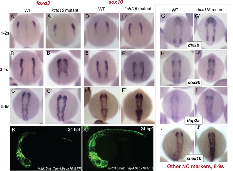
In zebrafish, the population of cells destined to become the NC localize to the neural plate border (NPB) at ~10.5 hpf, later delaminate and migrate to multiple locations where they differentiate to pigment cells, the craniofacial cartilage and bones, much of the peripheral nervous system, and other cell types (reviewed in [8, 9, 25]). The transcriptional regulatory networks underlying NC development have been well-characterized (reviewed in [26–28]). Previously we showed that Kctd15 overexpression inhibits NC formation as visualized by changes in gene marker expression patterns. We suggested that the role of Kctd15 was to delimit the early NC domain [6]. At a molecular level this effect on NC development is likely due to inhibition of Wnt signaling and of the function of transcription factor AP-2 [6, 7, 29, 30]; Wnt signaling and AP-2 function are essential for NC development [31, 32]. To further study the role of Kctd15 in NC formation, we examined expression of several NC markers in kctd15 mutants (Fig 1). The expression patterns of the early NC markers foxd3 and sox10 were unchanged in Kctd15 mutants at 1–2 somites (Fig 1A, 1A’, 1D and 1D’), but showed minor expansion at 3–4 somites (Fig 1B, 1B’, 1E and 1E’), which persists at least to 8–9 somites (Fig 1C, 1C’, 1F and 1F’). Additional markers, dlx3b, sox9b, tfap2a, and snail1b were examined at 8–9 somite stages, when all gave evidence of a modest level of expansion and up-regulation of expression (Fig 1G–1J’). This expansion of the NC domain suggests that kctd15 mutants contain more NC cells, and therefore are expected to generate an excess of NC derivatives. To test this, we crossed our kctd15 mutant into fish that contain a transgenic sox10-GFP reporter construct that labels most migrating NC cells [33]. An increase in GFP expression region and intensity was observed at 24 hpf in mutants compared to wild-type, supporting the view that the NC domain is expanded in kctd15 mutant embryos (Fig 1K and 1K’).
Fig 1. kctd15 mutants show up-regulation of several NC gene markers.
foxd3 (A-C) and sox10 (D-F) expression is indistinguishable between WT and mutants very early in NC development, but by 3-4s, expression of these markers shows both an expansion and up-regulation in expression, which persists at 8-9s. Other NC markers, including dlx3b (G), sox9b (H), tfap2a (I) and snail1b (J) also show up-regulation and expansion in our mutants at 8-9s. Additionally, an increase in NC cell number is apparent at 24hpf in kctd15 mutants, as visualized in a sox10-GFP reporter construct (K).
II. kctd15 mutants have increased pigmentation
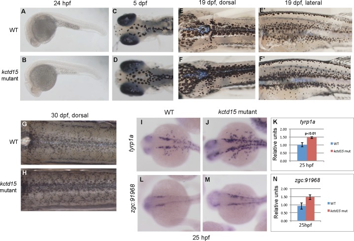
Pigment cell precursors that originate from the NC [34, 35] can be detected as early as ~24 hpf in zebrafish. These cells later differentiate into the three types of pigment cells found in adults—melanophores, xanthophores and iridophores. To check whether the apparent increase in the NC results in an increase in pigmentation we compared pigmentation patterns in wild type and kctd15 mutant embryos and larvae. No premature appearance of melanophores was seen by 24 hpf (Fig 2A and 2B) and no difference was apparent in melanophore pigmentation patterns at 48 hpf. However, sometimes at 3 dpf and always by 5 dpf, there are visibly more melanophores in the mutants (Fig 2C and 2D), a phenotype that is maintained through larval stages at 19 dpf (Fig 2E and 2F) and 30 dpf (Fig 2G and 2H). The observed increase in melanophores by 5 dpf suggests a possible up-regulation of genes involved in early melanophore development. Therefore, we examined expression levels of several genes involved in early melanophore development by in situ hybridization and quantitative PCR (qPCR; Fig 2I–2N, S2 Fig). We find up-regulation of typr1a [36] and another gene expressed in migrating NC cells destined to become melanophores, zgc:91968 ([37]; Fig 2I–2N). Somewhat surprisingly, we find that the “master regulator” of melanophore development, mitfa [38, 39], was not up-regulated at 25 hpf in the kctd15 mutants (S2A–S2C Fig), but was significantly up-regulated at 48 hpf (S2C Fig); similar results are seen with dct ([34, 40]; S2D Fig). Other genes known to be involved in melanophore development, kita [41], and tyr [42], were not significantly up-regulated at 25 hpf or 2 dpf (S2D and S2E Fig).
Fig 2. kctd15 mutants have more melanophores.
While there is no premature appearance of melanophores in our mutants (A,B), by 5 dpf there are visibly more on the dorsal side of the head (C,D), and by 19 dpf, there is a marked increase in melanophore pigment cells both on the dorsal and lateral sides of the larvae (E,F). This increase in pigmentation is still seen at 30 dpf (G,H). Examination of transcript levels and expression patterns in 25 hpf embryos revealed an up-regulation of the early melanophore markers tyrp1a (I-K) and zgc:91968 (L-N).
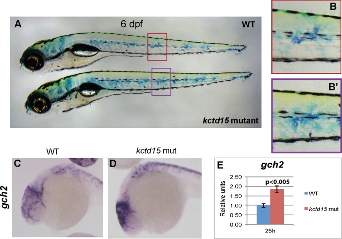
Xanthophore number, as visualized by immunostaining with Pax3/7 antibody, did not differ significantly between wild type and mutants at 30 hpf (S3A and S3B Fig; [43, 44]). However, we see an apparent increase in xanthophore number at 6 dpf by methylene blue staining (Fig 3A and 3B), while the appearance of these cells in mutant larvae is normal (Fig 3B and 3B’). There is also a more “gold” appearance on the dorsal side of mutant embryos, indicating a larger number of mature xanthophores. We examined the expression of several genes involved in early xanthophore development, including gch2 [45], xdh [46], csf1ra [47] and pax7b [48, 49] by ISH and qPCR (Fig 3C–3E; S3C–S3E Fig). Only gch2 is significantly up-regulated at 25 hpf when xanthophore development begins (Fig 3); the increase is seen primarily in the head region of the mutant embryos (Fig 3C and 3D).
Fig 3. Kctd15 mutant larvae have more xanthophores.
At 6 dpf, there are more mature xanthophores, as indicated by methylene blue staining (A); however, their appearance is just like WT (B). Examination of genes involved in xanthophore specification showed that there was an increase in abundance of gch2 transcripts at 25 hpf (C-E), most notably in the cranial pigment cells.
Finally, we inquired whether iridiphores are increased in the mutants by measuring expression levels of iridophore markers ednrab [50], pnp4a [44], ltk [51], and tfec [52] (S4 Fig). We found no significant change in expression of these markers at 25 or 48 hpf, suggesting that iridophore development is unaffected in kctd15 mutants at this time. Due to the increased number of melanophores, we could not quantify mature iridophores in the mutant larvae. Taken together, we find that in zebrafish, Kctd15 regulates NC derivatives destined to become melanophores and xanthophores.
III. Mutations in kctd15 do not affect glial cell formation
As reported above, we observe an increase in NC cell number and specific types of pigment cells. We wanted to know if other cell types derived from the NC were also affected, including glial cells of the developing peripheral nervous system [53, 54]. In situ hybridization of glial markers foxd3 and sox10 at 48 hpf showed no apparent change in number of cranial glial cells (S5A and S5B Fig) or trunk glial cells (S5C and S5D Fig) in our mutants compared to wild-type. These results suggest that loss of Kctd15 function does not result in a broad up-regulation of the NC, but is specific to certain cell populations.
IV. Kctd15 mutants exhibit craniofacial abnormalities as adults
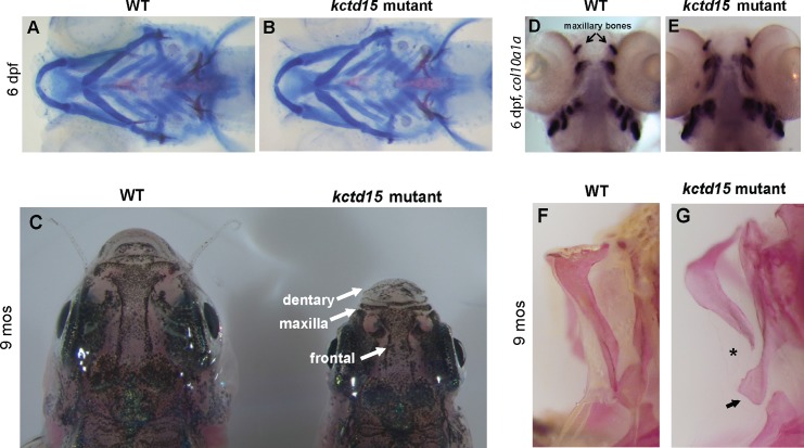
Much of the craniofacial skeleton and musculature in vertebrates is derived from the NC [9, 55]. Since there is mis-regulation of NC cell number and derivatives in kctd15 mutants, we checked for defects in the craniofacial skeleton [56]. Cranial cartilage and bone visualized by Alcian Blue and Alizarin Red staining, respectively, showed no abnormalities in mutant larvae compared to wild-type siblings (Fig 4A and 4B), but adult mutant fish exhibit a general “shortening” of the jaw and head elements, including the frontal and dental bones (Fig 4C), and are lacking a properly formed maxillary bone (Fig 4F and 4G). This malformation of the maxillary region likely is not due to an early mis-patterning of this region during embryogenesis, as staining with col10a1a mRNA, which is expressed in the developing craniofacial bones starting around 4–5 dpf [57], shows that the development of the maxillary bone is initiated in mutants and has comparable col10a1a expression to wild-type (Fig 4D and 4E). These results indicate that Kctd15 has a function in the formation of the maxilla at late larval stages.
Fig 4. Kctd15 is required for proper jaw formation later in development.
Alcian blue/Alizarin red staining does not reveal any early structural abnormalities in the patterning of kctd15 mutant jaws (A,B). However, adult mutants exhibit shortening of several jaw elements, including the dentary, maxillary and frontal regions (C). While mRNA staining at 6 dpf for col10a1a showed no difference in patterning of early craniofacial bones (D,E), adult double mutants lack a properly formed maxilla bone (F,G; abnormalities are indicated by an asterisk and arrow).
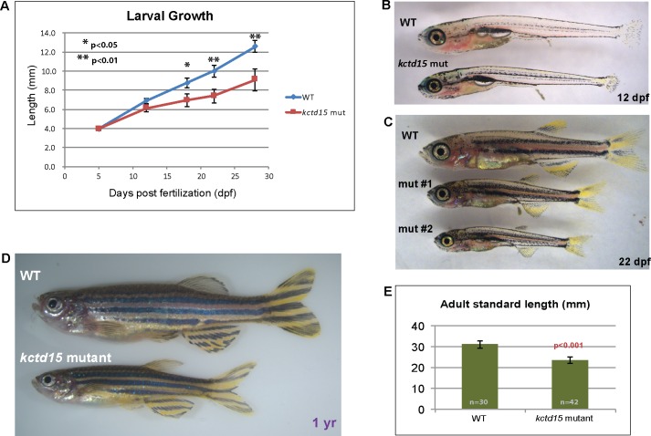
Interestingly, we also find that kctd15 mutants are missing all facial barbels, the sensory organs found on the face of fish, which contain many of the taste buds (S6 Fig). This is likely not correlated with the smaller size of kctd15 mutant fish, as barbels begin growing at standard length (SL) ~10–12 mm [58], and the adult mutant fish always surpass this length (Fig 5). Barbels may be missing in the mutants because the area where they normally develop on the maxillary segment isn’t properly formed (arrows, Fig 4G), and/or due to structural differences in the brain (see below).
Fig 5. Kctd15 mutants are smaller in size.
A) Growth of wild-type and mutant larvae, measured at 5, 12, 18, 22 and 28 dpf. While the difference in size is noticeable at 12 dpf (B), the difference is not significant until 18 dpf (C), when a range of sizes is seen, ranging from somewhat smaller (mut #1) to much smaller (mut #2). Adult mutants remain significantly smaller than wild-type siblings, even after a year (D,E).
Kctd15 mutants show a slow growth/small size phenotype
While kctd15 mutants are viable and fertile, they never reach the size of wild-type siblings (Fig 5) and take longer to reach sexual maturity. This size difference is not seen at 5 dpf, and while noticeable at 12 dpf (Fig 5B), is not significant until later in development (Fig 5A and 5C). Mutant larvae continue to remain smaller than wildtype siblings throughout larval development and into adulthood (Fig 5A, 5D and 5E). There are several possible explanations for the smaller size of the mutants, none of which are mutually exclusive. In addition to structural defects in the maxilla segment and missing barbels which might hinder proper feeding (Fig 4), additional possibilities include decreased growth hormone production [59], another brain abnormality that affects hormone production or distribution, or other mechanisms. We tested the level of growth hormone mRNA (gh) in kctd15 mutants by qPCR and in situ hybridization at 48 hpf, when the hormone is detected in the developing pituitary gland [60], and found that while there was a general trend towards lower gh levels, this difference was not significant (S7 Fig). Interestingly, whereas wild-type larva showed a rosette-like expression pattern of gh, kctd15 mutants had more variation in the number and organization of these cells (S7B Fig). However, while expression levels are not significantly lower, we have not excluded possible post-transcriptional effects on growth hormone levels in the kctd15 mutants.
Kctd15 is required for the formation of the torus lateralis in zebrafish
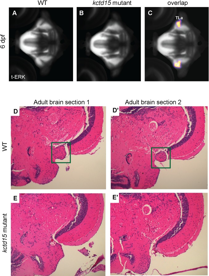
While kctd15 mutants lack gross morphological abnormalities early in development, it is possible more subtle structural defects exist. To search for subtle structural abnormalities in kctd15 mutant brains, we labeled larvae using a tERK antibody, which preferentially labels brain structure, and looked for differences in confocal image stacks aligned to a common reference and subjected to brain-wide voxel-wise analysis. Based on segmentations from Z-Brain [61] aligned to the Zebrafish Brain Browser [62], a region in the mid-brain that mapped to the torus lateralis (TLa) was identified as missing in kctd15 mutants (Fig 6A–6C). This absence is not due to a delay in growth or development of this region, as the TLa was missing in adult kctd15 mutant brains as well (Fig 6D and 6E). The TLa has been implicated in sensory functions in other fish [19–22], so it is possible that mutant zebrafish are impaired in taste or smell, inhibiting feeding.
Fig 6. Kctd15 mutants are missing the torus lateralis (TLa).
Total-ERK (tERK) antibody staining of (A) wild-type and (B) mutant 6 dpf larvae. C) Pixels whose intensity values are statistically significantly different between wildtype and mutant brains (p < 0.05, N = 14 per group, see Methods for statistical comparison procedure). Sectioning and H+E staining of a large region of adult (5 month old) wild-type (D) and mutant (E) brains revealed that this structure (green square in D) is absent throughout development. At least fifteen larval brains and five adult brains from each genotype were analyzed to confirm these results.
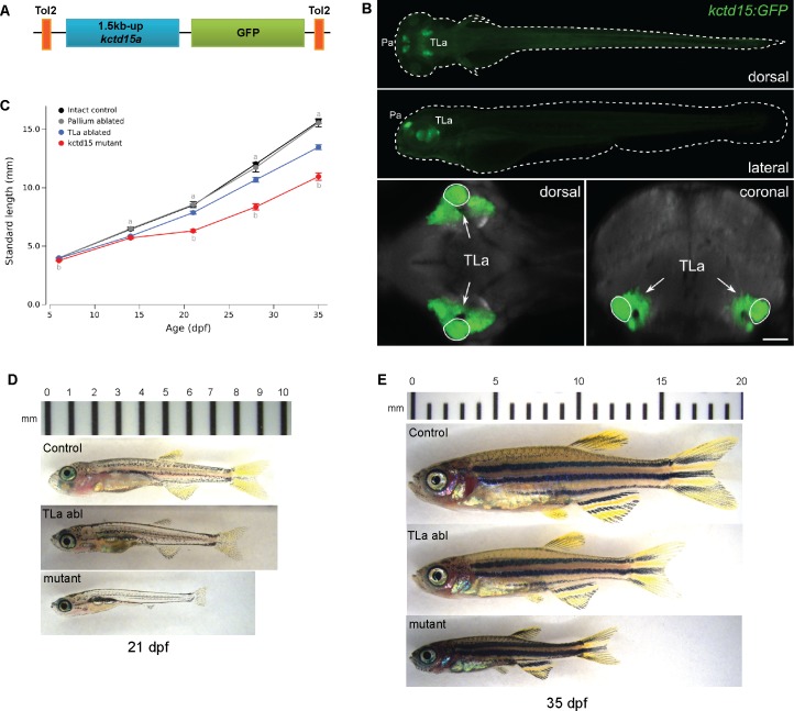
It is notable that the TLa produces growth hormone releasing hormone (GHRH) in the adult zebrafish brain [63]. GHRH functions by binding to the growth hormone releasing hormone receptor and stimulates the release of growth hormone [64]. We hypothesized that the smaller size of our kctd15 mutants could be due, at least in part, to a decreased production of GHRH and consequent reduction of GH availability. To investigate whether the TLa has a role in zebrafish growth we ablated this region at 5 dpf and measured growth for the subsequent 5 weeks. We generated a stable transgenic line expressing GFP driven by a 1.5kb region upstream of the kctd15a gene (Tg(1.5kb kctd15a-GFP); Fig 7A), which we found to be expressed in a region of the larval brain that overlaps with the TLa (Fig 7B, S8 Fig). The GFP label served as guidance in locating the TLa. Ablation of the TLa region, but not a control region in the pallium, resulted in larva that were on average smaller in size (Fig 7C–7E; two-way ANOVA for age and TLa ablation: significant main effect of ablation F[2,384] = 87.6, p < 0.001 and significant interaction effect F[8,384] = 13.0, p < 0.001; see Fig 7C for post-hoc tests). However, ablated fish were always larger than kctd15 mutants (Fig 7C). Together, these results suggest that kctd15 mutants are smaller in size at least in part due to the missing TLa, implicating the TLa in growth regulation in zebrafish.
Fig 7. TLa ablation resulted in larva of smaller size.
A) Construct design for generating a stable GFP transgenic line. 1.5kb upstream of the initiation codon of kctd15a was cloned upstream of GFP and inserted into the genome using Tol2. B) A dorsal maximum projection of an average representation of the transgenic line at 6 dpf (green) overlayed on vGLUT expression (gray) shows strong expression in the pallium region of the forebrain and torus lateralis in the midbrain. The superimposed heat map indicates the region missing in our kctd15 mutants. C) Growth measurements taken over the course of 5 weeks of non-ablated control larva (see methods; n = 39–55 per time point), control pallium ablated (n = 8 per time point), TLa ablated (n = 24–26 per time point) and kctd15 mutants (n = 28 per time point). At 6 dpf entire larval length was measured, at all other time points standard length (SL) was measured. See text for ANOVA results. For post-hoc t-tests: (a) indicates p < 0.05 for torus lateralis (TLa) ablated compared to intact control or pallium ablated controls. (b) indicates p < 0.05 for TLa compared to kctd15 mutants.
Discussion
In this work, we generated and characterized mutations in the zebrafish kctd15 paralogs to better understand the role of Kctd15 in development. Homozygous mutants in either paralog alone showed no obvious phenotype or delay in growth or fertility, but kctd15 double mutants, while viable and fertile, exhibit several phenotypes. These include defects in the NC domain and in NC derivatives, a growth delay and small size phenotype, and missing the torus lateralis, a region in the mid-brain. Among these phenotypes, the effect on the NC was expected on the basis of earlier work. Kctd15a/b morpholino injected embryos show an increase in NC markers and increased pigmentation, as also seen in the mutants. In addition the morphants show multiple morphological defects and larval lethality [6]; these defects are not seen in the mutants and thus are likely to be caused by non-specific effects of the morpholinos. Regarding the mechanistic basis of these phenotypes, we have shown that Kctd15 inhibits Wnt signaling and the activity of transcription factor AP-2 [6, 7]. These molecular effects are supported by the fact that Kctd1, a family member closely related to Kctd15, is likewise known to affect the Wnt and Ap-2 pathways [65, 66], and by the established requirement for these two pathways in NC formation and differentiation [31, 67–71]. We have suggested that Kctd15 functions to restrict the size of the NC domain. Its absence in the double kctd15a/b mutant likely leads to imbalance of NC cells rather than a whole-sale increase in the NC and its derivatives. This interpretation agrees with our observations that kctd15 mutants show selective defects in NC derivatives such as increases in melanophores and xantophores but apparently not iridophores, and in the malformation in the craniofacial skeleton that is restricted to the maxillary segment. Thus, Kctd15 function is required for the normal regulation of just some but not all NC derivatives. Whether this is due to strength and restricted location of Kctd15 in wild type embryos or a specific effect on some but not other precursor cells is presently not known.
Beyond the expected effects on NC development, novel phenotypes were observed in the kctd15 mutants. The generalized growth delay/small size phenotype may be secondary to the loss of the torus lateralis which is a site of GHRH production [63]. Moreover, the torus lateralis is immediately adjacent to the inferior lobe of the hypothalamus, which was recently implicated in visual control of feeding behavior [72]. The fact that TLa ablation only partially phenocopies the mutant growth delay may be due to contributions of other organs, incomplete ablation, or regeneration of the TLa in ablated wild type larvae. Finally, growth rates may also be affected by the loss of barbels which carry taste receptors [58, 73]; their absence may thus lead to reduced feeding. The loss of barbels may be due to the malformation of the maxillary bone (Fig 4F and 4G), and thus ultimately to the misregulation of the NC in kctd15 mutants. The molecular mechanism that leads to loss of the torus lateralis in the mutants is not understood. The Wnt signaling pathway is involved in many developmental processes, and thus its misregulation in the kctd15 mutants might be responsible for this phenotype. The AP-2 pathway is not quite as ubiquitously functioning in development as the Wnt pathway, yet AP-2 is known to interact with many proteins in the regulation of the expression of many genes (http://reactome.org/); however, no direct connection to midbrain development is apparent. In addition, it is possible that Kctd15 has molecular functions beyond the known effects on Wnt and Ap-2. Different members of the Kctd family have varied molecular functions, including as cofactors for E3 ubiquitin ligases and regulators of the hedgehog pathway [74, 75]. Thus, the range of molecular functions of Kctd15 may still not be fully explored, but the existence of mutations may help in future studies on this regulatory factor.
Materials and methods
Zebrafish maintenance and transgenic lines
Zebrafish (Danio rerio, AB strain) were maintained at 28.5°C [76] and embryos were staged according to [77] following all Animal Care Standard Operating Procedures. To prevent pigmentation in zebrafish larva, embryos were treated with 0.003% phenylthiourea (PTU) after 18 somites. Tg(1.5kb kctd15a-GFP) was generated through cloning the 1.5kb segment upstream of the kctd15a transcription start site upstream of a GFP reporter, and integrating this construct into the genome using Tol2.
TALEN construction and injection
TALENs were designed using the TAL Effector Nucleotide Targeter (http://tale-nt.cac.cornell.edu) and assembled using the Golden Gate vector system [23], using modified pCS2TAL3 vectors [78]. Capped mRNAs were synthesized using the mMESSAGE mMACHINE SP6 kit (Ambion), checked on formaldehyde gels, and injected into 1-cell embryos, each at a concentration of 100 pg/nl. TALEN efficiency was checked by pooling 10 injected embryos at 48 hpf, PCR-amplifying a 150bp region around the expected cut site, cloning into pGEMT and sequencing several individual clones. Embryos were grown to adulthood and outcrossed to wild-type AB fish to identify founders and establish mutant lines carrying single mutations.
Cell culture, protein expression and Western blotting
HEK293T cells were grown in Dulbecco’s modified Eagle’s medium supplemented with 10% FBS. Constructs containing the open reading frame of wild-type and mutant kctd15a and kctd15b sequences were cloned from 2-day cDNA of AB or homozygous mutant fish into pCS2+ or pCS2-Flag (N-terminal). These constructs were transfected into HEK293T cells using Xtreme-Gene HP (Roche) and treated according to [7]. Antibodies and dilutions used for Western Blot detection were: anti-Kctd15 (LSBio, 1:1000), anti-Flag (Sigma,1:5000), alpha-tubulin (Calbiochem, 1:1000); HRP-conjugated secondary antibodies were from Jackson (1:2000–1:5000).
Embryo fixation and whole mount in situ hybridization
Embryos were fixed in 4% paraformaldehyde for 2 hr at room temperature or overnight at 4°C, dehydrated in a series of methanol washes (25%, 50%, 75%, 100%), and incubated at -20°C overnight before use. foxd3, sox10, and dlx3b primers were from [6]. All other probe templates were amplified from cDNA sequences with a reverse primer containing the T7 promoter sequence. Dig-labeled (Roche) probes were generated using the appropriate RNA polymerase (SP6 or T7, Roche) and standard protocols. In situ hybridization (ISH) was performed as in [6]. Probes were detected using BM Purple (Roche). Embryos were photographed on a Leica MZ16F with a Leica DFC500 camera. All primer sequences for probes are available upon request.
qPCR and relative expression analysis
RNA was extracted from homogenized embryos and larva at the desired time using the TRIzol reagent and manufacturer’s protocol (Invitrogen). cDNA was synthesized using the QuantiTect Reverse Transcription Kit (Qiagen) and 25ng was used as a template. Primers were designed to amplify a region spanning an exon-exon border to avoid possible background from any genomic DNA contamination. e1fa was used as a control; all primer sequences are available upon request. qPCR was performed using SsoAdvanced Universal SYBR Green Supermix and protocol (Bio-Rad). All calculations for relative expression levels were done using the comparative CT method described by [79]. All wild-type expression levels were normalized to e1fa expression and set to 1.0; mutant expression levels were calculated relative to wild-type.
Alcian blue and Alizarin red stainings
For larval staining, cartilage (Alcian blue) and bone (Alizarin red) were stained simultaneously according to [56]. Adult craniofacial bones were stained using Alizarin red, followed by dissection of the anterior jaw elements and removal of remaining scales.
Torus lateralis (TLa) brain scanning and sectioning
Larvae at 6 dpf were fixed in 4% PFA and labeled using an anti-tERK primary antibody (1:500, Cell Signaling, 4696) and Alexa488 secondary antibody. Confocal scanning, followed by brain registration and analysis was performed as reported [62]. To detect statistically significant changes in brain structure we scanned 14 wildtype and 14 mutant brains, co-registered them to the Brain Browser atlas [62], then performed t-tests between all individual pixels within the brain. To control for multiple comparisons, we set significance thresholds by repeating this procedure 1000 times, each time randomly assigning each brain to one of two groups. For each such permutation, we recorded the most significant t-test result. We then ranked all 1000 results, and used the 95th percentile t-test result as our significance threshold for the comparison between the actual wildtype and mutant groups, thus representing a 5% false positive rate. Finally we gaussian smoothed the map of significant pixels to produce the image in Fig 6C. The region missing in our mutants was identified by alignment with an adult reference brain [61]. Adult brains were dissected from 5 month old AB and kctd15 mutant fish. Paraffin sectioning followed by H+E staining was performed on brain slices every 10 microns spanning the midbrain (HistoServ Inc, Germantown Maryland), in order to ensure that the presence or absence of the TLa could be ascertained.
TLa ablation studies
Tg(1.5kbup-kctd15a-GFP) 5 dpf larvae were anesthetized and embedded in 2.5% LMP agarose/tricaine and the TLa bilaterally ablated using a 2-photon laser (Spectra-Physics MaiTai DeepSee). As a control, cells in the pallium region were bilaterally ablated in another group. After ablation, larvae were freed from agarose and allowed to recover in E3 medium for 24 hours before transfer to tanks for ad libitum feeding. Measurements were taken weekly for 5 weeks; total length was measured at 6 dpf and standard length (SL) was measured from 14 dpf onwards.
Supporting information
A) Mutations generated in the second exon of the kctd15a locus using TALENS (targeted DNA sequences in red). Examples of mutations discovered in the germ line of different founder fish are listed. For results presented in this paper, we used the 23 bp deletion, which resulted in a premature stop codon after 11 amino acids. B) Mutations generated in the third exon of the kctd15b locus using TALENS targeting DNA sequences shown in red (splice acceptor site in bold italic). Examples of mutations discovered in the germ line of different founder fish are listed. For results presented in this paper, we used the 19 bp deletion, which resulted in a premature stop codon in the middle of the BTB domain. C) Western blot of protein samples from cell extracts after induction of wildtype and mutant transcript expression. Antibodies recognizing an epitope at the C-terminal end of Kctd15, an N-terminal FLAG tag, and alpha-tubulin were used (see Methods). No proteins from either mutant transcript were detected. D) Quantitative PCR (qPCR) of kctd15a and kctd15b transcript levels in double mutant embryos showed transcript upregulation compared to wild-type.
(TIF)
Expression of mitfa is unchanged at 25 hpf (A-C), and only shows up-regulation at 48 hpf (C), after establishment of melanophore cells. A similar pattern is seen with dct transcripts at 25 and 48 hfp (D). Expression levels of tyr (E) or kita (F) are unaffected in our mutants compared to wild-type.
(TIF)
There is no early up-regulation of mature xanthophore number in our mutants, as visualized by Pax7 antibody staining (A,B). Other gene markers known to be involved in the specification of xanthophores early in development, including csf1ra (C), xdh (D) and pax7b (E) are not up-regulated.
(TIF)
Expression levels of genes known to be involved in iridophore specification, including ednrba (A), ltk (B), pnp4a (C) and tfec (D) were unchanged at 25 and 48 hpf in kctd15 mutants compared to wildtype siblings.
(TIF)
Expression of foxd3 (A,B) and sox10 (C,D) transcripts at 48 hpf in kctd15 mutants shows no change in expression patterns of either cranial glia or trunk glia.
(TIF)
WT fish have 2 sets of facial barbels, nasal and maxillary (A), both of which are missing in kctd15 mutants (B,C). While WT fish may have fewer than 4 due to a loss for several reasons, kctd15 mutants never have any.
(TIF)
gh levels were examined by qPCR (A) and in situ hybridization (B) in WT and mutant embryos at 48 hpf. While there is a general trend towards lower gh levels, this difference is not significant. In ~60% of embryos, the staining pattern of gh transcripts appears more sparse (in fewer cells), when compared to the rosette pattern observed in WT.
(TIF)
Single horizontal (A) and coronal (B) z-stack images taken during confocal scanning of Tg(1.5kb-kctd15a-GFP) show GFP expression in the TLa. The heat maps in the single slices indicate the region missing in our kctd15 mutants.
(TIF)
Acknowledgments
We thank Valeria Zarelli and Minho Won for advice on TALENs, and Katie Grerup and Ben Feldman for various advice and suggestions.
Data Availability
All relevant data are within the paper and its Supporting Information files.
Funding Statement
This research has been funded in its entirety by the National Institute of Child Health and Human Development, Division of Intramural Research. The project number is HD008809. The funders had no role in study design, data collection and analysis, decision to publish, or preparation of the manuscript. However, the manuscript was subjected to the clearing process required for all articles and other communications from this Institute.
References
- 1.Skoblov M, Marakhonov A, Marakasova E, Guskova A, Chandhoke V, Birerdinc A, et al. Protein partners of KCTD proteins provide insights about their functional roles in cell differentiation and vertebrate development. BioEssays: news and reviews in molecular, cellular and developmental biology. 2013;35(7):586–96. Epub 2013/04/18. doi: 10.1002/bies.201300002 . [DOI] [PubMed] [Google Scholar]
- 2.Zollman S, Godt D, Prive GG, Couderc JL, Laski FA. The BTB domain, found primarily in zinc finger proteins, defines an evolutionarily conserved family that includes several developmentally regulated genes in Drosophila. Proc Natl Acad Sci U S A. 1994;91(22):10717–21. ; PubMed Central PMCID: PMCPMC45093. [DOI] [PMC free article] [PubMed] [Google Scholar]
- 3.Bardwell VJ, Treisman R. The POZ domain: a conserved protein-protein interaction motif. Genes Dev. 1994;8(14):1664–77. . [DOI] [PubMed] [Google Scholar]
- 4.Perez-Torrado R, Yamada D, Defossez PA. Born to bind: the BTB protein-protein interaction domain. BioEssays: news and reviews in molecular, cellular and developmental biology. 2006;28(12):1194–202. Epub 2006/11/23. doi: 10.1002/bies.20500 . [DOI] [PubMed] [Google Scholar]
- 5.Liu Z, Xiang Y, Sun G. The KCTD family of proteins: structure, function, disease relevance. Cell & bioscience. 2013;3(1):45 Epub 2013/11/26. doi: 10.1186/2045-3701-3-45 ; PubMed Central PMCID: PMC3882106. [DOI] [PMC free article] [PubMed] [Google Scholar]
- 6.Dutta S, Dawid IB. Kctd15 inhibits neural crest formation by attenuating Wnt/beta-catenin signaling output. Development. 2010;137(18):3013–8. Epub 2010/08/06. doi: dev.047548 [pii] doi: 10.1242/dev.047548 ; PubMed Central PMCID: PMC2926954. [DOI] [PMC free article] [PubMed] [Google Scholar]
- 7.Zarelli VE, Dawid IB. Inhibition of neural crest formation by Kctd15 involves regulation of transcription factor AP-2. Proceedings of the National Academy of Sciences of the United States of America. 2013;110(8):2870–5. Epub 2013/02/06. doi: 10.1073/pnas.1300203110 ; PubMed Central PMCID: PMC3581937. [DOI] [PMC free article] [PubMed] [Google Scholar]
- 8.Gammill LS, Bronner-Fraser M. Neural crest specification: migrating into genomics. Nat Rev Neurosci. 2003;4(10):795–805. doi: 10.1038/nrn1219 . [DOI] [PubMed] [Google Scholar]
- 9.Kaltschmidt B, Kaltschmidt C, Widera D. Adult craniofacial stem cells: sources and relation to the neural crest. Stem Cell Rev. 2012;8(3):658–71. doi: 10.1007/s12015-011-9340-9 . [DOI] [PubMed] [Google Scholar]
- 10.Williams MJ, Goergen P, Rajendran J, Klockars A, Kasagiannis A, Fredriksson R, et al. Regulation of aggression by obesity-linked genes TfAP-2 and Twz through octopamine signaling in Drosophila. Genetics. 2014;196(1):349–62. doi: 10.1534/genetics.113.158402 ; PubMed Central PMCID: PMCPMC3872196. [DOI] [PMC free article] [PubMed] [Google Scholar]
- 11.Williams MJ, Goergen P, Rajendran J, Zheleznyakova G, Hagglund MG, Perland E, et al. Obesity-linked homologues TfAP-2 and Twz establish meal frequency in Drosophila melanogaster. PLoS Genet. 2014;10(9):e1004499 doi: 10.1371/journal.pgen.1004499 ; PubMed Central PMCID: PMCPMC4154645. [DOI] [PMC free article] [PubMed] [Google Scholar]
- 12.Ng MC, Tam CH, So WY, Ho JS, Chan AW, Lee HM, et al. Implication of genetic variants near NEGR1, SEC16B, TMEM18, ETV5/DGKG, GNPDA2, LIN7C/BDNF, MTCH2, BCDIN3D/FAIM2, SH2B1, FTO, MC4R, and KCTD15 with obesity and type 2 diabetes in 7705 Chinese. J Clin Endocrinol Metab. 2010;95(5):2418–25. doi: 10.1210/jc.2009-2077 . [DOI] [PubMed] [Google Scholar]
- 13.Leon-Mimila P, Villamil-Ramirez H, Villalobos-Comparan M, Villarreal-Molina T, Romero-Hidalgo S, Lopez-Contreras B, et al. Contribution of common genetic variants to obesity and obesity-related traits in mexican children and adults. PloS one. 2013;8(8):e70640 Epub 2013/08/21. doi: 10.1371/journal.pone.0070640 ; PubMed Central PMCID: PMC3738539. [DOI] [PMC free article] [PubMed] [Google Scholar]
- 14.Zhao X, Xi B, Shen Y, Wu L, Hou D, Cheng H, et al. An obesity genetic risk score is associated with metabolic syndrome in Chinese children. Gene. 2014;535(2):299–302. doi: 10.1016/j.gene.2013.11.006 . [DOI] [PubMed] [Google Scholar]
- 15.Lv D, Zhang DD, Wang H, Zhang Y, Liang L, Fu JF, et al. Genetic variations in SEC16B, MC4R, MAP2K5 and KCTD15 were associated with childhood obesity and interacted with dietary behaviors in Chinese school-age population. Gene. 2015;560(2):149–55. doi: 10.1016/j.gene.2015.01.054 . [DOI] [PubMed] [Google Scholar]
- 16.Yoganathan P, Karunakaran S, Ho MM, Clee SM. Nutritional regulation of genome-wide association obesity genes in a tissue-dependent manner. Nutr Metab (Lond). 2012;9(1):65 doi: 10.1186/1743-7075-9-65 ; PubMed Central PMCID: PMCPMC3537611. [DOI] [PMC free article] [PubMed] [Google Scholar]
- 17.Liang SS, Ouyang HJ, Liu J, Chen B, Nie QH, Zhang XQ. Expression of variant transcripts of the potassium channel tetramerization domain-containing 15 (KCTD15) gene and their association with fatness traits in chickens. Domest Anim Endocrinol. 2015;50:65–71. doi: 10.1016/j.domaniend.2014.09.006 . [DOI] [PubMed] [Google Scholar]
- 18.Gharbi N, Zhao XF, Ellingsen S, Fjose A. Zebrafish enhancer trap line showing maternal and neural expression of kctd15a. Dev Growth Differ. 2012;54(2):241–52. Epub 2012/02/22. doi: 10.1111/j.1440-169X.2011.01326.x . [DOI] [PubMed] [Google Scholar]
- 19.Rink E, Wullimann MF. Some forebrain connections of the gustatory system in the goldfish Carassius auratus visualized by separate DiI application to the hypothalamic inferior lobe and the torus lateralis. J Comp Neurol. 1998;394(2):152–70. . [DOI] [PubMed] [Google Scholar]
- 20.Ahrens K, Wullimann MF. Hypothalamic inferior lobe and lateral torus connections in a percomorph teleost, the red cichlid (Hemichromis lifalili). J Comp Neurol. 2002;449(1):43–64. doi: 10.1002/cne.10264 . [DOI] [PubMed] [Google Scholar]
- 21.Folgueira M, Anadon R, Yanez J. Experimental study of the connections of the gustatory system in the rainbow trout, Oncorhynchus mykiss. J Comp Neurol. 2003;465(4):604–19. doi: 10.1002/cne.10879 . [DOI] [PubMed] [Google Scholar]
- 22.Kato T, Yamada Y, Yamamoto N. General visceral and gustatory connections of the posterior thalamic nucleus of goldfish. J Comp Neurol. 2011;519(15):3102–23. doi: 10.1002/cne.22669 . [DOI] [PubMed] [Google Scholar]
- 23.Cermak T, Doyle EL, Christian M, Wang L, Zhang Y, Schmidt C, et al. Efficient design and assembly of custom TALEN and other TAL effector-based constructs for DNA targeting. Nucleic acids research. 2011;39(12):e82 Epub 2011/04/16. doi: 10.1093/nar/gkr218 ; PubMed Central PMCID: PMC3130291. [DOI] [PMC free article] [PubMed] [Google Scholar]
- 24.Wong TC, Rebbert M, Wang C, Chen X, Heffer A, Zarelli VE, et al. Genes regulated by potassium channel tetramerization domain containing 15 (Kctd15) in the developing neural crest. Int J Dev Biol. 2016;60(4–6):159–66. doi: 10.1387/ijdb.160058id ; PubMed Central PMCID: PMCPMC4987960. [DOI] [PMC free article] [PubMed] [Google Scholar]
- 25.Knecht AK, Bronner-Fraser M. DBHR, a gene with homology to dopamine beta-hydroxylase, is expressed in the neural crest throughout early development. Dev Biol. 2001;234(2):365–75. doi: 10.1006/dbio.2001.0275 . [DOI] [PubMed] [Google Scholar]
- 26.LaBonne C, Bronner-Fraser M. Molecular mechanisms of neural crest formation. Annu Rev Cell Dev Biol. 1999;15:81–112. doi: 10.1146/annurev.cellbio.15.1.81 . [DOI] [PubMed] [Google Scholar]
- 27.Sauka-Spengler T, Bronner-Fraser M. A gene regulatory network orchestrates neural crest formation. Nat Rev Mol Cell Biol. 2008;9(7):557–68. Epub 2008/06/05. doi: nrm2428 [pii] doi: 10.1038/nrm2428 . [DOI] [PubMed] [Google Scholar]
- 28.Rogers CD, Jayasena CS, Nie S, Bronner ME. Neural crest specification: tissues, signals, and transcription factors. Wiley Interdiscip Rev Dev Biol. 2012;1(1):52–68. doi: 10.1002/wdev.8 . [DOI] [PubMed] [Google Scholar]
- 29.Dorsky RI, Moon RT, Raible DW. Control of neural crest cell fate by the Wnt signalling pathway. Nature. 1998;396(6709):370–3. doi: 10.1038/24620 . [DOI] [PubMed] [Google Scholar]
- 30.Zarelli VE, Dawid IB. The BTB-containing protein Kctd15 is SUMOylated in vivo. PloS one. 2013;8(9):e75016 Epub 2013/10/03. doi: 10.1371/journal.pone.0075016 ; PubMed Central PMCID: PMC3782465. [DOI] [PMC free article] [PubMed] [Google Scholar]
- 31.de Croze N, Maczkowiak F, Monsoro-Burq AH. Reiterative AP2a activity controls sequential steps in the neural crest gene regulatory network. Proceedings of the National Academy of Sciences of the United States of America. 2011;108(1):155–60. Epub 2010/12/21. doi: 1010740107 [pii] doi: 10.1073/pnas.1010740107 ; PubMed Central PMCID: PMC3017139. [DOI] [PMC free article] [PubMed] [Google Scholar]
- 32.Steventon B, Mayor R, Streit A. Mutual repression between Gbx2 and Otx2 in sensory placodes reveals a general mechanism for ectodermal patterning. Dev Biol. 2012;367(1):55–65. doi: 10.1016/j.ydbio.2012.04.025 ; PubMed Central PMCID: PMCPMC3384001. [DOI] [PMC free article] [PubMed] [Google Scholar]
- 33.Carney TJ, Dutton KA, Greenhill E, Delfino-Machin M, Dufourcq P, Blader P, et al. A direct role for Sox10 in specification of neural crest-derived sensory neurons. Development. 2006;133(23):4619–30. doi: 10.1242/dev.02668 . [DOI] [PubMed] [Google Scholar]
- 34.Kelsh RN, Brand M, Jiang YJ, Heisenberg CP, Lin S, Haffter P, et al. Zebrafish pigmentation mutations and the processes of neural crest development. Development. 1996;123:369–89. . [DOI] [PubMed] [Google Scholar]
- 35.Quigley IK, Parichy DM. Pigment pattern formation in zebrafish: a model for developmental genetics and the evolution of form. Microsc Res Tech. 2002;58(6):442–55. doi: 10.1002/jemt.10162 . [DOI] [PubMed] [Google Scholar]
- 36.Braasch I, Liedtke D, Volff JN, Schartl M. Pigmentary function and evolution of tyrp1 gene duplicates in fish. Pigment Cell Melanoma Res. 2009;22(6):839–50. doi: 10.1111/j.1755-148X.2009.00614.x . [DOI] [PubMed] [Google Scholar]
- 37.Thisse B, Thisse C. Fast Release Clones: A High Throughput Expression Analysis [ZFIN Direct Data Submission: (Unpublished]. 2004.
- 38.Lister JA, Robertson CP, Lepage T, Johnson SL, Raible DW. nacre encodes a zebrafish microphthalmia-related protein that regulates neural-crest-derived pigment cell fate. Development. 1999;126(17):3757–67. . [DOI] [PubMed] [Google Scholar]
- 39.Johnson SL, Nguyen AN, Lister JA. mitfa is required at multiple stages of melanocyte differentiation but not to establish the melanocyte stem cell. Dev Biol. 2011;350(2):405–13. doi: 10.1016/j.ydbio.2010.12.004 ; PubMed Central PMCID: PMCPMC3040983. [DOI] [PMC free article] [PubMed] [Google Scholar]
- 40.Kelsh RN, Schmid B, Eisen JS. Genetic analysis of melanophore development in zebrafish embryos. Dev Biol. 2000;225(2):277–93. doi: 10.1006/dbio.2000.9840 . [DOI] [PubMed] [Google Scholar]
- 41.Parichy DM, Rawls JF, Pratt SJ, Whitfield TT, Johnson SL. Zebrafish sparse corresponds to an orthologue of c-kit and is required for the morphogenesis of a subpopulation of melanocytes, but is not essential for hematopoiesis or primordial germ cell development. Development. 1999;126(15):3425–36. . [DOI] [PubMed] [Google Scholar]
- 42.Camp E, Lardelli M. Tyrosinase gene expression in zebrafish embryos. Dev Genes Evol. 2001;211(3):150–3. . [DOI] [PubMed] [Google Scholar]
- 43.Minchin JE, Hughes SM. Sequential actions of Pax3 and Pax7 drive xanthophore development in zebrafish neural crest. Dev Biol. 2008;317(2):508–22. doi: 10.1016/j.ydbio.2008.02.058 ; PubMed Central PMCID: PMCPMC3005709. [DOI] [PMC free article] [PubMed] [Google Scholar]
- 44.Curran K, Raible DW, Lister JA. Foxd3 controls melanophore specification in the zebrafish neural crest by regulation of Mitf. Dev Biol. 2009;332(2):408–17. doi: 10.1016/j.ydbio.2009.06.010 ; PubMed Central PMCID: PMCPMC2716409. [DOI] [PMC free article] [PubMed] [Google Scholar]
- 45.Pelletier I, Bally-Cuif L, Ziegler I. Cloning and developmental expression of zebrafish GTP cyclohydrolase I. Mech Dev. 2001;109(1):99–103. . [DOI] [PubMed] [Google Scholar]
- 46.Ziegler I. The pteridine pathway in zebrafish: regulation and specification during the determination of neural crest cell-fate. Pigment Cell Res. 2003;16(3):172–82. . [DOI] [PubMed] [Google Scholar]
- 47.Parichy DM, Ransom DG, Paw B, Zon LI, Johnson SL. An orthologue of the kit-related gene fms is required for development of neural crest-derived xanthophores and a subpopulation of adult melanocytes in the zebrafish, Danio rerio. Development. 2000;127(14):3031–44. . [DOI] [PubMed] [Google Scholar]
- 48.Lister JA, Cooper C, Nguyen K, Modrell M, Grant K, Raible DW. Zebrafish Foxd3 is required for development of a subset of neural crest derivatives. Dev Biol. 2006;290(1):92–104. doi: 10.1016/j.ydbio.2005.11.014 . [DOI] [PubMed] [Google Scholar]
- 49.Nord H, Dennhag N, Muck J, von Hofsten J. Pax7 is required for establishment of the xanthophore lineage in zebrafish embryos. Mol Biol Cell. 2016;27(11):1853–62. doi: 10.1091/mbc.E15-12-0821 ; PubMed Central PMCID: PMCPMC4884075. [DOI] [PMC free article] [PubMed] [Google Scholar]
- 50.Parichy DM, Mellgren EM, Rawls JF, Lopes SS, Kelsh RN, Johnson SL. Mutational analysis of endothelin receptor b1 (rose) during neural crest and pigment pattern development in the zebrafish Danio rerio. Dev Biol. 2000;227(2):294–306. doi: 10.1006/dbio.2000.9899 . [DOI] [PubMed] [Google Scholar]
- 51.Lopes SS, Yang X, Muller J, Carney TJ, McAdow AR, Rauch GJ, et al. Leukocyte tyrosine kinase functions in pigment cell development. PLoS Genet. 2008;4(3):e1000026 doi: 10.1371/journal.pgen.1000026 ; PubMed Central PMCID: PMCPMC2265441. [DOI] [PMC free article] [PubMed] [Google Scholar]
- 52.Lister JA, Lane BM, Nguyen A, Lunney K. Embryonic expression of zebrafish MiT family genes tfe3b, tfeb, and tfec. Dev Dyn. 2011;240(11):2529–38. doi: 10.1002/dvdy.22743 ; PubMed Central PMCID: PMCPMC3197887. [DOI] [PMC free article] [PubMed] [Google Scholar]
- 53.Le Douarin N, Dulac C, Dupin E, Cameron-Curry P. Glial cell lineages in the neural crest. Glia. 1991;4(2):175–84. doi: 10.1002/glia.440040209 . [DOI] [PubMed] [Google Scholar]
- 54.Jessen KR, Mirsky R. The origin and development of glial cells in peripheral nerves. Nat Rev Neurosci. 2005;6(9):671–82. doi: 10.1038/nrn1746 . [DOI] [PubMed] [Google Scholar]
- 55.Mork L, Crump G. Zebrafish Craniofacial Development: A Window into Early Patterning. Curr Top Dev Biol. 2015;115:235–69. doi: 10.1016/bs.ctdb.2015.07.001 ; PubMed Central PMCID: PMCPMC4758817. [DOI] [PMC free article] [PubMed] [Google Scholar]
- 56.Walker MB, Kimmel CB. A two-color acid-free cartilage and bone stain for zebrafish larvae. Biotech Histochem. 2007;82(1):23–8. doi: 10.1080/10520290701333558 . [DOI] [PubMed] [Google Scholar]
- 57.Chen Y, Yang Z, Meng M, Zhao Y, Dong N, Yan H, et al. Cullin mediates degradation of RhoA through evolutionarily conserved BTB adaptors to control actin cytoskeleton structure and cell movement. Molecular cell. 2009;35(6):841–55. Epub 2009/09/29. doi: 10.1016/j.molcel.2009.09.004 . [DOI] [PubMed] [Google Scholar]
- 58.LeClair EE, Topczewski J. Development and regeneration of the zebrafish maxillary barbel: a novel study system for vertebrate tissue growth and repair. PLoS One. 2010;5(1):e8737 doi: 10.1371/journal.pone.0008737 ; PubMed Central PMCID: PMCPMC2806924. [DOI] [PMC free article] [PubMed] [Google Scholar]
- 59.McMenamin SK, Minchin JE, Gordon TN, Rawls JF, Parichy DM. Dwarfism and increased adiposity in the gh1 mutant zebrafish vizzini. Endocrinology. 2013;154(4):1476–87. doi: 10.1210/en.2012-1734 ; PubMed Central PMCID: PMCPMC3602633. [DOI] [PMC free article] [PubMed] [Google Scholar]
- 60.Herzog W, Zeng X, Lele Z, Sonntag C, Ting JW, Chang CY, et al. Adenohypophysis formation in the zebrafish and its dependence on sonic hedgehog. Dev Biol. 2003;254(1):36–49. . [DOI] [PubMed] [Google Scholar]
- 61.Randlett O, Wee CL, Naumann EA, Nnaemeka O, Schoppik D, Fitzgerald JE, et al. Whole-brain activity mapping onto a zebrafish brain atlas. Nat Methods. 2015;12(11):1039–46. doi: 10.1038/nmeth.3581 ; PubMed Central PMCID: PMCPMC4710481. [DOI] [PMC free article] [PubMed] [Google Scholar]
- 62.Marquart GD, Tabor KM, Brown M, Strykowski JL, Varshney GK, LaFave MC, et al. A 3D Searchable Database of Transgenic Zebrafish Gal4 and Cre Lines for Functional Neuroanatomy Studies. Front Neural Circuits. 2015;9:78 doi: 10.3389/fncir.2015.00078 ; PubMed Central PMCID: PMCPMC4656851. [DOI] [PMC free article] [PubMed] [Google Scholar]
- 63.Castro A, Becerra M, Manso MJ, Tello J, Sherwood NM, Anadon R. Distribution of growth hormone-releasing hormone-like peptide: Immunoreactivity in the central nervous system of the adult zebrafish (Danio rerio). J Comp Neurol. 2009;513(6):685–701. doi: 10.1002/cne.21977 . [DOI] [PubMed] [Google Scholar]
- 64.Vance ML. Growth-hormone-releasing hormone. Clin Chem. 1990;36(3):415–20. . [PubMed] [Google Scholar]
- 65.Ding X, Luo C, Zhou J, Zhong Y, Hu X, Zhou F, et al. The interaction of KCTD1 with transcription factor AP-2alpha inhibits its transactivation. Journal of cellular biochemistry. 2009;106(2):285–95. Epub 2008/12/31. doi: 10.1002/jcb.22002 . [DOI] [PubMed] [Google Scholar]
- 66.Li X, Chen C, Wang F, Huang W, Liang Z, Xiao Y, et al. KCTD1 suppresses canonical Wnt signaling pathway by enhancing beta-catenin degradation. PloS one. 2014;9(4):e94343 Epub 2014/04/17. doi: 10.1371/journal.pone.0094343 ; PubMed Central PMCID: PMC3988066. [DOI] [PMC free article] [PubMed] [Google Scholar] [Retracted]
- 67.Ikeya M, Lee SM, Johnson JE, McMahon AP, Takada S. Wnt signalling required for expansion of neural crest and CNS progenitors. Nature. 1997;389(6654):966–70. Epub 1997/11/14. doi: 10.1038/40146 . [DOI] [PubMed] [Google Scholar]
- 68.Luo T, Lee YH, Saint-Jeannet JP, Sargent TD. Induction of neural crest in Xenopus by transcription factor AP2alpha. Proceedings of the National Academy of Sciences of the United States of America. 2003;100(2):532–7. Epub 2003/01/04. doi: 10.1073/pnas.0237226100 ; PubMed Central PMCID: PMC141030. [DOI] [PMC free article] [PubMed] [Google Scholar]
- 69.Saint-Jeannet JP, He X, Varmus HE, Dawid IB. Regulation of dorsal fate in the neuraxis by Wnt-1 and Wnt-3a. Proceedings of the National Academy of Sciences of the United States of America. 1997;94(25):13713–8. Epub 1998/02/12. ; PubMed Central PMCID: PMC28371. [DOI] [PMC free article] [PubMed] [Google Scholar]
- 70.Schorle H, Meier P, Buchert M, Jaenisch R, Mitchell PJ. Transcription factor AP-2 essential for cranial closure and craniofacial development. Nature. 1996;381(6579):235–8. Epub 1996/05/16. doi: 10.1038/381235a0 . [DOI] [PubMed] [Google Scholar]
- 71.Simoes-Costa M, Bronner ME. Establishing neural crest identity: a gene regulatory recipe. Development. 2015;142(2):242–57. Epub 2015/01/08. doi: 10.1242/dev.105445 ; PubMed Central PMCID: PMC4302844. [DOI] [PMC free article] [PubMed] [Google Scholar]
- 72.Muto A, Lal P, Ailani D, Abe G, Itoh M, Kawakami K. Activation of the hypothalamic feeding centre upon visual prey detection. Nat Commun. 2017;8:15029 doi: 10.1038/ncomms15029 . [DOI] [PMC free article] [PubMed] [Google Scholar]
- 73.Hansen A, Reutter K, Zeiske E. Taste bud development in the zebrafish, Danio rerio. Dev Dyn. 2002;223(4):483–96. doi: 10.1002/dvdy.10074 . [DOI] [PubMed] [Google Scholar]
- 74.Bayon Y, Trinidad AG, de la Puerta ML, Del Carmen Rodriguez M, Bogetz J, Rojas A, et al. KCTD5, a putative substrate adaptor for cullin3 ubiquitin ligases. The FEBS journal. 2008;275(15):3900–10. Epub 2008/06/25. doi: EJB6537 [pii] doi: 10.1111/j.1742-4658.2008.06537.x . [DOI] [PubMed] [Google Scholar]
- 75.Canettieri G, Di Marcotullio L, Greco A, Coni S, Antonucci L, Infante P, et al. Histone deacetylase and Cullin3-REN(KCTD11) ubiquitin ligase interplay regulates Hedgehog signalling through Gli acetylation. Nature cell biology. 2010;12(2):132–42. Epub 2010/01/19. doi: ncb2013 [pii] doi: 10.1038/ncb2013 . [DOI] [PubMed] [Google Scholar]
- 76.Westerfield M. The Zebrafish Book. A Guide for the Laboratory Use of Zebrafish (Danio rerio), 4th Edition2000.
- 77.Kimmel CB, Ballard WW, Kimmel SR, Ullmann B, Schilling TF. Stages of embryonic development of the zebrafish. Developmental dynamics: an official publication of the American Association of Anatomists. 1995;203(3):253–310. Epub 1995/07/01. doi: 10.1002/aja.1002030302 . [DOI] [PubMed] [Google Scholar]
- 78.Won M, Ro H, Dawid IB. Lnx2 ubiquitin ligase is essential for exocrine cell differentiation in the early zebrafish pancreas. Proc Natl Acad Sci U S A. 2015;112(40):12426–31. doi: 10.1073/pnas.1517033112 ; PubMed Central PMCID: PMCPMC4603503. [DOI] [PMC free article] [PubMed] [Google Scholar]
- 79.Schmittgen TD, Livak KJ. Analyzing real-time PCR data by the comparative C(T) method. Nat Protoc. 2008;3(6):1101–8. . [DOI] [PubMed] [Google Scholar]
Associated Data
This section collects any data citations, data availability statements, or supplementary materials included in this article.
Supplementary Materials
A) Mutations generated in the second exon of the kctd15a locus using TALENS (targeted DNA sequences in red). Examples of mutations discovered in the germ line of different founder fish are listed. For results presented in this paper, we used the 23 bp deletion, which resulted in a premature stop codon after 11 amino acids. B) Mutations generated in the third exon of the kctd15b locus using TALENS targeting DNA sequences shown in red (splice acceptor site in bold italic). Examples of mutations discovered in the germ line of different founder fish are listed. For results presented in this paper, we used the 19 bp deletion, which resulted in a premature stop codon in the middle of the BTB domain. C) Western blot of protein samples from cell extracts after induction of wildtype and mutant transcript expression. Antibodies recognizing an epitope at the C-terminal end of Kctd15, an N-terminal FLAG tag, and alpha-tubulin were used (see Methods). No proteins from either mutant transcript were detected. D) Quantitative PCR (qPCR) of kctd15a and kctd15b transcript levels in double mutant embryos showed transcript upregulation compared to wild-type.
(TIF)
Expression of mitfa is unchanged at 25 hpf (A-C), and only shows up-regulation at 48 hpf (C), after establishment of melanophore cells. A similar pattern is seen with dct transcripts at 25 and 48 hfp (D). Expression levels of tyr (E) or kita (F) are unaffected in our mutants compared to wild-type.
(TIF)
There is no early up-regulation of mature xanthophore number in our mutants, as visualized by Pax7 antibody staining (A,B). Other gene markers known to be involved in the specification of xanthophores early in development, including csf1ra (C), xdh (D) and pax7b (E) are not up-regulated.
(TIF)
Expression levels of genes known to be involved in iridophore specification, including ednrba (A), ltk (B), pnp4a (C) and tfec (D) were unchanged at 25 and 48 hpf in kctd15 mutants compared to wildtype siblings.
(TIF)
Expression of foxd3 (A,B) and sox10 (C,D) transcripts at 48 hpf in kctd15 mutants shows no change in expression patterns of either cranial glia or trunk glia.
(TIF)
WT fish have 2 sets of facial barbels, nasal and maxillary (A), both of which are missing in kctd15 mutants (B,C). While WT fish may have fewer than 4 due to a loss for several reasons, kctd15 mutants never have any.
(TIF)
gh levels were examined by qPCR (A) and in situ hybridization (B) in WT and mutant embryos at 48 hpf. While there is a general trend towards lower gh levels, this difference is not significant. In ~60% of embryos, the staining pattern of gh transcripts appears more sparse (in fewer cells), when compared to the rosette pattern observed in WT.
(TIF)
Single horizontal (A) and coronal (B) z-stack images taken during confocal scanning of Tg(1.5kb-kctd15a-GFP) show GFP expression in the TLa. The heat maps in the single slices indicate the region missing in our kctd15 mutants.
(TIF)
Data Availability Statement
All relevant data are within the paper and its Supporting Information files.


