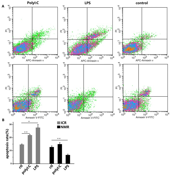
Figure 4. PolyI:C and LPS induce apoptosis in mouse and naked mole rat macrophages.
(A) Macrophages were treated with 10 ng/ml polyI:C or 1 ng/ml LPS for 12 h. The negative control was treated with PBS. Apoptosis was analyzed with PI and annexin V staining by flow cytometry. The upper panels represent the results for mice, and the lower panels correspond to naked mole rats. (B) The graph indicates the percentage of apoptotic cells (means ± SD). *P<0.05, **P<0.01.

