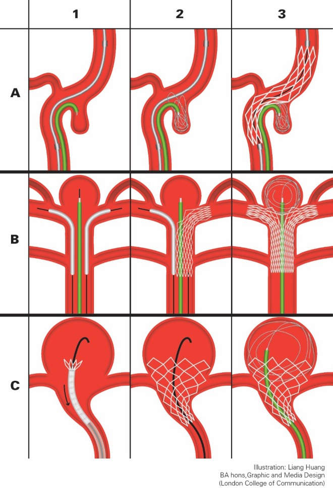
Figure 6.

Schematic diagrams of special SAC techniques. A, stent jack: (A1) self-expandable stent navigated across the aneurysmal neck and microcatheter into the aneurysmal sac; (A2) first coil deployed; (A3) Stent deployed fully (or partially) bridging the neck, pushing the coil into the sac. B, Y-stenting: (B1) microcatheter navigated into the aneurysmal sac while 2 Neuroform stents navigated through the BA and respective PCA via exchange wires; (B2) 1 stent deployed; (B3) contralateral stent deployed in a “kissing” fashion. C, Waffle-cone technique: (C1) Enterprise stent and exchange-wire positioned; (C2) stent deployed; (C3) microcatheter positioned, coils deployed in waffle-cone configuration. SAC indicates stent-assisted coiling; BA, basilar artery; PCA; posterior cerebral artery.
