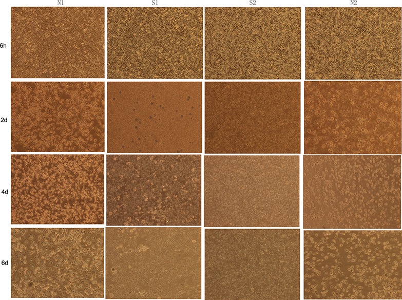
Figure 2. The differentiation state of monocytes isolated from clinical chicken.

Images of chicken monocytes were taken every 2 d (magnification: 150 ×). N1 and N2 represented uninfected chickens; S1 and S2 represented sick chickens infected with ALV.
