Abstract
Woolly hair nevus is a rare condition characterized by a structural anomaly of the hair, restricted to certain areas of the scalp. The hair becomes coiled and slightly hypopigmented. The term woolly hair refers to changes that affect all the scalp and has a hereditary character. We present a case of woolly hair nevus, that developed at the age of 2 years, associated with dental diastema and verrucous epidermal nevus.
Keywords: Hair diseases, Diastema, Nevus
INTRODUCTION
Woolly hair nevus is a benign, uncommon condition that is characterized by hair changes, that become coiled and slightly hypopigmented.1,2 It affects both sexes equally, and usually develop in the first years of life, although there are reports of development in adolescence.3 Hutchinson et al classified woolly hair in three groups: two generalized and genetically transmitted forms (1-autosomal dominant inheritance; 2-autosomal recessive inheritance) and a localized, non-hereditary form (woolly hair nevus).1-3 In Post’s classification, wooly hair nevus can be subdivided in three types: Type 1-no cutaneous involvement; Type 2-associated to linear verrucous epidermal nevus; Type 3-acquired, in young adult patients, where scalp hair has the same features of pubic hair.1,3,4 It is estimated that 50% of the cases of woolly hair nevus are associated to linear verrucous epidermal nevus on the ipsilateral upper limb, face or neck.1,5 The authors describe a case of woolly hair nevus Post’s type 2, developed during childhood.
CASE REPORT
Nine-year-old male child, sought medical care with the complaint of curled and light hair, restricted to the right parietal and occipital regions since he was 2 years old. Developmental history was unremarkable. There was no report of consanguinity in the family. On physical examination, we could observe well defined areas with coiled, lighter and shinier hair on the parietal and occipital regions, with no changes on the skin of the scalp on the affected areas (Figures 1 and 2). There was also widening of the spaces between the central incisors (diastema) and linear verrucous epidermal nevus on the posterior cervical region (Figures 3 and 4). Histology of the scalp showed usual density of hair follicles, with 3-5 hairs per follicular unit, being most of them terminal and anagen. There was no significant inflammatory infiltrate and some hair shafts in the sections were slightly oval-shaped (Figure 5). Anagen trichogram and the examination of the hair shafts did not show any abnormalities. Laboratory tests, echocardiogram, electrocardiogram, abdominal ultrasound and ophthalmological exam were all normal. The patient was advised that the condition was benign and is undergoing clinical follow-up.
Figure 1.
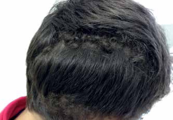
Well defined areas with coiled hair on the right parietal and occipital regions, with no changes on the scalp
Figure 2.
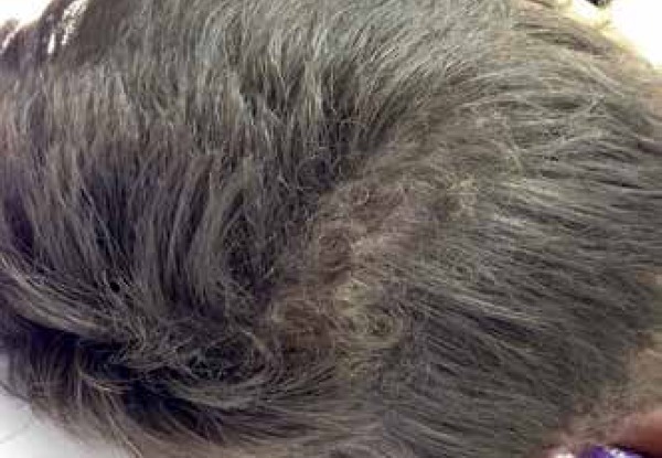
Lighter and shinier coiled hair on the parietal region
Figure 3.
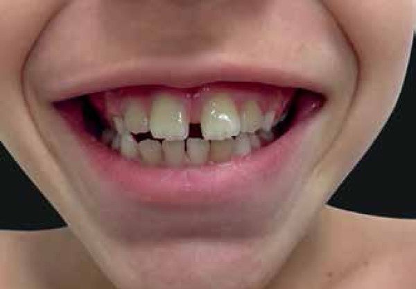
Widening of the spaces between the central incisors (diastema)
Figure 4.
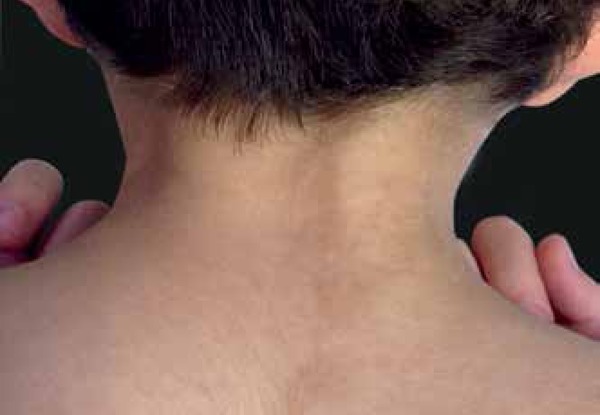
Linear epidermal nevus on the posterior cervical region
Figure 5.
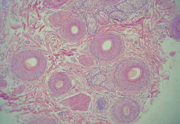
Histopathology of the scalp (Hematoxylin & eosin, X40): usual density of hair follicles, with 3-5 hairs per follicular unit, most of them terminal and anagen. Some hair shafts were slightly oval-shaped
DISCUSSION
The term woolly refers to an abnormal change in the hair structure, that becomes coiled and, in some cases, slightly hypopigmented. There are generalized forms that are usually associated with systemic conditions, and a localized form.2 In 1907, Gossage was the first to describe woolly hair in an European family, comparing this abnormality with the hair of patients of African descent.1 Subsequently, Hutchinson et al classified woolly hair in 3 variants.1,2,6 The generalized form is further subdivided in: hereditary form, autosomal dominant, characterized by the appearance of extremely coiled hairs in the first weeks of life; and the familial form, with autosomal recessive inheritance, characterized by thin, lighter hair compared to non-affected family members. Body hair and the lateral portion of the eyebrows can be affected in some cases. The localized form is not hereditary and is also known as woolly hair nevus.1 In 1927, Wise was the first to describe woolly hair nevus in a 2-year-old girl, associated with epidermal nevus on the neck.1,2 Subsequently, Post divided woolly hair in 3 types (Type 1-no association with scalp disorders or loss of body hair; Type 2-associated with linear verrucous epidermal nevus; Type 3-acquired, in young adult patients; the hair is curly, dark and short, similar to pubic hair).1,3,4,6 Woolly hair nevus is a rare, non-hereditary form of woolly hair. It is easily distinguished from the generalized form, for it affects only a few well defined areas of the scalp.2 In 50% of cases, it is associated with verrucous epidermal nevus, which may or may not overlap the affected area of the scalp.2,5 It is very important to refer these patients to an ophthalmological evaluation due to the association with persistent pupillary membrane.2 Dental abnormalities were also reported, such as recurrent cavities and diastema.7 As a rule, the hair does not become weaker, only if associated with trichorrhexis nodosa. There is no effective treatment but, in some patients, the hair can become less coiled with age.2 Woolly hair nevus syndrome is characterized by the association of the hair abnormality with verrucous epidermal nevus and other extracutaneous changes such as bone, neurological, ophthalmological and, less frequently, cardiac or renal abnormalities. Other cutaneous changes can also be seen, such as sebaceous nevus, acanthosis nigricans and hemangioma.1 Generalized woolly hair affects the whole scalp and can happen in association with other cutaneous or extracutaneous changes. The presence of facial dysmorphism characterizes other conditions associated with wooly hair, such as Noonan syndrome and cardiofaciocutaneous syndrome.2 In the reported case, the patient had woolly hair associated with linear verrucous epidermal nevus on the posterior cervical region. With the work-up, any systemic abnormality or association with any syndrome was ruled out. Therefore, the diagnosis of woolly hair nevus was made.
Footnotes
Conflict of Interests: None.
Study conducted at Hospital Naval Marcílio Dias (HNMD) - Rio de Janeiro (RJ), Brazil.
Financial Support: None.
REFERENCES
- 1.Martín-González T, del Boz-González J, Vera-Casaño A. Woolly hair nevus associated with an ipsilateral linear epidermal nevus. Actas Dermosifiliogr. 2007;98:198–201. [PubMed] [Google Scholar]
- 2.Torres T, Machado S, Selores M. Woolly hair generalizado: caso clínico e revisão da literatura. An Bras Dermatol. 2010;85:97–100. doi: 10.1590/s0365-05962010000100016. [DOI] [PubMed] [Google Scholar]
- 3.Oliveira JR, Mazocco VT, Arruda LH. Síndrome do nevo de cabelo lanoso. An Bras Dermatol. 2004;79:103–106. [Google Scholar]
- 4.Grant PW. A case of wooly hair naevus. Arch Dis Child. 1960;35:512–514. doi: 10.1136/adc.35.183.512. [DOI] [PMC free article] [PubMed] [Google Scholar]
- 5.Hong H, Lee WS. Woolly hair nevus involving entire occipital and temporal scalp. Ann Dermatol. 2013;25:396–397. doi: 10.5021/ad.2013.25.3.396. [DOI] [PMC free article] [PubMed] [Google Scholar]
- 6.Singh SK, Manchanda K, Kumar A, Verma A. Familial woolly hair: a rare entity. Int J Trichology. 2012;4:288–289. doi: 10.4103/0974-7753.111214. [DOI] [PMC free article] [PubMed] [Google Scholar]
- 7.Vasudevan B, Verma R, Pragasam V, Badad A. A rare case of woolly hair with unusual associations. Indian Dermatol Online J. 2013;4:222–224. doi: 10.4103/2229-5178.115524. [DOI] [PMC free article] [PubMed] [Google Scholar]


