Abstract
Although we often see patients with neck tumors, neck schwannomas are quite rare. We should keep schwannnoma in mind when we identified tumors on the side of the neck.

Keywords: neck tumor, schwannoma, thyroid tumor
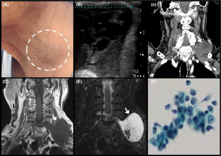
An 87‐year‐old Japanese woman with no underlying diseases noticed a growing soft tumor on the left side of her neck (Figure 1A). Ultrasonography detected a low‐echoic mass (Figure 1B). The mass included multiple low‐density spots in the intervertebral foramen as determined by enhanced computed tomography (Figure 1C). Neck magnetic resonance imaging indicated a solid low‐intensified (Figure 1D) and high‐intensified (Figure 1E) tumor by T1‐ and T2‐images, respectively. The tumor was connected to the cervical spinal cord, suggesting a neurogenic origin. Aspiration cytology indicated a class III (Figure 1F). Tumor resection and tissue biopsy were waived, as the patient complained of radiating pain after aspiration. Although core needle biopsy is useful in soft tissue tumor like schwannoma,1 it may cause persistent and difficult‐to‐treat pain.2 We diagnosed the tumor as a schwannoma based on the imaging and cytological results.
Figure 1.

(A) is a macroscopic image. You can see a mass in the circle, (B) is an ultrasonographic image of schwannoma, (C) is an image of enhanced CT. The arrow indicates schwannoma, (D) and (E) are T1‐ and T2‐weighted images of MRI. The arrows also indicate schwannoma, (F) is an image of cytological diagnosis
Neck schwannomas are rare compared to thyroid tumors. Although infrequent, we must keep this disease entity in mind when we identified neck tumor. Neck schwannoma can be distinguished by image inspection and cytology.
CONFLICT OF INTEREST
The authors have stated explicitly that there are no conflicts of interest in connection with this article.
DISCLOSURE
None of the authors has any financial relationships relevant to this publication to disclose.
Oka K, Iwamuro M, Otsuka F. Neck schwannoma mimicking a thyroid tumor. J Gen Fam Med. 2017;18:473–474. https://doi.org/10.1002/jgf2.121
REFERENCES
- 1. Nasrollah N, Trimboli P, Bianchi D, Taccogna S. Neck schwannoma diagnosed by core needle biopsy: a case report. J Ultrasound. 2014;18:407–10. [DOI] [PMC free article] [PubMed] [Google Scholar]
- 2. Pianta M, Chock E, Schlicht S, McCombe D. Accuracy and complications of CT‐guided core needle biopsy of peripheral nerve sheath tumours. Skeletal Radiol. 2015;44:1341–9. [DOI] [PubMed] [Google Scholar]


