Crystal structures of hydrogen-bonded 1:2 compounds of chloranilic acid with 2-pyridone, 3-hydroxypyridine and 4-hyroxypyridine have been determined at 120 K. In each crystal structure, the acid and base molecules are linked by short O—H⋯O and N—H⋯O hydrogen bonds.
Keywords: crystal structure, chloranilic acid, 2-pyridone, 3-hydroxypyridine, 4-hyroxypyridine, hydrogen bond
Abstract
The crystal structures of the 1:2 compounds of chloranilic acid (systematic name: 2,5-dichloro-3,6-dihydroxy-1,4-benzoquinone) with 2-pyridone, 3-hydroxypyridine and 4-hyroxypyridine, namely, bis(2-pyridone) chloranilic acid, 2C5H5NO·C6H2Cl2O4, (I), bis(3-hydroxypyridinium) chloranilate, 2C5H6NO+·C6Cl2O4 2−, (II), and bis(4-hydroxypyridinium) chloranilate, 2C5H6NO+·C6Cl2O4 2−, (III), have been determined at 120 K. In the crystal of (I), the base molecule is in the lactam form and no acid–base interaction involving H-atom transfer is observed. The acid molecule lies on an inversion centre and the asymmetric unit consists of one half-molecule of chloranilic acid and one 2-pyridone molecule, which are linked via a short O—H⋯O hydrogen bond. 2-Pyridone molecules form a head-to-head dimer via a pair of N—H⋯O hydrogen bonds, resulting in a tape structure along [201]. In the crystals of (II) and (III), acid–base interactions involving H-atom transfer are observed and the divalent cations lie on an inversion centre. The asymmetric unit of (II) consists of one half of a chloranilate anion and one 3-hydroxypyridinium cation, while that of (III) comprises two independent halves of anions and two 4-hydroxypyridinium cations. The primary intermolecular interaction in (II) is a bifurcated O—H⋯(O,O) hydrogen bond between the cation and the anion. The hydrogen-bonded units are further linked via N—H⋯O hydrogen bonds, forming a layer parallel to the bc plane. In (III), one anion is surrounded by four cations via O—H⋯O and C—H⋯O hydrogen bonds, while the other is surrounded by four cations via N—H⋯O and C—H⋯Cl hydrogen bonds. These interactions link the cations and the anions into a layer parallel to (301).
Chemical context
Chloranilic acid, a dibasic acid with hydrogen-bond donor and acceptor groups, appears particularly attractive as a template for generating tightly bound self-assemblies with various pyridine derivatives, as well as a model compound for investigating hydrogen transfer motions in O—H⋯N and N—H⋯O hydrogen-bond systems (Zaman et al., 2004 ▸; Seliger et al., 2009 ▸; Asaji et al. 2010 ▸). In the present study, we have prepared three 1:2 compounds of chloranilic acid with 2-pyridone, 3-hydroxypyridine and 4-hydroxypyridine in order to extend our study of D—H⋯A hydrogen bonding (D = N, O or C; A = N, O or Cl) in chloranilic acid–substituted-pyridine systems (Gotoh et al., 2009a
▸,b
▸, 2010 ▸). The crystal structure of the 1:1 compound of chloranilic acid with 3-hydroxypyridine, namely, 3-hydroxypyridinium hydrogen chloranilate monohydrate, has been reported (Gotoh & Ishida, 2009 ▸).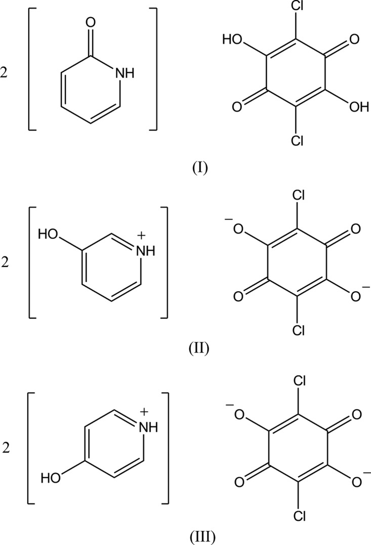
Structural commentary
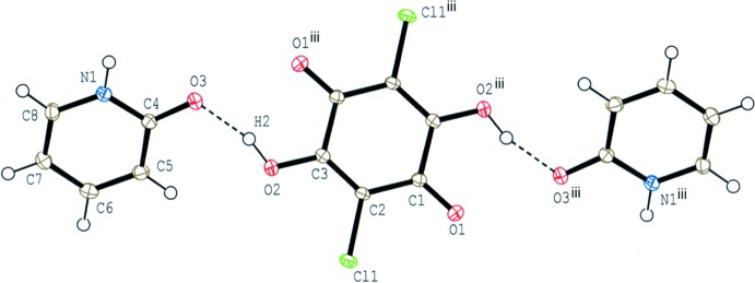
In compound (I), the base molecule is in the lactam form and no acid-base interaction involving H-atom transfer is observed (Fig. 1 ▸). The acid molecule lies on an inversion centre and the asymmetric unit consists of one-half acid molecule and one base molecule, which are linked via a short O—H⋯O hydrogen bond (O2—H2⋯O3; Table 1 ▸). The dihedral angle between the acid ring and the base ring is 37.82 (5)°.
Figure 1.
The molecular structure of compound (I), showing the atom-numbering scheme. Displacement ellipsoids are drawn at the 50% probability level and H atoms are shown as small spheres of arbitrary radii. O—H⋯O hydrogen bonds are shown as dashed lines. [Symmetry code: (iii) −x + 1, −y + 1, −z + 1.]
Table 1. Hydrogen-bond geometry (Å, °) for (I) .
| D—H⋯A | D—H | H⋯A | D⋯A | D—H⋯A |
|---|---|---|---|---|
| O2—H2⋯O3 | 0.901 (15) | 1.627 (16) | 2.4989 (11) | 161.9 (19) |
| N1—H1⋯O3i | 0.893 (16) | 1.996 (17) | 2.8743 (12) | 167.6 (16) |
| C7—H7⋯Cl1ii | 0.95 | 2.79 | 3.5122 (13) | 134 |
Symmetry codes: (i)  ; (ii)
  .
.
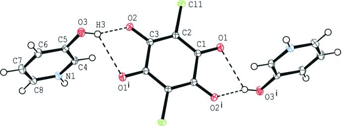
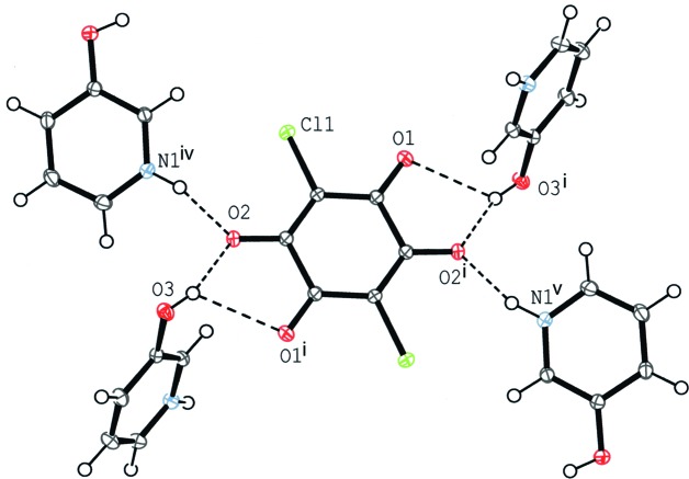
In compound (II), an acid–base interaction involving H-atom transfer is observed. The chloranilate anion is located on an inversion centre and the asymmetric unit contains one-half anion molecule and one cation molecule. The primary intermolecular interaction between the cation and the anion is a bifurcated O—H⋯(O,O) hydrogen bond (O3—H3⋯O2 and O3—H3⋯O1i; symmetry code as in Table 2 ▸) to afford a centrosymmetric 1:2 aggregate of the anion and the cation (Fig. 2 ▸). The dihedral angle between the acid ring and the base ring is 72.69 (5)°.
Table 2. Hydrogen-bond geometry (Å, °) for (II) .
| D—H⋯A | D—H | H⋯A | D⋯A | D—H⋯A |
|---|---|---|---|---|
| O3—H3⋯O2 | 0.852 (17) | 1.803 (17) | 2.6277 (12) | 162.5 (16) |
| O3—H3⋯O1i | 0.852 (17) | 2.438 (17) | 2.9738 (12) | 121.6 (14) |
| N1—H1⋯O2ii | 0.889 (17) | 1.807 (17) | 2.6684 (12) | 162.6 (16) |
| C8—H8⋯O1iii | 0.95 | 2.45 | 3.1481 (13) | 130 |
Symmetry codes: (i)  ; (ii)
  ; (iii)
  .
.
Figure 2.
The molecular structure of compound (II), showing the atom-numbering scheme. Displacement ellipsoids are drawn at the 50% probability level and H atoms are shown as small spheres of arbitrary radii. O—H⋯O hydrogen bonds are shown as dashed lines. [Symmetry code: (i) −x + 2, −y + 1, −z + 1.]
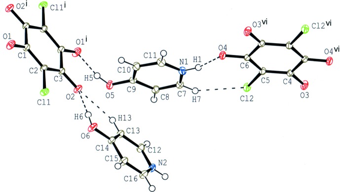
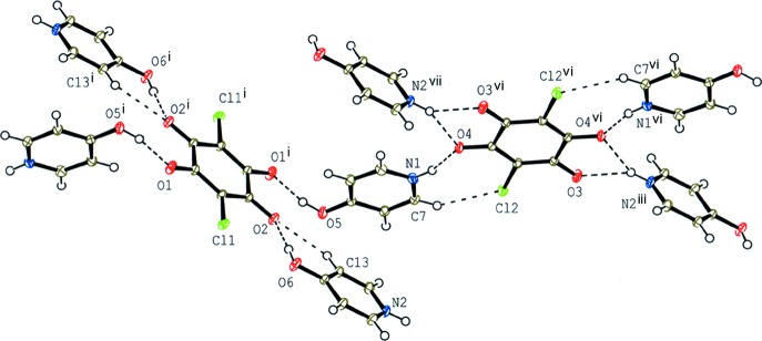
The compound (III) crystallizes with two independent halves of chloranilate anions and two 4-hydroxypyridinium cations in the asymmetric unit (Fig. 3 ▸). Although both anions lie on an inversion centre, the hydrogen-bonding schemes around the anions are quite different (Fig. 4 ▸); one anion is surrounded by four cations via O—H⋯O and C—H⋯O hydrogen bonds (O5—H5⋯O1i, O6—H6⋯O2 and C13—H13⋯O2; symmetry code as in Table 3 ▸), while the other is surrounded by four cation via N—H⋯O and C—H⋯Cl hydrogen bonds (N1—H1⋯O4, N2—H2⋯O4ii, N2—H2⋯O3iii and C7—H7⋯Cl2; Table 3 ▸).
Figure 3.
The molecular structure of compound (III), showing the atom-numbering scheme. Displacement ellipsoids are drawn at the 50% probability level and H atoms are shown as small spheres of arbitrary radii. O—H⋯O, N—H⋯O, C—H⋯Cl and C—H⋯O hydrogen bonds are shown as dashed lines. [Symmetry codes: (i) −x, −y, −z; (vi) −x + 3, −y, −z + 1.]
Figure 4.
A partial packing diagram for compound (III) around two independent chloranilate anions. O—H⋯O, N—H⋯O, C—H⋯Cl and C—H⋯O hydrogen bonds are shown as dashed lines. [Symmetry codes: (i) −x, −y, −z; (iii) −x + 3, −y + 1, −z + 1; (vi) −x + 3, −y, −z + 1; (vii) x, y − 1, z.]
Table 3. Hydrogen-bond geometry (Å, °) for (III) .
| D—H⋯A | D—H | H⋯A | D⋯A | D—H⋯A |
|---|---|---|---|---|
| O5—H5⋯O1i | 0.905 (17) | 1.640 (17) | 2.5208 (13) | 163.2 (17) |
| O6—H6⋯O2 | 0.852 (17) | 1.800 (17) | 2.6510 (13) | 177.3 (18) |
| N1—H1⋯O4 | 0.919 (17) | 1.810 (17) | 2.7000 (13) | 162.3 (16) |
| N2—H2⋯O4ii | 0.876 (18) | 2.156 (17) | 2.9603 (14) | 152.3 (15) |
| N2—H2⋯O3iii | 0.876 (18) | 2.176 (17) | 2.8384 (14) | 132.1 (14) |
| C7—H7⋯Cl2 | 0.95 | 2.81 | 3.4540 (12) | 126 |
| C12—H12⋯O3iv | 0.95 | 2.32 | 3.1541 (14) | 146 |
| C13—H13⋯O2 | 0.95 | 2.49 | 3.1685 (14) | 128 |
| C16—H16⋯Cl1v | 0.95 | 2.77 | 3.4427 (12) | 128 |
Symmetry codes: (i)  ; (ii)
  ; (iii)
  ; (iv)
  ; (v)
; (v)  .
.
Supramolecular features
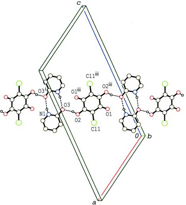
In the crystal of compound (I), two adjacent 2-pyridone molecules, which are related by a twofold rotation axis, form a head-to-head dimer via a pair of N—H⋯O hydrogen bonds (N1—H1⋯O3i; symmetry code as in Table 1 ▸), as observed in various cocrystals of 2-pyridone (Odani & Matsumoto, 2002 ▸). The acid and base molecules form an undulating tape structure running along [201] through the above-mentioned O—H⋯O and N—H⋯O hydrogen bonds (Fig. 5 ▸). The tapes are stacked along the b axis into a layer structure through a π–π interaction between the pyridine rings [centroid-to-centroid distance = 3.7005 (6) Å and interplanar spacing = 3.4239 (4) Å] and a short C⋯C contact [C2⋯C3iv = 3.3056 (13) Å; symmetry code: (iv) x, y + 1, z]. A weak C—H⋯Cl interaction formed between the acid and base molecules (C7—H7⋯Cl1ii; Table 1 ▸) links the layers. The O—H⋯O hydrogen bond between the acid and base molecules is short [O2⋯O3 = 2.4989 (11) Å], suggesting possible disorder of the H atom in the hydrogen bond, but no distinct evidence of the disorder was observed in the difference Fourier map, nor from the molecular geometry.
Figure 5.
A packing diagram for compound (I), showing the tape structure formed by O—H⋯O and N—H⋯O hydrogen bonds (dashed lines). H atoms not involved in the interactions have been omitted. [Symmetry codes: (i) −x + 2, y, −z +  ; (iii) −x + 1, −y + 1, −z + 1.]
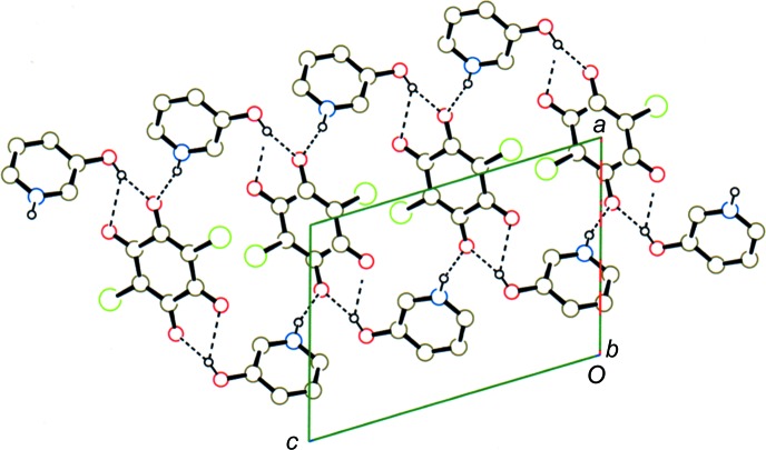
In the crystal of (II), the cation–anion units are further connected by N—H⋯O (N1—H1⋯O2ii; symmetry code as in Table 2 ▸ and Fig. 6 ▸), forming a layer expanding parallel to the bc plane (Fig. 7 ▸). Adjacent layers are connected to each other with a C—H⋯O hydrogen bond (C8—H8⋯O1iii; Table 2 ▸) and a short O⋯N contact [O3⋯N1vi = 3.0430 (12) Å; symmetry code: (vi) −x + 1, y −  , −z +
  ].
].
Figure 6.
A partial packing diagram for compound (II) around the chloranilate anion. O—H⋯O and N—H⋯O hydrogen bonds are shown as dashed lines. [Symmetry codes: (i) −x + 2, −y + 1, −z + 1; (iv) x, −y +  , z +
, z +  ; (v) −x + 2, y −
  , −z +
  .]
.]
Figure 7.
A packing diagram for compound (II), viewed along the b axis, showing the layer structure formed by O—H⋯O and N—H⋯O hydrogen bonds (dashed lines). H atoms not involved in the interactions have been omitted.
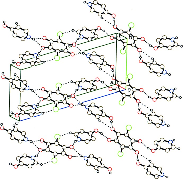
In the crystal of (III), the above-mentioned O—H⋯O, N—H⋯O, C—H⋯O and C—H⋯Cl hydrogen bonds link the cations and anions into a layer parallel to (301) (Fig. 8 ▸). Adjacent layers are further linked via weak C—H⋯O and C—H⋯Cl interactions (C12—H12⋯O3iv and C16—H16⋯Cl1v; symmetry codes as given in Table 3 ▸).
Figure 8.
A packing diagram for compound (III), showing the hydrogen-bonded network in the layer. O—H⋯O, N—H⋯O, C—H⋯Cl and C—H⋯O hydrogen bonds are shown as dashed lines. H atoms not involved in the hydrogen bonds have been omitted.
Database survey
A search of the Cambridge Structural Database (Version 5.38, last update May 2017; Groom et al., 2016 ▸) for organic crystals of chloranilic acid with substituted pyridines (except for di-, tri- and tetrapyridine derivatives) gave 32 hits. Of these, crystal structures of 16 compounds of chloranilic acid with methyl-substituted pyridines (Adam et al., 2010 ▸; Łuczyńska et al., 2016 ▸; Molčanov & Kojić-Prodić, 2010 ▸, and references therein), three compounds of carbamoyl-substituted pyridines (Gotoh et al., 2009a ▸), three compounds of carboxy-substituted pyridines (Gotoh et al., 2009b ▸, and references therein) and three compounds of cyano-substituted pyridines (Gotoh & Ishida, 2012 ▸, and references therein) were reported.
Synthesis and crystallization
Single crystals of compound (I) were obtained by slow evaporation from an ethanol solution (120 ml) of chloranilic acid (350 mg) with 2-hydroxypyridine (340 mg) at room temperature. Crystals of compound (II) were obtained by slow evaporation from a methanol solution (400 ml) of chloranilic acid (170 mg) with 3-hydroxypyridine (160 mg) at room temperature. Crystals of compound (III) were obtained by slow diffusion of a methanol solution (20 ml) of 4-hydroxypyridine (160 mg) into an acetonitrile solution (200 ml) of chloranilic acid (170 mg) at room temperature.
Refinement
Crystal data, data collection and structure refinement details are summarized in Table 4 ▸. All H atoms in compounds (I)–(III) were found in difference Fourier maps. The positions of O- and N-bound H atoms were refined freely, with U iso(H) = 1.5U eq(O or N). C-bound H atoms were positioned geometrically (C—H = 0.95 Å) and were treated as riding with U iso(H) = 1.2U eq(C).
Table 4. Experimental details.
| (I) | (II) | (III) | |
|---|---|---|---|
| Crystal data | |||
| Chemical formula | 2C5H5NO·C6H2Cl2O4 | 2C5H6NO+·C6Cl2O4 2− | 2C5H6NO+·C6Cl2O4 2− |
| M r | 399.19 | 399.19 | 399.19 |
| Crystal system, space group | Monoclinic, P2/c | Monoclinic, P21/c | Triclinic, P

|
| Temperature (K) | 120 | 120 | 120 |
| a, b, c (Å) | 11.9402 (7), 3.7005 (2), 21.7919 (13) | 8.3659 (6), 8.5492 (6), 11.7087 (8) | 5.49136 (13), 8.2195 (4), 18.1382 (9) |
| α, β, γ (°) | 90, 121.278 (2), 90 | 90, 106.968 (3), 90 | 102.177 (3), 93.952 (3), 95.316 (4) |
| V (Å3) | 822.92 (9) | 800.98 (9) | 793.52 (6) |
| Z | 2 | 2 | 2 |
| Radiation type | Mo Kα | Mo Kα | Mo Kα |
| μ (mm−1) | 0.43 | 0.45 | 0.45 |
| Crystal size (mm) | 0.39 × 0.36 × 0.21 | 0.21 × 0.20 × 0.12 | 0.35 × 0.25 × 0.12 |
| Data collection | |||
| Diffractometer | Rigaku R-AXIS RAPIDII | Rigaku R-AXIS RAPIDII | Rigaku R-AXIS RAPIDII |
| Absorption correction | Multi-scan (ABSCOR; Higashi, 1995 ▸) | Numerical (NUMABS; Higashi, 1999 ▸) | Numerical (NUMABS; Higashi, 1999 ▸) |
| T min, T max | 0.804, 0.913 | 0.903, 0.948 | 0.890, 0.948 |
| No. of measured, independent and observed [I > 2σ(I)] reflections | 22767, 2401, 2316 | 15315, 2329, 2166 | 12373, 4597, 4124 |
| R int | 0.013 | 0.017 | 0.036 |
| (sin θ/λ)max (Å−1) | 0.703 | 0.703 | 0.703 |
| Refinement | |||
| R[F 2 > 2σ(F 2)], wR(F 2), S | 0.027, 0.076, 1.06 | 0.028, 0.075, 1.07 | 0.030, 0.080, 1.07 |
| No. of reflections | 2401 | 2329 | 4597 |
| No. of parameters | 124 | 124 | 247 |
| H-atom treatment | H atoms treated by a mixture of independent and constrained refinement | H atoms treated by a mixture of independent and constrained refinement | H atoms treated by a mixture of independent and constrained refinement |
| Δρmax, Δρmin (e Å−3) | 0.49, −0.24 | 0.52, −0.20 | 0.75, −0.34 |
Supplementary Material
Crystal structure: contains datablock(s) I, II, III, global. DOI: 10.1107/S2056989017013536/lh5855sup1.cif
Structure factors: contains datablock(s) I. DOI: 10.1107/S2056989017013536/lh5855Isup2.hkl
Structure factors: contains datablock(s) II. DOI: 10.1107/S2056989017013536/lh5855IIsup3.hkl
Structure factors: contains datablock(s) III. DOI: 10.1107/S2056989017013536/lh5855IIIsup4.hkl
Additional supporting information: crystallographic information; 3D view; checkCIF report
supplementary crystallographic information
Bis[pyridin-2(1H)-one] 2,5-dichloro-3,6-dihydroxy-1,4-bebzoquinone (I) . Crystal data
| 2C5H5NO·C6H2Cl2O4 | F(000) = 408.00 |
| Mr = 399.19 | Dx = 1.611 Mg m−3 |
| Monoclinic, P2/c | Mo Kα radiation, λ = 0.71075 Å |
| a = 11.9402 (7) Å | Cell parameters from 20111 reflections |
| b = 3.7005 (2) Å | θ = 3.3–30.1° |
| c = 21.7919 (13) Å | µ = 0.43 mm−1 |
| β = 121.278 (2)° | T = 120 K |
| V = 822.92 (9) Å3 | Block, brown |
| Z = 2 | 0.39 × 0.36 × 0.21 mm |
Bis[pyridin-2(1H)-one] 2,5-dichloro-3,6-dihydroxy-1,4-bebzoquinone (I) . Data collection
| Rigaku R-AXIS RAPIDII diffractometer | 2316 reflections with I > 2σ(I) |
| Detector resolution: 10.000 pixels mm-1 | Rint = 0.013 |
| ω scans | θmax = 30.0°, θmin = 3.4° |
| Absorption correction: multi-scan (ABSCOR; Higashi, 1995) | h = −16→16 |
| Tmin = 0.804, Tmax = 0.913 | k = −4→5 |
| 22767 measured reflections | l = −30→30 |
| 2401 independent reflections |
Bis[pyridin-2(1H)-one] 2,5-dichloro-3,6-dihydroxy-1,4-bebzoquinone (I) . Refinement
| Refinement on F2 | Primary atom site location: structure-invariant direct methods |
| Least-squares matrix: full | Secondary atom site location: difference Fourier map |
| R[F2 > 2σ(F2)] = 0.027 | Hydrogen site location: mixed |
| wR(F2) = 0.076 | H atoms treated by a mixture of independent and constrained refinement |
| S = 1.06 | w = 1/[σ2(Fo2) + (0.0443P)2 + 0.3557P] where P = (Fo2 + 2Fc2)/3 |
| 2401 reflections | (Δ/σ)max = 0.001 |
| 124 parameters | Δρmax = 0.49 e Å−3 |
| 0 restraints | Δρmin = −0.24 e Å−3 |
Bis[pyridin-2(1H)-one] 2,5-dichloro-3,6-dihydroxy-1,4-bebzoquinone (I) . Special details
| Geometry. All esds (except the esd in the dihedral angle between two l.s. planes) are estimated using the full covariance matrix. The cell esds are taken into account individually in the estimation of esds in distances, angles and torsion angles; correlations between esds in cell parameters are only used when they are defined by crystal symmetry. An approximate (isotropic) treatment of cell esds is used for estimating esds involving l.s. planes. |
Bis[pyridin-2(1H)-one] 2,5-dichloro-3,6-dihydroxy-1,4-bebzoquinone (I) . Fractional atomic coordinates and isotropic or equivalent isotropic displacement parameters (Å2)
| x | y | z | Uiso*/Ueq | ||
| Cl1 | 0.60869 (2) | 0.78653 (6) | 0.40403 (2) | 0.01704 (8) | |
| O1 | 0.33921 (7) | 0.8229 (2) | 0.37552 (4) | 0.02149 (16) | |
| O2 | 0.75999 (6) | 0.4050 (2) | 0.54453 (4) | 0.01794 (15) | |
| H2 | 0.8039 (15) | 0.289 (4) | 0.5870 (8) | 0.027* | |
| O3 | 0.92219 (7) | 0.1199 (2) | 0.66164 (4) | 0.02094 (16) | |
| N1 | 1.13199 (8) | −0.0736 (2) | 0.72858 (4) | 0.01604 (16) | |
| H1 | 1.1281 (13) | −0.016 (4) | 0.7672 (8) | 0.024* | |
| C1 | 0.41591 (9) | 0.6765 (2) | 0.43248 (5) | 0.01361 (17) | |
| C2 | 0.55361 (8) | 0.6274 (3) | 0.45806 (5) | 0.01311 (16) | |
| C3 | 0.63639 (8) | 0.4587 (2) | 0.52136 (5) | 0.01342 (17) | |
| C4 | 1.02174 (9) | −0.0153 (3) | 0.66317 (5) | 0.01563 (17) | |
| C5 | 1.02800 (9) | −0.1116 (3) | 0.60168 (5) | 0.01779 (18) | |
| H5 | 0.953363 | −0.080221 | 0.554948 | 0.021* | |
| C6 | 1.14171 (10) | −0.2497 (3) | 0.60990 (5) | 0.01837 (19) | |
| H6 | 1.145376 | −0.312374 | 0.568705 | 0.022* | |
| C7 | 1.25330 (10) | −0.2995 (3) | 0.67902 (5) | 0.01870 (19) | |
| H7 | 1.332337 | −0.392760 | 0.684778 | 0.022* | |
| C8 | 1.24515 (9) | −0.2109 (3) | 0.73732 (5) | 0.01775 (18) | |
| H8 | 1.318846 | −0.245048 | 0.784277 | 0.021* |
Bis[pyridin-2(1H)-one] 2,5-dichloro-3,6-dihydroxy-1,4-bebzoquinone (I) . Atomic displacement parameters (Å2)
| U11 | U22 | U33 | U12 | U13 | U23 | |
| Cl1 | 0.02123 (12) | 0.01778 (12) | 0.01883 (12) | 0.00121 (8) | 0.01512 (10) | 0.00259 (7) |
| O1 | 0.0172 (3) | 0.0304 (4) | 0.0166 (3) | 0.0046 (3) | 0.0086 (3) | 0.0080 (3) |
| O2 | 0.0124 (3) | 0.0244 (4) | 0.0172 (3) | 0.0022 (3) | 0.0079 (2) | 0.0040 (3) |
| O3 | 0.0149 (3) | 0.0307 (4) | 0.0184 (3) | 0.0048 (3) | 0.0095 (3) | 0.0034 (3) |
| N1 | 0.0158 (3) | 0.0189 (4) | 0.0145 (3) | 0.0021 (3) | 0.0086 (3) | 0.0000 (3) |
| C1 | 0.0150 (4) | 0.0138 (4) | 0.0134 (4) | 0.0000 (3) | 0.0083 (3) | −0.0005 (3) |
| C2 | 0.0150 (4) | 0.0139 (4) | 0.0139 (4) | 0.0000 (3) | 0.0098 (3) | 0.0002 (3) |
| C3 | 0.0139 (4) | 0.0137 (4) | 0.0143 (4) | −0.0005 (3) | 0.0084 (3) | −0.0010 (3) |
| C4 | 0.0142 (4) | 0.0170 (4) | 0.0162 (4) | −0.0005 (3) | 0.0083 (3) | 0.0012 (3) |
| C5 | 0.0178 (4) | 0.0207 (4) | 0.0146 (4) | −0.0011 (3) | 0.0082 (3) | 0.0004 (3) |
| C6 | 0.0221 (4) | 0.0189 (4) | 0.0179 (4) | −0.0015 (3) | 0.0131 (4) | −0.0018 (3) |
| C7 | 0.0184 (4) | 0.0190 (4) | 0.0211 (4) | 0.0023 (3) | 0.0119 (4) | −0.0009 (3) |
| C8 | 0.0157 (4) | 0.0191 (4) | 0.0174 (4) | 0.0033 (3) | 0.0078 (3) | 0.0002 (3) |
Bis[pyridin-2(1H)-one] 2,5-dichloro-3,6-dihydroxy-1,4-bebzoquinone (I) . Geometric parameters (Å, º)
| Cl1—C2 | 1.7233 (9) | C2—C3 | 1.3614 (12) |
| O1—C1 | 1.2217 (11) | C4—C5 | 1.4256 (13) |
| O2—C3 | 1.3033 (10) | C5—C6 | 1.3726 (14) |
| O2—H2 | 0.901 (16) | C5—H5 | 0.9500 |
| O3—C4 | 1.2739 (11) | C6—C7 | 1.4120 (14) |
| N1—C8 | 1.3611 (12) | C6—H6 | 0.9500 |
| N1—C4 | 1.3648 (11) | C7—C8 | 1.3636 (14) |
| N1—H1 | 0.892 (15) | C7—H7 | 0.9500 |
| C1—C2 | 1.4475 (12) | C8—H8 | 0.9500 |
| C1—C3i | 1.5182 (12) | ||
| C3—O2—H2 | 114.1 (10) | O3—C4—C5 | 125.24 (8) |
| C8—N1—C4 | 123.66 (8) | N1—C4—C5 | 116.69 (8) |
| C8—N1—H1 | 119.5 (9) | C6—C5—C4 | 120.10 (9) |
| C4—N1—H1 | 116.9 (9) | C6—C5—H5 | 120.0 |
| O1—C1—C2 | 123.32 (8) | C4—C5—H5 | 120.0 |
| O1—C1—C3i | 118.05 (8) | C5—C6—C7 | 120.62 (9) |
| C2—C1—C3i | 118.63 (7) | C5—C6—H6 | 119.7 |
| C3—C2—C1 | 121.99 (8) | C7—C6—H6 | 119.7 |
| C3—C2—Cl1 | 121.00 (7) | C8—C7—C6 | 118.57 (9) |
| C1—C2—Cl1 | 117.01 (7) | C8—C7—H7 | 120.7 |
| O2—C3—C2 | 122.88 (8) | C6—C7—H7 | 120.7 |
| O2—C3—C1i | 117.75 (8) | N1—C8—C7 | 120.35 (9) |
| C2—C3—C1i | 119.37 (8) | N1—C8—H8 | 119.8 |
| O3—C4—N1 | 118.06 (8) | C7—C8—H8 | 119.8 |
| O1—C1—C2—C3 | −179.08 (9) | C8—N1—C4—O3 | −178.90 (9) |
| C3i—C1—C2—C3 | 1.18 (15) | C8—N1—C4—C5 | 1.04 (15) |
| O1—C1—C2—Cl1 | −0.05 (13) | O3—C4—C5—C6 | 178.84 (10) |
| C3i—C1—C2—Cl1 | −179.79 (6) | N1—C4—C5—C6 | −1.09 (15) |
| C1—C2—C3—O2 | 178.46 (9) | C4—C5—C6—C7 | 0.28 (16) |
| Cl1—C2—C3—O2 | −0.53 (14) | C5—C6—C7—C8 | 0.64 (16) |
| C1—C2—C3—C1i | −1.19 (15) | C4—N1—C8—C7 | −0.13 (16) |
| Cl1—C2—C3—C1i | 179.82 (7) | C6—C7—C8—N1 | −0.73 (15) |
Symmetry code: (i) −x+1, −y+1, −z+1.
Bis[pyridin-2(1H)-one] 2,5-dichloro-3,6-dihydroxy-1,4-bebzoquinone (I) . Hydrogen-bond geometry (Å, º)
| D—H···A | D—H | H···A | D···A | D—H···A |
| O2—H2···O3 | 0.901 (15) | 1.627 (16) | 2.4989 (11) | 161.9 (19) |
| N1—H1···O3ii | 0.893 (16) | 1.996 (17) | 2.8743 (12) | 167.6 (16) |
| C7—H7···Cl1iii | 0.95 | 2.79 | 3.5122 (13) | 134 |
Symmetry codes: (ii) −x+2, y, −z+3/2; (iii) −x+2, −y, −z+1.
Bis(3-hyroxypyridinium) 2,5-dichloro-3,6-dioxocyclohexa-1,4-diene-1,4-diolate (II). Crystal data
| 2C5H6NO+·C6Cl2O42− | F(000) = 408.00 |
| Mr = 399.19 | Dx = 1.655 Mg m−3 |
| Monoclinic, P21/c | Mo Kα radiation, λ = 0.71075 Å |
| a = 8.3659 (6) Å | Cell parameters from 13386 reflections |
| b = 8.5492 (6) Å | θ = 3.0–30.0° |
| c = 11.7087 (8) Å | µ = 0.45 mm−1 |
| β = 106.968 (3)° | T = 120 K |
| V = 800.98 (9) Å3 | Block, brown |
| Z = 2 | 0.21 × 0.20 × 0.12 mm |
Bis(3-hyroxypyridinium) 2,5-dichloro-3,6-dioxocyclohexa-1,4-diene-1,4-diolate (II). Data collection
| Rigaku R-AXIS RAPIDII diffractometer | 2166 reflections with I > 2σ(I) |
| Detector resolution: 10.000 pixels mm-1 | Rint = 0.017 |
| ω scans | θmax = 30.0°, θmin = 3.0° |
| Absorption correction: numerical (NUMABS; Higashi, 1999) | h = −11→11 |
| Tmin = 0.903, Tmax = 0.948 | k = −12→12 |
| 15315 measured reflections | l = −15→16 |
| 2329 independent reflections |
Bis(3-hyroxypyridinium) 2,5-dichloro-3,6-dioxocyclohexa-1,4-diene-1,4-diolate (II). Refinement
| Refinement on F2 | Primary atom site location: structure-invariant direct methods |
| Least-squares matrix: full | Secondary atom site location: difference Fourier map |
| R[F2 > 2σ(F2)] = 0.028 | Hydrogen site location: mixed |
| wR(F2) = 0.075 | H atoms treated by a mixture of independent and constrained refinement |
| S = 1.07 | w = 1/[σ2(Fo2) + (0.0411P)2 + 0.357P] where P = (Fo2 + 2Fc2)/3 |
| 2329 reflections | (Δ/σ)max = 0.001 |
| 124 parameters | Δρmax = 0.52 e Å−3 |
| 0 restraints | Δρmin = −0.20 e Å−3 |
Bis(3-hyroxypyridinium) 2,5-dichloro-3,6-dioxocyclohexa-1,4-diene-1,4-diolate (II). Special details
| Geometry. All esds (except the esd in the dihedral angle between two l.s. planes) are estimated using the full covariance matrix. The cell esds are taken into account individually in the estimation of esds in distances, angles and torsion angles; correlations between esds in cell parameters are only used when they are defined by crystal symmetry. An approximate (isotropic) treatment of cell esds is used for estimating esds involving l.s. planes. |
Bis(3-hyroxypyridinium) 2,5-dichloro-3,6-dioxocyclohexa-1,4-diene-1,4-diolate (II). Fractional atomic coordinates and isotropic or equivalent isotropic displacement parameters (Å2)
| x | y | z | Uiso*/Ueq | ||
| Cl1 | 0.93157 (3) | 0.73912 (3) | 0.68834 (2) | 0.01680 (8) | |
| O1 | 1.24731 (9) | 0.58136 (9) | 0.69329 (7) | 0.01740 (16) | |
| O2 | 0.68396 (9) | 0.61601 (9) | 0.46095 (7) | 0.01689 (16) | |
| O3 | 0.41012 (10) | 0.51422 (9) | 0.30475 (7) | 0.01640 (16) | |
| H3 | 0.509 (2) | 0.5340 (19) | 0.3482 (15) | 0.025* | |
| N1 | 0.48820 (11) | 0.71217 (11) | 0.05541 (8) | 0.01529 (17) | |
| H1 | 0.566 (2) | 0.7718 (19) | 0.0393 (15) | 0.023* | |
| C1 | 1.13017 (12) | 0.54789 (11) | 0.60534 (9) | 0.01283 (18) | |
| C2 | 0.96428 (12) | 0.60823 (12) | 0.58348 (9) | 0.01358 (18) | |
| C3 | 0.83396 (12) | 0.56708 (12) | 0.48551 (9) | 0.01327 (18) | |
| C4 | 0.51167 (12) | 0.66180 (12) | 0.16734 (9) | 0.01383 (18) | |
| H4 | 0.609229 | 0.691448 | 0.228545 | 0.017* | |
| C5 | 0.39288 (12) | 0.56593 (11) | 0.19388 (9) | 0.01309 (18) | |
| C6 | 0.25163 (13) | 0.52430 (13) | 0.10152 (10) | 0.0171 (2) | |
| H6 | 0.169400 | 0.457667 | 0.116909 | 0.021* | |
| C7 | 0.23181 (14) | 0.58069 (13) | −0.01295 (10) | 0.0192 (2) | |
| H7 | 0.135178 | 0.553857 | −0.075971 | 0.023* | |
| C8 | 0.35290 (14) | 0.67593 (13) | −0.03506 (9) | 0.0180 (2) | |
| H8 | 0.340534 | 0.715121 | −0.113094 | 0.022* |
Bis(3-hyroxypyridinium) 2,5-dichloro-3,6-dioxocyclohexa-1,4-diene-1,4-diolate (II). Atomic displacement parameters (Å2)
| U11 | U22 | U33 | U12 | U13 | U23 | |
| Cl1 | 0.01596 (13) | 0.01889 (13) | 0.01530 (13) | 0.00248 (8) | 0.00420 (9) | −0.00478 (8) |
| O1 | 0.0143 (3) | 0.0210 (4) | 0.0144 (3) | 0.0017 (3) | 0.0004 (3) | −0.0030 (3) |
| O2 | 0.0118 (3) | 0.0224 (4) | 0.0151 (3) | 0.0047 (3) | 0.0018 (3) | −0.0023 (3) |
| O3 | 0.0141 (3) | 0.0203 (4) | 0.0142 (3) | −0.0010 (3) | 0.0032 (3) | 0.0034 (3) |
| N1 | 0.0148 (4) | 0.0160 (4) | 0.0155 (4) | −0.0020 (3) | 0.0050 (3) | 0.0005 (3) |
| C1 | 0.0129 (4) | 0.0133 (4) | 0.0121 (4) | 0.0010 (3) | 0.0034 (3) | 0.0006 (3) |
| C2 | 0.0134 (4) | 0.0151 (4) | 0.0122 (4) | 0.0021 (3) | 0.0036 (3) | −0.0021 (3) |
| C3 | 0.0132 (4) | 0.0144 (4) | 0.0120 (4) | 0.0016 (3) | 0.0034 (3) | 0.0006 (3) |
| C4 | 0.0122 (4) | 0.0142 (4) | 0.0142 (4) | −0.0007 (3) | 0.0025 (3) | 0.0001 (3) |
| C5 | 0.0121 (4) | 0.0127 (4) | 0.0144 (4) | 0.0010 (3) | 0.0037 (3) | 0.0001 (3) |
| C6 | 0.0129 (4) | 0.0180 (5) | 0.0194 (5) | −0.0033 (3) | 0.0031 (4) | 0.0000 (4) |
| C7 | 0.0167 (5) | 0.0209 (5) | 0.0169 (5) | −0.0031 (4) | −0.0001 (4) | −0.0019 (4) |
| C8 | 0.0198 (5) | 0.0199 (5) | 0.0132 (4) | −0.0014 (4) | 0.0031 (4) | −0.0005 (4) |
Bis(3-hyroxypyridinium) 2,5-dichloro-3,6-dioxocyclohexa-1,4-diene-1,4-diolate (II). Geometric parameters (Å, º)
| Cl1—C2 | 1.7409 (10) | C2—C3 | 1.3778 (13) |
| O1—C1 | 1.2302 (12) | C4—C5 | 1.3915 (13) |
| O2—C3 | 1.2735 (12) | C4—H4 | 0.9500 |
| O3—C5 | 1.3387 (12) | C5—C6 | 1.3952 (14) |
| O3—H3 | 0.850 (18) | C6—C7 | 1.3880 (15) |
| N1—C4 | 1.3384 (13) | C6—H6 | 0.9500 |
| N1—C8 | 1.3414 (14) | C7—C8 | 1.3818 (15) |
| N1—H1 | 0.891 (17) | C7—H7 | 0.9500 |
| C1—C2 | 1.4319 (13) | C8—H8 | 0.9500 |
| C1—C3i | 1.5409 (14) | ||
| C5—O3—H3 | 109.0 (11) | N1—C4—H4 | 120.1 |
| C4—N1—C8 | 123.22 (9) | C5—C4—H4 | 120.1 |
| C4—N1—H1 | 119.0 (11) | O3—C5—C4 | 121.95 (9) |
| C8—N1—H1 | 117.8 (11) | O3—C5—C6 | 119.64 (9) |
| O1—C1—C2 | 124.03 (9) | C4—C5—C6 | 118.41 (9) |
| O1—C1—C3i | 117.27 (8) | C7—C6—C5 | 119.69 (9) |
| C2—C1—C3i | 118.70 (8) | C7—C6—H6 | 120.2 |
| C3—C2—C1 | 123.17 (9) | C5—C6—H6 | 120.2 |
| C3—C2—Cl1 | 120.15 (7) | C8—C7—C6 | 119.88 (9) |
| C1—C2—Cl1 | 116.69 (7) | C8—C7—H7 | 120.1 |
| O2—C3—C2 | 126.33 (9) | C6—C7—H7 | 120.1 |
| O2—C3—C1i | 115.54 (8) | N1—C8—C7 | 118.94 (9) |
| C2—C3—C1i | 118.13 (8) | N1—C8—H8 | 120.5 |
| N1—C4—C5 | 119.86 (9) | C7—C8—H8 | 120.5 |
| O1—C1—C2—C3 | −178.76 (10) | C8—N1—C4—C5 | 0.55 (16) |
| C3i—C1—C2—C3 | 0.78 (16) | N1—C4—C5—O3 | −179.23 (9) |
| O1—C1—C2—Cl1 | 1.08 (14) | N1—C4—C5—C6 | 0.30 (15) |
| C3i—C1—C2—Cl1 | −179.38 (7) | O3—C5—C6—C7 | 178.57 (10) |
| C1—C2—C3—O2 | 179.87 (10) | C4—C5—C6—C7 | −0.97 (15) |
| Cl1—C2—C3—O2 | 0.03 (15) | C5—C6—C7—C8 | 0.83 (17) |
| C1—C2—C3—C1i | −0.78 (16) | C4—N1—C8—C7 | −0.70 (16) |
| Cl1—C2—C3—C1i | 179.39 (7) | C6—C7—C8—N1 | −0.01 (17) |
Symmetry code: (i) −x+2, −y+1, −z+1.
Bis(3-hyroxypyridinium) 2,5-dichloro-3,6-dioxocyclohexa-1,4-diene-1,4-diolate (II). Hydrogen-bond geometry (Å, º)
| D—H···A | D—H | H···A | D···A | D—H···A |
| O3—H3···O2 | 0.852 (17) | 1.803 (17) | 2.6277 (12) | 162.5 (16) |
| O3—H3···O1i | 0.852 (17) | 2.438 (17) | 2.9738 (12) | 121.6 (14) |
| N1—H1···O2ii | 0.889 (17) | 1.807 (17) | 2.6684 (12) | 162.6 (16) |
| C8—H8···O1iii | 0.95 | 2.45 | 3.1481 (13) | 130 |
Symmetry codes: (i) −x+2, −y+1, −z+1; (ii) x, −y+3/2, z−1/2; (iii) x−1, y, z−1.
Bis(4-hyroxypyridinium) 2,5-dichloro-3,6-dioxocyclohexa-1,4-diene-1,4-diolate (III). Crystal data
| 2C5H6NO+·C6Cl2O42− | Z = 2 |
| Mr = 399.19 | F(000) = 408.00 |
| Triclinic, P1 | Dx = 1.671 Mg m−3 |
| a = 5.49136 (13) Å | Mo Kα radiation, λ = 0.71075 Å |
| b = 8.2195 (4) Å | Cell parameters from 11077 reflections |
| c = 18.1382 (9) Å | θ = 3.0–30.1° |
| α = 102.177 (3)° | µ = 0.45 mm−1 |
| β = 93.952 (3)° | T = 120 K |
| γ = 95.316 (4)° | Platelet, brown |
| V = 793.52 (6) Å3 | 0.35 × 0.25 × 0.12 mm |
Bis(4-hyroxypyridinium) 2,5-dichloro-3,6-dioxocyclohexa-1,4-diene-1,4-diolate (III). Data collection
| Rigaku R-AXIS RAPIDII diffractometer | 4124 reflections with I > 2σ(I) |
| Detector resolution: 10.000 pixels mm-1 | Rint = 0.036 |
| ω scans | θmax = 30.0°, θmin = 3.0° |
| Absorption correction: numerical (NUMABS; Higashi, 1999) | h = −7→7 |
| Tmin = 0.890, Tmax = 0.948 | k = −11→11 |
| 12373 measured reflections | l = −25→25 |
| 4597 independent reflections |
Bis(4-hyroxypyridinium) 2,5-dichloro-3,6-dioxocyclohexa-1,4-diene-1,4-diolate (III). Refinement
| Refinement on F2 | Primary atom site location: structure-invariant direct methods |
| Least-squares matrix: full | Secondary atom site location: difference Fourier map |
| R[F2 > 2σ(F2)] = 0.030 | Hydrogen site location: mixed |
| wR(F2) = 0.080 | H atoms treated by a mixture of independent and constrained refinement |
| S = 1.07 | w = 1/[σ2(Fo2) + (0.0406P)2 + 0.3136P] where P = (Fo2 + 2Fc2)/3 |
| 4597 reflections | (Δ/σ)max = 0.001 |
| 247 parameters | Δρmax = 0.75 e Å−3 |
| 0 restraints | Δρmin = −0.34 e Å−3 |
Bis(4-hyroxypyridinium) 2,5-dichloro-3,6-dioxocyclohexa-1,4-diene-1,4-diolate (III). Special details
| Geometry. All esds (except the esd in the dihedral angle between two l.s. planes) are estimated using the full covariance matrix. The cell esds are taken into account individually in the estimation of esds in distances, angles and torsion angles; correlations between esds in cell parameters are only used when they are defined by crystal symmetry. An approximate (isotropic) treatment of cell esds is used for estimating esds involving l.s. planes. |
Bis(4-hyroxypyridinium) 2,5-dichloro-3,6-dioxocyclohexa-1,4-diene-1,4-diolate (III). Fractional atomic coordinates and isotropic or equivalent isotropic displacement parameters (Å2)
| x | y | z | Uiso*/Ueq | ||
| Cl1 | 0.42856 (5) | 0.16233 (3) | −0.07860 (2) | 0.01668 (7) | |
| Cl2 | 1.13911 (5) | 0.28348 (3) | 0.52483 (2) | 0.01730 (7) | |
| O1 | 0.03685 (16) | −0.10701 (11) | −0.14946 (5) | 0.02014 (18) | |
| O2 | 0.33010 (15) | 0.24516 (10) | 0.08473 (5) | 0.01729 (17) | |
| O3 | 1.55337 (16) | 0.23416 (11) | 0.62981 (5) | 0.01865 (17) | |
| O4 | 1.14201 (15) | 0.00027 (10) | 0.38854 (5) | 0.01586 (16) | |
| O5 | 0.18017 (16) | 0.33437 (10) | 0.25526 (5) | 0.01818 (17) | |
| H5 | 0.118 (3) | 0.263 (2) | 0.2115 (10) | 0.027* | |
| O6 | 0.74923 (17) | 0.40347 (11) | 0.05992 (5) | 0.02044 (18) | |
| H6 | 0.617 (3) | 0.350 (2) | 0.0681 (11) | 0.031* | |
| N1 | 0.76255 (18) | 0.16991 (12) | 0.35832 (6) | 0.01695 (19) | |
| H1 | 0.896 (3) | 0.128 (2) | 0.3779 (10) | 0.025* | |
| N2 | 1.04584 (18) | 0.71260 (12) | 0.25721 (6) | 0.01696 (19) | |
| H2 | 1.114 (3) | 0.777 (2) | 0.2998 (10) | 0.025* | |
| C1 | 0.02618 (19) | −0.05358 (13) | −0.08002 (6) | 0.01355 (19) | |
| C2 | 0.19204 (19) | 0.07509 (13) | −0.03554 (6) | 0.01332 (19) | |
| C3 | 0.18242 (19) | 0.13365 (13) | 0.04220 (6) | 0.01289 (19) | |
| C4 | 1.52060 (19) | 0.12916 (13) | 0.56903 (6) | 0.01293 (19) | |
| C5 | 1.33437 (19) | 0.12624 (13) | 0.51055 (6) | 0.01346 (19) | |
| C6 | 1.30221 (19) | 0.00728 (13) | 0.44252 (6) | 0.01271 (19) | |
| C7 | 0.6571 (2) | 0.30150 (14) | 0.39505 (7) | 0.0178 (2) | |
| H7 | 0.719110 | 0.355184 | 0.445342 | 0.021* | |
| C8 | 0.4617 (2) | 0.35945 (14) | 0.36116 (7) | 0.0169 (2) | |
| H8 | 0.390102 | 0.453256 | 0.387366 | 0.020* | |
| C9 | 0.3693 (2) | 0.27801 (13) | 0.28719 (6) | 0.0141 (2) | |
| C10 | 0.4863 (2) | 0.14337 (14) | 0.24949 (6) | 0.0169 (2) | |
| H10 | 0.430463 | 0.088006 | 0.198898 | 0.020* | |
| C11 | 0.6819 (2) | 0.09297 (15) | 0.28659 (7) | 0.0180 (2) | |
| H11 | 0.761693 | 0.002289 | 0.261273 | 0.022* | |
| C12 | 0.8247 (2) | 0.62477 (14) | 0.25631 (6) | 0.0169 (2) | |
| H12 | 0.743357 | 0.635815 | 0.301370 | 0.020* | |
| C13 | 0.7170 (2) | 0.51997 (14) | 0.19096 (6) | 0.0152 (2) | |
| H13 | 0.560916 | 0.459117 | 0.190415 | 0.018* | |
| C14 | 0.8392 (2) | 0.50334 (13) | 0.12492 (6) | 0.0145 (2) | |
| C15 | 1.0695 (2) | 0.59750 (15) | 0.12741 (7) | 0.0178 (2) | |
| H15 | 1.155223 | 0.590054 | 0.083294 | 0.021* | |
| C16 | 1.1663 (2) | 0.69955 (15) | 0.19462 (7) | 0.0181 (2) | |
| H16 | 1.321913 | 0.762460 | 0.197088 | 0.022* |
Bis(4-hyroxypyridinium) 2,5-dichloro-3,6-dioxocyclohexa-1,4-diene-1,4-diolate (III). Atomic displacement parameters (Å2)
| U11 | U22 | U33 | U12 | U13 | U23 | |
| Cl1 | 0.01602 (12) | 0.01837 (13) | 0.01388 (12) | −0.00454 (9) | 0.00310 (9) | 0.00181 (9) |
| Cl2 | 0.01776 (13) | 0.01555 (12) | 0.01668 (13) | 0.00527 (9) | −0.00194 (9) | −0.00109 (9) |
| O1 | 0.0204 (4) | 0.0252 (4) | 0.0108 (4) | −0.0062 (3) | 0.0022 (3) | −0.0016 (3) |
| O2 | 0.0165 (4) | 0.0179 (4) | 0.0136 (4) | −0.0054 (3) | −0.0007 (3) | −0.0014 (3) |
| O3 | 0.0202 (4) | 0.0181 (4) | 0.0140 (4) | 0.0038 (3) | −0.0031 (3) | −0.0038 (3) |
| O4 | 0.0164 (4) | 0.0164 (4) | 0.0129 (4) | 0.0021 (3) | −0.0034 (3) | 0.0005 (3) |
| O5 | 0.0183 (4) | 0.0175 (4) | 0.0162 (4) | 0.0037 (3) | −0.0050 (3) | −0.0004 (3) |
| O6 | 0.0196 (4) | 0.0230 (4) | 0.0135 (4) | −0.0083 (3) | 0.0008 (3) | −0.0029 (3) |
| N1 | 0.0157 (4) | 0.0177 (4) | 0.0181 (5) | 0.0022 (3) | −0.0003 (4) | 0.0057 (4) |
| N2 | 0.0177 (4) | 0.0173 (4) | 0.0128 (4) | −0.0016 (3) | −0.0028 (4) | −0.0006 (3) |
| C1 | 0.0126 (4) | 0.0151 (5) | 0.0117 (5) | −0.0008 (4) | 0.0003 (4) | 0.0014 (4) |
| C2 | 0.0118 (4) | 0.0151 (4) | 0.0121 (5) | −0.0025 (4) | 0.0018 (4) | 0.0023 (4) |
| C3 | 0.0120 (4) | 0.0131 (4) | 0.0126 (5) | −0.0004 (3) | 0.0000 (4) | 0.0019 (4) |
| C4 | 0.0134 (4) | 0.0129 (4) | 0.0116 (5) | 0.0001 (4) | 0.0011 (4) | 0.0012 (4) |
| C5 | 0.0135 (4) | 0.0128 (4) | 0.0133 (5) | 0.0028 (4) | −0.0001 (4) | 0.0007 (4) |
| C6 | 0.0122 (4) | 0.0124 (4) | 0.0126 (5) | −0.0005 (3) | −0.0001 (4) | 0.0020 (4) |
| C7 | 0.0200 (5) | 0.0153 (5) | 0.0162 (5) | 0.0000 (4) | −0.0032 (4) | 0.0016 (4) |
| C8 | 0.0196 (5) | 0.0141 (5) | 0.0154 (5) | 0.0020 (4) | −0.0011 (4) | 0.0003 (4) |
| C9 | 0.0144 (4) | 0.0131 (4) | 0.0146 (5) | 0.0000 (4) | 0.0006 (4) | 0.0032 (4) |
| C10 | 0.0201 (5) | 0.0171 (5) | 0.0125 (5) | 0.0027 (4) | 0.0010 (4) | 0.0012 (4) |
| C11 | 0.0197 (5) | 0.0180 (5) | 0.0170 (5) | 0.0045 (4) | 0.0043 (4) | 0.0034 (4) |
| C12 | 0.0170 (5) | 0.0198 (5) | 0.0129 (5) | 0.0010 (4) | 0.0016 (4) | 0.0015 (4) |
| C13 | 0.0132 (4) | 0.0164 (5) | 0.0147 (5) | −0.0011 (4) | 0.0008 (4) | 0.0019 (4) |
| C14 | 0.0149 (5) | 0.0137 (4) | 0.0129 (5) | −0.0006 (4) | −0.0006 (4) | 0.0002 (4) |
| C15 | 0.0156 (5) | 0.0205 (5) | 0.0148 (5) | −0.0039 (4) | 0.0023 (4) | 0.0001 (4) |
| C16 | 0.0146 (5) | 0.0191 (5) | 0.0181 (5) | −0.0032 (4) | −0.0001 (4) | 0.0010 (4) |
Bis(4-hyroxypyridinium) 2,5-dichloro-3,6-dioxocyclohexa-1,4-diene-1,4-diolate (III). Geometric parameters (Å, º)
| Cl1—C2 | 1.7363 (11) | C4—C5 | 1.4165 (14) |
| Cl2—C5 | 1.7438 (11) | C4—C6ii | 1.5426 (15) |
| O1—C1 | 1.2508 (13) | C5—C6 | 1.3924 (15) |
| O2—C3 | 1.2532 (12) | C7—C8 | 1.3722 (16) |
| O3—C4 | 1.2390 (13) | C7—H7 | 0.9500 |
| O4—C6 | 1.2586 (13) | C8—C9 | 1.4035 (15) |
| O5—C9 | 1.3223 (13) | C8—H8 | 0.9500 |
| O5—H5 | 0.907 (18) | C9—C10 | 1.4065 (16) |
| O6—C14 | 1.3208 (13) | C10—C11 | 1.3703 (16) |
| O6—H6 | 0.852 (19) | C10—H10 | 0.9500 |
| N1—C11 | 1.3456 (15) | C11—H11 | 0.9500 |
| N1—C7 | 1.3469 (15) | C12—C13 | 1.3693 (15) |
| N1—H1 | 0.918 (18) | C12—H12 | 0.9500 |
| N2—C16 | 1.3430 (16) | C13—C14 | 1.4009 (16) |
| N2—C12 | 1.3511 (15) | C13—H13 | 0.9500 |
| N2—H2 | 0.877 (18) | C14—C15 | 1.4126 (15) |
| C1—C2 | 1.3997 (14) | C15—C16 | 1.3671 (15) |
| C1—C3i | 1.5410 (15) | C15—H15 | 0.9500 |
| C2—C3 | 1.3970 (15) | C16—H16 | 0.9500 |
| C9—O5—H5 | 111.3 (11) | C8—C7—H7 | 119.4 |
| C14—O6—H6 | 107.1 (13) | C7—C8—C9 | 118.99 (11) |
| C11—N1—C7 | 120.85 (10) | C7—C8—H8 | 120.5 |
| C11—N1—H1 | 114.5 (11) | C9—C8—H8 | 120.5 |
| C7—N1—H1 | 124.6 (11) | O5—C9—C8 | 118.46 (10) |
| C16—N2—C12 | 121.30 (10) | O5—C9—C10 | 122.89 (10) |
| C16—N2—H2 | 119.3 (12) | C8—C9—C10 | 118.62 (10) |
| C12—N2—H2 | 119.4 (12) | C11—C10—C9 | 119.23 (10) |
| O1—C1—C2 | 123.43 (10) | C11—C10—H10 | 120.4 |
| O1—C1—C3i | 117.79 (9) | C9—C10—H10 | 120.4 |
| C2—C1—C3i | 118.79 (9) | N1—C11—C10 | 121.03 (11) |
| C3—C2—C1 | 123.56 (10) | N1—C11—H11 | 119.5 |
| C3—C2—Cl1 | 118.10 (8) | C10—C11—H11 | 119.5 |
| C1—C2—Cl1 | 118.31 (8) | N2—C12—C13 | 120.52 (11) |
| O2—C3—C2 | 125.93 (10) | N2—C12—H12 | 119.7 |
| O2—C3—C1i | 116.43 (9) | C13—C12—H12 | 119.7 |
| C2—C3—C1i | 117.64 (9) | C12—C13—C14 | 119.37 (10) |
| O3—C4—C5 | 124.83 (10) | C12—C13—H13 | 120.3 |
| O3—C4—C6ii | 116.58 (9) | C14—C13—H13 | 120.3 |
| C5—C4—C6ii | 118.59 (9) | O6—C14—C13 | 123.08 (10) |
| C6—C5—C4 | 123.72 (10) | O6—C14—C15 | 118.05 (10) |
| C6—C5—Cl2 | 118.75 (8) | C13—C14—C15 | 118.87 (10) |
| C4—C5—Cl2 | 117.52 (8) | C16—C15—C14 | 118.63 (11) |
| O4—C6—C5 | 126.22 (10) | C16—C15—H15 | 120.7 |
| O4—C6—C4ii | 116.09 (9) | C14—C15—H15 | 120.7 |
| C5—C6—C4ii | 117.69 (9) | N2—C16—C15 | 121.29 (10) |
| N1—C7—C8 | 121.23 (11) | N2—C16—H16 | 119.4 |
| N1—C7—H7 | 119.4 | C15—C16—H16 | 119.4 |
| O1—C1—C2—C3 | −179.19 (11) | C11—N1—C7—C8 | 1.17 (18) |
| C3i—C1—C2—C3 | 1.29 (18) | N1—C7—C8—C9 | 0.81 (18) |
| O1—C1—C2—Cl1 | −1.30 (16) | C7—C8—C9—O5 | 179.64 (10) |
| C3i—C1—C2—Cl1 | 179.17 (7) | C7—C8—C9—C10 | −2.23 (17) |
| C1—C2—C3—O2 | 178.00 (11) | O5—C9—C10—C11 | 179.80 (10) |
| Cl1—C2—C3—O2 | 0.11 (16) | C8—C9—C10—C11 | 1.75 (17) |
| C1—C2—C3—C1i | −1.27 (17) | C7—N1—C11—C10 | −1.68 (17) |
| Cl1—C2—C3—C1i | −179.16 (7) | C9—C10—C11—N1 | 0.18 (17) |
| O3—C4—C5—C6 | 179.64 (11) | C16—N2—C12—C13 | −0.23 (17) |
| C6ii—C4—C5—C6 | −0.41 (17) | N2—C12—C13—C14 | 0.54 (17) |
| O3—C4—C5—Cl2 | 0.75 (16) | C12—C13—C14—O6 | 179.17 (11) |
| C6ii—C4—C5—Cl2 | −179.29 (7) | C12—C13—C14—C15 | −0.92 (17) |
| C4—C5—C6—O4 | −179.11 (10) | O6—C14—C15—C16 | −179.08 (11) |
| Cl2—C5—C6—O4 | −0.23 (16) | C13—C14—C15—C16 | 1.01 (17) |
| C4—C5—C6—C4ii | 0.40 (17) | C12—N2—C16—C15 | 0.33 (18) |
| Cl2—C5—C6—C4ii | 179.28 (7) | C14—C15—C16—N2 | −0.72 (18) |
Symmetry codes: (i) −x, −y, −z; (ii) −x+3, −y, −z+1.
Bis(4-hyroxypyridinium) 2,5-dichloro-3,6-dioxocyclohexa-1,4-diene-1,4-diolate (III). Hydrogen-bond geometry (Å, º)
| D—H···A | D—H | H···A | D···A | D—H···A |
| O5—H5···O1i | 0.905 (17) | 1.640 (17) | 2.5208 (13) | 163.2 (17) |
| O6—H6···O2 | 0.852 (17) | 1.800 (17) | 2.6510 (13) | 177.3 (18) |
| N1—H1···O4 | 0.919 (17) | 1.810 (17) | 2.7000 (13) | 162.3 (16) |
| N2—H2···O4iii | 0.876 (18) | 2.156 (17) | 2.9603 (14) | 152.3 (15) |
| N2—H2···O3iv | 0.876 (18) | 2.176 (17) | 2.8384 (14) | 132.1 (14) |
| C7—H7···Cl2 | 0.95 | 2.81 | 3.4540 (12) | 126 |
| C12—H12···O3v | 0.95 | 2.32 | 3.1541 (14) | 146 |
| C13—H13···O2 | 0.95 | 2.49 | 3.1685 (14) | 128 |
| C16—H16···Cl1vi | 0.95 | 2.77 | 3.4427 (12) | 128 |
Symmetry codes: (i) −x, −y, −z; (iii) x, y+1, z; (iv) −x+3, −y+1, −z+1; (v) −x+2, −y+1, −z+1; (vi) −x+2, −y+1, −z.
References
- Adam, M. S., Parkin, A., Thomas, L. H. & Wilson, C. C. (2010). CrystEngComm, 12, 917–924.
- Asaji, T., Seliger, J., Žagar, V. & Ishida, H. (2010). Magn. Reson. Chem. 48, 531–536. [DOI] [PubMed]
- Farrugia, L. J. (2012). J. Appl. Cryst. 45, 849–854.
- Gotoh, K., Asaji, T. & Ishida, H. (2010). Acta Cryst. C66, o114–o118. [DOI] [PMC free article] [PubMed]
- Gotoh, K. & Ishida, H. (2009). Acta Cryst. E65, o3060. [DOI] [PMC free article] [PubMed]
- Gotoh, K. & Ishida, H. (2012). Acta Cryst. E68, o2830. [DOI] [PMC free article] [PubMed]
- Gotoh, K., Nagoshi, H. & Ishida, H. (2009a). Acta Cryst. C65, o273–o277. [DOI] [PubMed]
- Gotoh, K., Nagoshi, H. & Ishida, H. (2009b). Acta Cryst. E65, o614. [DOI] [PMC free article] [PubMed]
- Groom, C. R., Bruno, I. J., Lightfoot, M. P. & Ward, S. C. (2016). Acta Cryst. B72, 171–179. [DOI] [PMC free article] [PubMed]
- Higashi, T. (1995). ABCSOR. Rigaku Corporation, Tokyo, Japan.
- Higashi, T. (1999). NUMABS. Rigaku Corporation, Tokyo, Japan.
- Łuczyńska, K., Drużbicki, K., Lyczko, K. & Dobrowolski, J. Cz. (2016). Cryst. Growth Des. 16, 6069–6083.
- Molčanov, K. & Kojić-Prodić, B. (2010). CrystEngComm, 12, 925–939.
- Odani, T. & Matsumoto, A. (2002). CrystEngComm, 4, 467–471.
- Rigaku (2006). RAPID-AUTO. Rigaku Corporation, Tokyo, Japan.
- Rigaku (2010). CrystalStructure. Rigaku Corporation, Tokyo, Japan.
- Seliger, J., Žagar, V., Gotoh, K., Ishida, H., Konnai, A., Amino, D. & Asaji, T. (2009). Phys. Chem. Chem. Phys. 11, 2281–2286. [DOI] [PubMed]
- Sheldrick, G. M. (2008). Acta Cryst. A64, 112–122. [DOI] [PubMed]
- Sheldrick, G. M. (2015). Acta Cryst. C71, 3–8.
- Spek, A. L. (2009). Acta Cryst. D65, 148–155. [DOI] [PMC free article] [PubMed]
- Zaman, Md. B., Udachin, K. A. & Ripmeester, J. A. (2004). Cryst. Growth Des. 4, 585–589.
Associated Data
This section collects any data citations, data availability statements, or supplementary materials included in this article.
Supplementary Materials
Crystal structure: contains datablock(s) I, II, III, global. DOI: 10.1107/S2056989017013536/lh5855sup1.cif
Structure factors: contains datablock(s) I. DOI: 10.1107/S2056989017013536/lh5855Isup2.hkl
Structure factors: contains datablock(s) II. DOI: 10.1107/S2056989017013536/lh5855IIsup3.hkl
Structure factors: contains datablock(s) III. DOI: 10.1107/S2056989017013536/lh5855IIIsup4.hkl
Additional supporting information: crystallographic information; 3D view; checkCIF report


