Abstract
We standardised a Ventilation Mechanical Test (VMT) after video-assisted thoracoscopic surgery (VATS) lobectomy that classifies intraoperative alveolar air leaks (IOAALs) in mild, moderate and severe. We assumed that mild IOAALs (<100 mL/min) are self-limiting, whereas severe IOAALs (>400 mL/min) must be treated. An IOAAL between 100 and 400 mL/min was defined moderate and constituted the study population of a prospective multicentre randomised trial on the use of a polymeric biodegradable sealant (ProgelTM Pleural Air Leak Sealant, Bard Davol, USA) in case of moderate IOAAL compared with no treatment. We assumed that the standardised VMT allows to accurately selected patients needing treatment, thus limiting unnecessary sealant use. We analysed data of the randomised trial to assess the cost-effectiveness of Progel treatment in VMT selected patients. This is a multicenter randomised controlled trial. Patients with moderate IOAAL were randomised to Progel (group A) or “no treatment” (group B).The primary efficacy endpoint of the study was the postoperative duration of air leakage. The secondary outcome measures included: mean time to chest drain removal, mean length of hospitalisation, the percentage of postoperative complications occurring within two months, and cost of treatment. Between January 2015 and January 2017, 255 VATS lobectomies were performed in 4 centres, 55 met the inclusion criteria, and they were randomly assigned to 2 different groups (28 in the Progel and 27 in the control group). The mean air leakage duration was statistically different between the two groups: in the group A was 1.60 vs. 5.04 days in group B (P<0.001). The average duration of chest drainage was statistically shorter in group A than in the control group (4.1 vs. 6.74 days; P=0.008). The mean time to hospital discharge was also statistically shorter in group A than in group B (5.75 vs. 7.85 days, P=0.026). In the Progel group, a statistically significant reduction of hospitalisation costs compared with the control group was observed (Progel group =12,905₤, Control group =39,690₤; P<0.001). Our standardised VMT helps in reducing the length of hospital stay after VATS lobectomy because in case of IOAALs between 100 and 400 mL/min the use of ProgelTM significantly reduces postoperative air leak, time to drain removal and length of hospitalisation compared with no treatment. This shorter hospital stays results in significant cost saving benefits. Selection of patients with standardised VMT is essential to limit unnecessary intraoperative sealant treatments, thus contributing to limit the costs.
Keywords: Air leak, video-assisted thoracoscopic surgery lobectomy (VATS lobectomy), lung tissue sealing
Introduction
Intraoperative alveolar air leak (IOAAL) is one of the most common complications after video-assisted thoracoscopic surgery (VATS) lobectomy despite the adoption of preventive surgical techniques such as “fissure-last” lobectomy and the use of stapling devices to divide fissures. Although many air leaks will resolve spontaneously within 48 hours, some will persist. According to the Italian VATS group registry, the incidence of prolonged air leak (PAL), defined as air leakage lasting more than seven days, is 7.99% in 4,068 cases. Patients with PAL have longer drainage time and a higher incidence of postoperative complications (1-3) and therefore prolonged length of stay (LOS) and increased medical costs. Several risk factors of PAL have been identified, including age, body mass index (BMI), surgeon experience, surgical site (upper lobectomy and bilobectomy), reduced pulmonary function and pleural adhesions (4-6). The management of IOALs is best done during surgery. The adopted techniques are over-sewing or stapling areas of air leakage and using sealing agents to treat leaks (7). In VATS stitching is very troublesome and time-consuming, whereas using a spray sealant is much more comfortable and faster. Its use increases, of course, the cost factors. In the literature, it is not well defined which amount of IOAAL will be self-limiting without treatment and therefore when the use of a lung sealant is indicated.
We standardised a Ventilation Mechanical Test (VMT) after VATS lobectomy that classifies IOAAL in mild, moderate and severe. We assumed that mild IOAALs (<100 mL/min) are self-limiting, whereas severe IOAALs (>400 mL/min) must be treated. An IOAAL between 100 and 400 mL/min was defined moderate and constituted the study population of a prospective multicentre randomised trial on the use of a polymeric biodegradable sealant (ProgelTM Pleural Air Leak Sealant, Bard Davol, USA) in case of moderate IOAAL compared with no treatment.
We assumed that the standardised VMT allows to accurately selected patients needing treatment, thus limiting unnecessary sealant use. We analysed data of the randomised trial to assess the cost-effectiveness of Progel treatment in VMT selected patients.
Methods
Trial design and participants
This is a multicenter randomised controlled, parallel group study with balanced allocation ratio 1:1. The study is spontaneous, independent and no profit; the customer is the Italian VATS group. The trial population comprises patients older than 18 years of age who were scheduled for elective lobectomy (n=53) or bilobectomy (n=2) performed by VATS for proven or suspected primary lung cancer or centrally located pulmonary metastasis of extra-pulmonary malignancies.
The study was conducted according to the ethical principles of the Declaration of Helsinki and by good clinical practice. The schedule for inclusion was determined by the patient’s admission to hospital. All patients were required to provide written approved consent before enrolment. The study protocol was first approved by the local ethics committee of the coordinating centre of Bolzano (N.1-2014bis, 30/07/2014) and then from each local ethics committee of all participating centres. The protocol can be fully accessed at the Department of Vascular and Thoracic Surgery, General Hospital of Bolzano (South Tyrol, Italy).
The patients were included in the study and allocated through a Web-based computer program to either the Progel group or “no treatment” group. The nature of the treatment precluded blinding the surgeon administering the intervention. Randomization was performed intraoperatively according to the following criteria: at the end of VATS lobectomy, air leak was demonstrated by saline submersion test and re-ventilation of the lung, applying a peak pressure of 22 cmH2O. In the presence of IOAAL, leakage was graded according to a VMT as mild (leak <100 mL/min) (Figure 1), moderate (leak between 100 and 400 mL/min) (Figure 2) and severe (leak >400 mL/min) (Figure 3). The VMT was standardised: duration time was one minute of volumetric ventilation with the constant flow with a peak pressure of 22 cmH2O, 12 respiratory rates per minute and a PEEP of 5 cmH2O. After double-lumen intubation and before surgery a ventilator check and a VMT were also performed, to exclude leak due to ventilator system or endotracheal tube. Patients with moderate air leak were randomised to Progel (group A) (Figure 4) or “no treatment” (group B). Patients with severe leak underwent further treatment using sealant (Figure 5), conventional procedures including parenchymal stapling or suturing followed by another VMT until the leak was downgraded. Patients with mild leak were excluded from the study because we assumed that an IOAAL <100 mL/min is self-limiting in most cases.
Figure 1.
Mild IOAAL graded according to a VMT: leak 60 mL/min (8). IOAAL, intraoperative alveolar air leak; VMT, Ventilation Mechanical Test. Available online: http://www.asvide.com/articles/1854
Figure 2.
Moderate IOAAL graded according to a VMT: leak 180 mL/min (9). IOAAL, intraoperative alveolar air leak; VMT, Ventilation Mechanical Test. Available online: http://www.asvide.com/articles/1855
Figure 3.
Severe IOAAL graded according to a VMT: leak 520 mL/min (10). IOAAL, intraoperative alveolar air leak; VMT, Ventilation Mechanical Test. Available online: http://www.asvide.com/articles/1856
Figure 4.
Moderate IOAAL graded according to a VMT: leak 260 mL/min treated with Progel application. Control after treatment (11). IOAAL, intraoperative alveolar air leak; VMT, Ventilation Mechanical Test. Available online: http://www.asvide.com/articles/1857
Figure 5.
Severe IOAAL graded according to a VMT: leak 520 mL/min treated with Progel application. Control after treatment (12). IOAAL, intraoperative alveolar air leak; VMT, Ventilation Mechanical Test. Available online: http://www.asvide.com/articles/1858
The anaesthetist performed the first intraoperative measurement immediately after the submersion test and before the treatment; a second intraoperative measurement followed the treatment of air leakage. Postoperatively, air leakage volume (mL/min) was measured using a digital mass airflow sensor device (DrentechTM Palmevo, Redax®, Mirandola-Italy) connected to the chest drain-suction unit with portable vacuum unit and rechargeable battery. The real-time air leak data were digitally recorded postoperatively, stored for 100 hours and downloaded through a mini USB-port. This multicenter, randomized controlled study enrolled patients from 4 institutions: the Department of Vascular and Thoracic Surgery, Regional Hospital Bolzano (South Tyrol-Italy); the Thoracic Surgery Unit, University of L’ Aquila, G. Mazzini Hospital (Teramo-Italy); the Department of Morphology, Experimental Medicine and Surgery, Section of General and Thoracic Surgery, University of Ferrara (Ferrara-Italy) and the Thoracic Surgery Unit, S. Orsola Malpighi Hospital (Bologna-Italy).
Interventions
Patients who were randomised to the Progel group (group A) were treated with a sealant. After application of the sealant to each of the identified air leaks, ventilation to the treated lung was suspended for two min; a second VMT was then repeated and recorded. If an air leak was still present, the investigator was permitted to reapply sealant up to two more times.
Kits containing the sealant were stored at 2 to 8 °C before usage. Each kit included two glass cartridges (one with human serum albumin-USP and the other with powdered crosslinker PEG), a syringe, and a vial of sterile water to rehydrate the powdered crosslinker just before usage. A double-barreled applicator was used for housing the two cartridges with a special tip to facilitate mixing of the components and to spray the mixture onto the lung. Each kit was supplied sterile and contained 4 mL of sealant.
Patients who were randomly allocated to group B were not treated.
At the end of the operation, a 24-Fr. coaxial smart drain tube (Round Coaxial Drain; Redax®, Mirandola, Italy) was inserted through the anterior-inferior port (the camera port) in all cases; the tip of the tube was positioned at the apex of the chest cavity, regardless of the type of lobectomy performed.
In all patients, immediately after surgery, the chest tube was collected to an active suction system with a continuous negative pressure of 20 cmH2O for 24 h, after which they were placed to water seal. In case of tension pneumothorax or increasing subcutaneous emphysema, active suction was applied again at the discretion of each investigator. Digital, regulated intra-pleural pressure drains were utilised. The tube was removed when there was no residual air leak following the switch to water seal, the lung had expanded sufficiently, or in the investigator’s opinion, there was no significant increase in the size of a pneumothorax that would prevent removal, and amount of drained fluid <250 mL/24 h. The duration of post-operative air leak (POAL) was measured from the day of surgery until the chest tube was removed. Study investigators of each centre assessed air leaks. Chest roentgenograms were obtained soon after surgery, within 24 h before and after chest tube removal, and at 1- and 2-month follow-up. Patients of both groups were followed for adverse events after allocation at 1- and 2-month.
Outcomes
The primary efficacy endpoint of the study was the postoperative duration of air leakage through digital measurement. The duration of postoperative air leakage was calculated from soon after surgery to the last moment when air leakage was digitally recorded.
The secondary outcome measures included: mean time to chest drain removal, mean length of hospitalisation, the percentage of postoperative complications occurring within two months, and cost of treatment. Complications were considered as a composite rate of the device- and procedure-related events.
Economic evaluation
The following hospital resources were evaluated: duration of surgery, the price of the sealant, and cost of one day of hospital stay. The average price of the medical device used (ProgelTM) was obtained from each centre and was 445€. Mean hospitalisation cost per day was calculated with the same method and was about 750€. We conducted an economic evaluation to assess the cost-effectiveness of the Progel utilised in case of moderate IOAAL analysing the reduction of cost of hospitalisation.
Sample size
For sample size calculations, to detect a reduction in postoperative air leakage of 2 days (from 3.21 to 1.19, SD 2.14 days) between the 2 groups, according to the Cochrane Database Review (13) and the study of Klijian et al. (14), with a two-sided 5% significance level and a power of 80%, a sample size of 24 patients per group was needed (rounded to 25 patients per group). To recruit this number of patients a 24-month inclusion period was anticipated.
Randomization
For allocation of the participants, a computer-generated randomisation list was used.
The randomisation sequence was created using PHP, Apache e MySql statistical software and was stratified by centre with a 1:1 allocation using random block sizes of 2 (1= Progel treatment; 2= no treatment).
Patients were randomised through a centralised web-based server that was available for access in the operating rooms, through a username and password for each centre. Randomization was performed only after moderate IOAAL detection.
Statistical methods
Outcomes analysis was performed for the modified intention-to-treat (ITT) population.
The binomial outcome measures were tested using chi-square test (Fischer test and Pearson test); the statistical outcome measures were tested using Student’s T-test or Mann-Whitney test as appropriate. Differences were considered significant at the level of P<0.05. The software Stata 9.0 (Stata Corp, College Station, TX, USA) was used for the statistical analysis.
Statistics for economic evaluation
We performed economic analyses for the entire ITT population. Dichotomous variables were compared using the χ2 test or Fisher exact test, and continuous variables reported as mean (SD) values were assessed using the Student’s T-test. The software Stata 9.0 (Stata Corp, College Station, TX) was used for the statistical analysis.
Results
Participants and recruitment
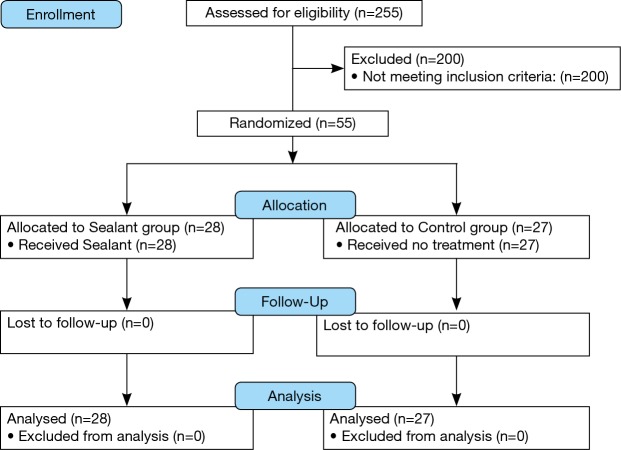
Between January 2015 and January 2017, written informed consent was obtained from 255 patients, who underwent VATS lobectomy, 55 met the inclusion criteria and they were randomly assigned to 2 different groups. All patients were included in the ITT population (28 in the Progel group and 27 in the no treatment group), and all participants were followed-up at 1- and 2-month. Figure 6 depicts the flow of participants through each arm of the trial. Among the 200 non-randomized patients, the reason for exclusion was the absence of moderate air leak during the underwater air-tightness test.
Figure 6.
Participant flow diagram.
Baseline data and clinical characteristics
The baseline demographic, surgical, and pathological characteristics of randomised patients in the two study groups are shown in Tables 1,2. As would be expected, the two arms were well balanced.
Table 1. Patient baseline demographics and clinical characteristics (55 patients).
| Variables | Progel group, n=28 | “No treatment” group, n=27 | P |
|---|---|---|---|
| Sex | 0.38 | ||
| Male | 8 (28.6%) | 5 (18.5%) | |
| Female | 20 (71.4%) | 22 (81.5%) | |
| Age (years) | 69.8±9.0 | 69.6±7.7 | 0.92 |
| COPD | 0.93 | ||
| No | 20 (71.4%) | 19 (70.4%) | |
| Yes | 8 (28.6%) | 8 (29.6%) | |
| BMI >30 | 0.15 | ||
| No | 25 (92.6%) | 27 (100%) | |
| YES | 2 (7.4%) | 0 (0%) | |
| Tobacco | 0.69 | ||
| No | 11 (39.3%) | 12 (44.4%) | |
| Yes | 17 (60.7%) | 15 (55.6%) | |
| Preoperative chemotherapy | 0.32 | ||
| No | 27 (96.4%) | 27 (100%) | |
| Yes | 1 (3.6%) | 0 (0%) | |
| Previous homolateral surgery | 0.32 | ||
| No | 27 (96.4%) | 27 (100%) | |
| Yes | 1 (3.6%) | 0 (0%) | |
| FEV1 liter | 2.58±0.69 | 2.68±0.74 | 0.61 |
| PaCO2 mmHg | 38±3.9 | 38.4±3.12 | 0.71 |
| PaO2 mmHg | 81±9.1 | 83±7.5 | 0.6 |
COPD, chronic obstructive pulmonary disease; BMI, body mass index; FEV1, forced expiratory volume in one second; PaCO2, partial pressure of carbon dioxide; mmHg, Millimeter of Mercury; PaO2, partial pressure of oxygen.
Table 2. Surgical and pathological data (55 patients).
| Variables | Progel group, n=28 | “No treatment” group, n=27 | P |
|---|---|---|---|
| Pleural adhesions | 0.73 | ||
| No | 21 (77.8%) | 22 (81.5%) | |
| Yes | 6 (22.2%) | 5 (18.5%) | |
| Fissure completeness | 0.73 | ||
| Grade 1 | 3 (13.0%) | 7 (28.0%) | |
| Grade 2 | 11 (47.9%) | 9 (36.0%) | |
| Grade 3 | 7 (30.4%) | 8 (32.0%) | |
| Grade 4 | 2 (8.7%) | 1 (4.0%) | |
| Intraoperative air leak locations | |||
| Stapler line | 0.89 | ||
| No | 14 (50%) | 14 (51.8%) | |
| Yes | 14 (50%) | 13 (48.2%) | |
| Parenchyma | 0.28 | ||
| No | 4 (14.3%) | 7 (25.9%) | |
| Yes | 24 (85.7%) | 20 (74.1%) | |
| Intraoperative air leak measure (mL/min) | 253±101 | 222±86 | 0.23 |
| Resection | 0.16 | ||
| Bilobectomy | 2 (7.1%) | 0 (0%) | |
| Lobectomy | 26 (92.9%) | 27 (100%) | |
| Side | 0.64 | ||
| Right | 17 (65.4%) | 11 (40.7%) | |
| Left | 9 (34.6%) | 16 (59.3%) | |
| Resection | 0.67 | ||
| Upper | 13 (46.4%) | 11 (40.7%) | |
| Lower | 15 (43.6%) | 16 (59.3%) | |
| Histology | 0.28 | ||
| Adenocarcinoma | 19 (70.4%) | 15 (55.6%) | |
| Squamous | 4 (14.8%) | 3 (11.1%) | |
| Other | 4 (14.8%) | 9 (33.3%) |
The indication for pulmonary resection was primary lung cancer in 66.7% of the cases, with an equal distribution in the two patient groups. In 100% of patients, a VATS procedure was performed. Completeness of the fissure was graded in four stages according to Craig and Walker classification (15): grade 1—complete fissure with entirely separate lobes; grade 2—complete visceral cleft but parenchymal fusion at the base of the fissure; grade 3—visceral cleft evident for part of the fissure; grade 4—complete fusion of the lobes with no evident fissure line.
After lung resection, 55 (21.6%) patients had mild IOAALs. 96.4% of patients (27/28) in the group A were treated with application of one Progel, and in only one patient (3.4%) Progel application was repeated once, while in the control group (n=27) no further intervention was performed. No sealant application of any type was used in in the control group. There were no differences in the postoperative management of patients in either group.
One chest tube was inserted in all patients in both groups to manage air leaks and for drainage. The location and quantity of IOAAL in both groups (Table 2) were not significantly different. Also regarding the other surgical and pathological data, including intraoperative risk factors for IOAAL, the two arms were well balanced (Table 2).
Outcomes and estimations
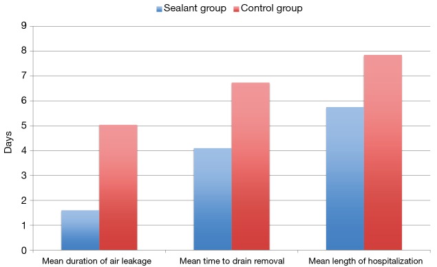
At randomisation, the degree of IOAAL was similar in the two groups. Based on an ITT analysis, the mean air leakage duration was statistically different between the two groups (Figure 7 and Table 3): in the Progel group was 1.60 vs. 5.04 days in the “no treatment” group (P<0.001). The average duration of chest drainage was statistically different in the two groups: 4.1 days in the interventional group and 6.74 days in the control group (P=0.008).
Figure 7.
Mean time to air leak cessation, chest drain removal and hospital discharge in the Progel and “no treatment” groups.
Table 3. Outcome data (primary and secondary endpoints).
| Variables | Progel group, n=28 mean (SD) | “No treatment” group, n=27 mean (SD) | Mean difference (95% CI) | P |
|---|---|---|---|---|
| Duration of air leak | 1.60 (1.13) | 5.04 (3.63) | 3.42 (1.98–4.87) | <0.001 |
| Time to drain removal | 4.10 (3.42) | 6.74 (4.69) | 2.63 (0.73–4.54) | 0.008 |
| Length of hospitalization | 5.75 (4.89) | 7.85 (6.15) | 2.10 (0.26–3.93) | 0.026 |
| Peri- and post-operative complications/mortality | 0 | 0 | – | – |
The mean time to hospital discharge was also statistically shorter in the group A than in the group B (5.75 vs. 7.85 days, P=0.026).
Economic evaluation
There was no significant difference in the mean operation time between the two groups (Progel group =142±37 minutes vs. Control group =151±50 minutes, P=0.49) and therefore no difference in the costs related to the use of operating theatre. Of course, costs related to using the medical devices were revealed only in the group A. The mean hospital stay was 2.1 days shorter in the Progel group. Therefore, there was a significant difference in the costs related to the hospitalisation days: in the Progel group, the costs related to the sealant were of 12.905€ (29 pieces) while in the group B the costs related to the prolonged hospital stay were of 39.690€ (P<0.001).
Harms
We did not observe patients requiring reoperation nor repeat chest drain placements in the two groups at 2 months follow-up. There were no observed device-related adverse events. The overall complication rate, in-hospital mortality and 30-day mortality for the entire treated group were nil.
Discussion
The clinical and economic impact of IOAALs after primary pulmonary resection is well known and significant (1-3). The reduction of these air leaks indeed decreases LOS, postoperative complications, and related costs. The management of IOAALs is best done during surgery, but standard techniques are usually inadequate (13), troublesome and time-consuming during VATS. A Cochrane Database Review demonstrated that surgical sealants after open pulmonary resection reduce postoperative air leaks and time to chest drain removal, but it failed to demonstrate a reduction in LOS (13). An exact measurement of the IOAAL, however, was not performed in these studies, and it may represent a potential bias.
Many air leaks will resolve spontaneously within 48 hours; some will persist.
A correct intraoperative evaluation of IOAALs is essential and challenging to achieve. Many Authors adopted the visual grading of air leaks according to Macchiarini’s scale as 0 (no leakage), 1 (mild, countable bubbles), 2 (moderate, stream of bubbles), or 3 (severe, coalescent bubbles) (1). We standardised a VMT to obtain a quantitative grading of IOAALs. The exact and objective classification of IOAALs represents the strength of our study. We focused our attention on the patients with moderate IOAAL where the need of intraoperative use of a lung sealant was still controversial. We empirically defined as moderate an air leak between 100 and 400 mL/min, on the bases of our experience. Clinical experience shows us that mild air leakages (<100 mL/min) are usually self-limiting; whereas treatment of severe air leaks (>400 mL/min) is mandatory. As data of patients with mild and severe air leaks were not prospectively recorded, we can not demonstrate this statement. This is a limitation of our study.
The current randomised controlled trial was designed to exclude patients with mild or severe IOAALs who would not benefit from the sealant. We standardised a VMT, to accurately and reliably identify patients with mild IOAAL. To achieve intraoperative air leak sealing following VATS lobectomy, a spray sealant appeared in our opinion to be the best solution. To the best of our knowledge, this is the first report of a randomised controlled trial analysing the clinical and economic outcomes of a lung sealant in case of IOAAL following VATS lobectomy. A recent study analysed the use of a biodegradable spray polymer (Progel) for the closure of IOAAL after minimally invasive pulmonary resection, but it was not a randomised controlled trial (16). Other RCTs analysing lung sealant efficacy were almost exclusively in patients undergoing thoracotomy (17-19).
We demonstrated that the use of Progel significantly reduces air leakage duration, time to chest drain removal and time to hospital discharge, in patients with an IOAAL between 100 and 400 mL/min (moderate air leakage) following VATS lobectomy or bi-lobectomy.
We experienced in our series a moderate IOAAL in 21.6% of patients (55/255). The spray sealant represents during VATS procedure the ideal device, easy and quick to use. We did not observe a significant difference of operation time in the two groups. Progel polymerises to form a definite, flexible hydrogel matrix that adheres to the lung tissue within 15 seconds and forms a flexible seal that can withstand 30 mmHg air pressure within 2 minutes of application and a maximum burst pressure of greater than 90 mmHg in less than 10 min. The material is wholly reabsorbed within one month postoperatively. We did not observe device-related complications.
The thoracic drainage system utilised from each centre (DrenetechTM Palmevo, Redax) allowed us to record air leak data digitally and to download the results through a mini USB-key port. We could accurately analyse time of air leak cessation.
Cost saving analysis showed obviously a significant reduction of hospitalisation costs, although we showed any significant differences in postoperative complications. The costs of 2 days of hospitalisation abundantly exceed the price of the lung sealing.
Conclusions
Our standardised VMT helps in reducing the length of hospital stay after VATS lobectomy because in case of IOAALs between 100 and 400 mL/min the use of ProgelTM significantly reduces postoperative air leak, time to drain removal and length of hospitalisation compared with no treatment. This shorter hospital stays results in significant cost saving benefits. Selection of patients with standardised VMT is essential to limit unnecessary intraoperative sealant treatments, thus contributing to limit the costs.
Acknowledgements
We thank Dr. Andrea Ponzoni for his assistance to the bio-statistical elaboration.
Footnotes
Conflicts of Interest: The authors have no conflicts of interest to declare.
References
- 1.Brunelli A, Monteverde M, Borri A, et al. Predictors of prolonged air leak after pulmonary lobectomy. Ann Thorac Surg 2004;77:1205-10. 10.1016/j.athoracsur.2003.10.082 [DOI] [PubMed] [Google Scholar]
- 2.Brunelli A, Xiume F, Al Refai M, et al. Air leaks after lobectomy increase the risk of empyema but not of cardiopulmonary complications: a case-matched analysis. Chest 2006;130:1150-6. 10.1378/chest.130.4.1150 [DOI] [PubMed] [Google Scholar]
- 3.Stolz AJ, Schützner J, Lischke R, et al. Predictors of prolonged air leak following pulmonary lobectomy. Eur J Cardiothorac Surg 2005;27:334-6. 10.1016/j.ejcts.2004.11.004 [DOI] [PubMed] [Google Scholar]
- 4.Okereke I, Murthy SC, Alster JM, et al. Characterization and importance of air leak after lobectomy. Ann Thorac Surg 2005;79:1167-73. 10.1016/j.athoracsur.2004.08.069 [DOI] [PubMed] [Google Scholar]
- 5.Rivera C, Bernard A, Falcoz PE, et al. Characterization and prediction of prolonged air leak after pulmonary resection: a nationwide study setting up the index of prolonged air leak. Ann Thorac Surg 2011;92:1062-8. 10.1016/j.athoracsur.2011.04.033 [DOI] [PubMed] [Google Scholar]
- 6.Zhao K, Mei J, Xia C, et al. Prolonged air leak after video-assisted thoracic surgery lung cancer resection: risk factors and its effect on postoperative clinical recovery. J Thorac Dis 2017;9:1219-25. 10.21037/jtd.2017.04.31 [DOI] [PMC free article] [PubMed] [Google Scholar]
- 7.Merritt RE, Singhal S, Shrager JB. Evidence-based suggestions for management of air leaks. Thorac Surg Clin 2010;20:435-48. 10.1016/j.thorsurg.2010.03.005 [DOI] [PubMed] [Google Scholar]
- 8.Zaraca F, Vaccarili M, Zaccagna G, et al. Mild IOAAL graded according to a VMT: leak 60 mL/min. Asvide 2017;534. Available online: http://www.asvide.com/articles/1854
- 9.Zaraca F, Vaccarili M, Zaccagna G, et al. Moderate IOAAL graded according to a VMT: leak 180 mL/min. Asvide 2017;535. Available online: Available online: http://www.asvide.com/articles/1855
- 10.Zaraca F, Vaccarili M, Zaccagna G, et al. Severe IOAAL graded according to a VMT: leak 520 mL/min. Asvide 2017;536. Available online: Available online: http://www.asvide.com/articles/1856
- 11.Zaraca F, Vaccarili M, Zaccagna G, et al. Moderate IOAAL graded according to a VMT: leak 260 mL/min treated with Progel application. Control after treatment. Asvide 2017;537. Available online: Available online: http://www.asvide.com/articles/1857
- 12.Zaraca F, Vaccarili M, Zaccagna G, et al. Severe IOAAL graded according to a VMT: leak 520 mL/min treated with Progel application. Control after treatment. Asvide 2017;538. Available online: Available online: http://www.asvide.com/articles/1858
- 13.Belda-Sanchis J, Serra-Mitjans M, Iglesias Sentis M, et al. Surgical sealant for preventing air leaks after pulmonary resections in patients with lung cancer. Cochrane Database Syst Rev 2010;1:CD003051. [DOI] [PMC free article] [PubMed] [Google Scholar]
- 14.Klijian A. A novel approach to control air leaks in complex lung surgery: a retrospective review. J Cardiothorac Surg 2012;7:49. 10.1186/1749-8090-7-49 [DOI] [PMC free article] [PubMed] [Google Scholar]
- 15.Craig SR, Walker WS. A proposed anatomical classification of the pulmonary fissures. J R Coll Surg Edinb 1997;42:233-4. [PubMed] [Google Scholar]
- 16.Park BJ, Snider JM, Bates NR, et al. Prospective evaluation of biodegradable polymeric sealant for intraoperative air leaks. J Cardiothorac Surg 2016;11:168-75. 10.1186/s13019-016-0563-3 [DOI] [PMC free article] [PubMed] [Google Scholar]
- 17.D’Andrilli A, Andreetti C, Ibrahim M, et al. A prospective randomized study to assess the efficacy of a surgical sealant to treat air leaks in lung surgery. Eur J Cardiothorac Surg 2009;35:817-20. 10.1016/j.ejcts.2009.01.027 [DOI] [PubMed] [Google Scholar]
- 18.Lequaglie C, Giudice G, Marasco R, et al. Use of a sealant to prevent prolonged air leaks after lung resection: a prospective randomized study. J Cardiothorac Surg 2012;7:106. 10.1186/1749-8090-7-106 [DOI] [PMC free article] [PubMed] [Google Scholar]
- 19.Anegg U, Lindenmann J, Matzi V, et al. Efficiency of fleece-bound sealing (TachoSil) of air leaks in lung surgery: a prospective randomised trial. Eur J Cardiothorac Surg 2007;31:198-202. 10.1016/j.ejcts.2006.11.033 [DOI] [PubMed] [Google Scholar]


