Abstract
Introduction:
Supernumerary teeth are the presence of more number of teeth over the normal dental formula and may occur in permanent as well as early mixed dentition. This study determined the prevalence, characteristics, and complications caused by supernumerary teeth in nonsyndromic South Indian pediatric population.
Materials and Methods:
Characteristics of supernumerary teeth determined by clinical and radiographic examination were recorded. The age, sex, number of supernumerary teeth, eruption status, morphology, position, orientation, and complications (if any) associated with supernumerary teeth were recorded for each patient who had supernumerary teeth. The data collected were statistically analyzed.
Results:
Supernumerary teeth were detected in 45 subjects (1.1%), of which 34 (75.6%) were male and 11 (24.4%) were female. There was no association between the number of supernumerary teeth and the gender of the patient. The total number of supernumerary teeth among the affected 45 patients was 54. The average number of supernumerary teeth per person was 1.2. The number of supernumerary teeth was one in 35 cases, two in 8 cases, and 3 in 1 case. Of the 45 patients, 8 patients with supernumerary teeth were in deciduous dentition stage, 29 patients were in mixed dentition stage, and 8 patients were in permanent dentition stage. Most supernumerary teeth presented in the anterior maxilla. Morphologically, conical-shaped supernumerary teeth were the most common finding. 68.5% of supernumerary teeth presented with straight orientation and inverted orientation was seen in 24.1%. Complications seen in patients with supernumerary teeth were delayed or noneruption of adjacent tooth malposition or rotation of adjacent teeth, diastema formation, and formation of dentigerous cyst.
Conclusions:
Supernumerary teeth have an incidence of 1.1% in South Indian population and can cause many complications that can harm the developing occlusion. Knowledge about supernumerary teeth may help the dentist in early diagnosis and early intervention.
Keywords: Hyperdontia, mesiodens, supernumerary teeth
INTRODUCTION
Supernumerary teeth are significant dental anomaly that may occur in permanent as well as early mixed dentition. These may be revealed in routine radiographic examinations or during investigations of impacted teeth or may have erupted into the oral cavity.[1]
Supernumerary teeth also known as hyperdontia is the presence of more number of teeth over the normal dental formula (32 in permanent dentition and 20 in deciduous dentition). Supernumerary teeth have a prevalence of 0.1%–3.8% in permanent and 0.35%–0.6% in deciduous dentition. Supernumerary teeth may have erupted into the oral cavity or may remain unerupted for years without creating any disturbances or clinical complications. However, sometimes, supernumerary teeth may result in delay in eruption or impaction of permanent teeth, malalignment of teeth, crowding of teeth, diastema particularly midline diastema, root resorption of teeth in contact with the supernumerary teeth, and cyst formation.
This study determined the prevalence, characteristics, and complications caused by supernumerary teeth in nonsyndromic South Indian pediatric population.
MATERIALS AND METHODS
The data for the study was collected from 3936 patients who attended the Department of Pedodontics and Preventive Dentistry in Pushpagiri College of Dental Sciences, Tiruvalla, from January 2015 to June 2015. A skilled pedodontist examined each patient who visited the Department during the study period intraorally with mouth mirror under adequate lighting.
A tooth was considered an impacted tooth if it was prevented from erupting by a physical barrier, usually other teeth or because of nonvertical orientation of the teeth within the periodontal structures remaining in the jaw for more than 2 years after its mean age of eruption. A supernumerary tooth is an erupted or unerupted extra-tooth, resembling or unlike other teeth in its group.[2]
Radiographic examination of clinically suspicious cases of supernumerary teeth was undertaken for confirmation. Characteristics of supernumerary teeth determined by clinical and radiographic examination were recorded using a printed pro forma. The age, sex, number of supernumerary teeth, eruption status, morphology, position, orientation, and complications (if any) associated with supernumerary teeth were recorded for each patient who had supernumerary teeth.
Exclusion criteria for the study were patients with one or more of the following pathological situations;
Patients with disease, trauma, or fracture of teeth that may have affected growth of dentition; and
Patients with hereditary diseases or syndromes (e.g., Down's syndrome or cleidocranial dysostosis).
The data collected were statistically analyzed using SPSS Version 16 software (SPSS Inc, Chicago, Illinois, USA). Age was summarized as means and standard deviation and all other variables were calculated as frequencies and percentages. The prevalence among males and females was compared using Chi-square test (P < 0.05).
RESULTS
During the above-mentioned time, 3936 children were examined. Of this, 2145 patients were female (54.49%) and 1791 were male (45.51%).
Supernumerary teeth were detected in 45 subjects (1.1%), of which 34 (75.6%) were males and 11 (24.4%) were females with a male female ratio of 3.1:1 [Table 1 and Figure 1]. The incidence of impacted teeth was not related to the sex of the patient (P = 0.82), i.e., there was no association between the number of supernumerary teeth and the gender of the patient (P > 0.05).
Table 1.
Distribution of supernumerary teeth according to gender

Figure 1.
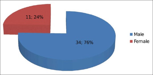
Pie-Diagram representing distribution of supernumerary teeth according to gender
Total number of supernumerary teeth among the affected 45 patients was 54. The average number of supernumerary teeth per person was 1.2. The number of supernumerary teeth was one in 35 cases (77.8%), two in 8 cases (20%), and 3 in 1 case (2.2%) [Table 2 and Figure 2].
Table 2.
Distribution according to number of supernumerary teeth

Figure 2.
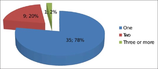
Pie-Diagram representing distribution according to number of supernumerary teeth
11.1% had a positive family history of occurrence of supernumerary teeth, but 88.9% presented with a negative family history.
The mean age of detection of supernumerary teeth was 8.6 years (standard deviation of 2.17). Eight patients (17.8%) were below 6 years (Group 1), 20 patients (44.4%) were between 6 and 9 years (Group 2), and 17 patients (37.8%) were between 9 and 12 years (Group 3).
Of the 45 patients, 8 patients (17.8%) with supernumerary teeth were in deciduous dentition stage, 29 (64.4%) patients were in mixed dentition stage and 8 (17.8%) patients were in permanent dentition stage [Table 3 and Figure 3].
Table 3.
Distribution of supernumerary teeth according to type of dentition

Figure 3.
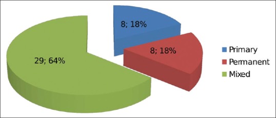
Pie-Diagram showing distribution of supernumerary teeth according to type of dentition
Most supernumerary teeth presented in the anterior maxilla. In 42 (93.3%) patients, supernumerary teeth were in the anterior maxilla. Supernumerary teeth were located in posterior mandible in 2 patients (4.4%) and in anterior mandible in 1 patient (2.2%) [Table 4 and Figure 4].
Table 4.
Distribution of supernumerary teeth according to its position

Figure 4.
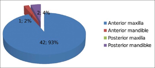
Pie-Diagram showing distribution of supernumerary teeth according to its position
Morphologically, conical-shaped supernumerary teeth were the most common finding. 61.1% (33) of the total sample size were conical, other types being supplemental 20.4% (11), tuberculate 14.8% (8), and odontome type 3.7% (2) [Table 5 and Figure 5].
Table 5.
Distribution of supernumerary teeth according to type and morphology

Figure 5.
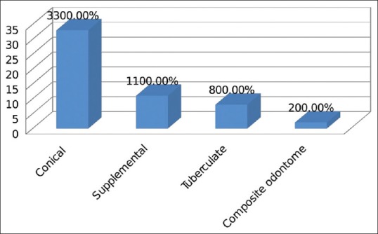
Bar-Diagram showing distribution of supernumerary teeth according to type and morphology
In the 45 patients, 17 patients had (48.6%) the supernumerary teeth erupted into the oral cavity. 12 patients (34.3%) presented with supernumerary teeth that were impacted and remaining six patients (17.1%) presented with both erupted as well as impacted teeth [Table 6 and Figure 6].
Table 6.
Distribution of supernumerary teeth according to its eruption status

Figure 6.
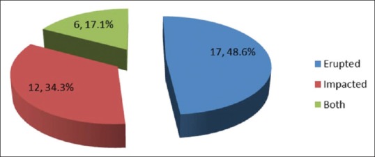
Pie-Diagram representing distribution of supernumerary teeth according to its eruption status
Among the 56 supernumerary teeth detected, 37 (68.5%) supernumerary teeth presented with straight orientation and inverted orientation was seen in 13 (24.1%) [Table 7 and Figure 7].
Table 7.
Distribution of supernumerary teeth according to its orientation

Figure 7.
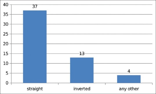
Bar-Diagram representing distribution of supernumerary teeth according to its orientation
Regarding the sagittal position of supernumerary teeth, the majority of them were placed on the arch 51.7% (29), 39.2% (22) were placed palatally, and only 9.1% (5) were placed labial to the arch.
Complications seen in patients with supernumerary teeth were delayed or noneruption of adjacent tooth (24.4%), malposition or rotation of adjacent teeth (37.8%), diastema formation (22.2%), formation of dentigerous cyst (2.2%), and in remaining 13.4%, no dental abnormalities were detected in association with supernumerary teeth [Table 8 and Figure 8].
Table 8.
Distribution of supernumerary teeth according to its complications
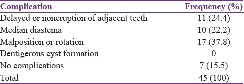
Figure 8.
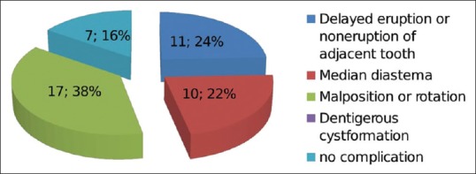
Bar-Diagram showing distribution of supernumerary teeth according to its complications
DISCUSSION
Teeth that are found in addition to the normal series that may be located anywhere in the dental arch are called supernumerary teeth.[3] These alterations in development may present in any area of dental arch and can affect both primary and permanent dentitions. Higher incidence of supernumerary teeth is found in permanent dentition and may be found in syndromic as well as nonsyndromic patients.
Various theories explain the etiology of different types of supernumerary teeth. As per Liu J, dichotomy of the tooth bud results in supernumerary teeth. According to hyperactivity theory, local hyperactivity of the dental lamina that is both independent and conditioned results in supernumerary teeth.[3]
Supernumerary teeth have a prevalence of 0.1%–3.8% in the general population. Patients with cleft lip and cleft palate have very high incidence of supernumerary teeth up to even 28%.[4,5,6]
The prevalence of supernumerary teeth was found to be 1.1% in the present study. Celikoglu et al. have also reported similar results.[2]
Male-to-female ratio of patients with supernumerary was found to be 3.1:1 in the current study which was in close the 2:8 ratio obtained by Asaumi et al.[7] and 4:1 ratio obtained by Kim and Lee.[8]
Supernumerary teeth present most frequently as single tooth but may also present as multiple teeth.[9,10] In the present study, one supernumerary teeth was seen in 35 cases (77.8%), two in 8 cases (20%), and 3 in 1 case (2.2%). Results similar to this were obtained in earlier studies also.
The mean age of detection of supernumerary teeth was 8.6 years in the current study. Similar result was seen in a study conducted in Indian population by Mukhopadhyay.[1] This finding may be because this is the normal eruption time of maxillary permanent central incisors.[1]
Of 45 patients, 8 patients (17.8%) with supernumerary teeth were in primary dentition stage, 8 (17.8%) patients in permanent dentition stage, and 29 (64.4%) patients in mixed dentition stage. This incidence of supernumerary teeth detected in mixed dentition stages is similar to that obtained by Anegundi et al.[11]
Regarding the location of supernumerary teeth, 93.3% (42) were located in anterior maxilla, while 2.2% (1) located in anterior mandible and posterior mandible accounted for 4.4% (2) of supernumerary teeth. De Oliveira et al. have also reported that 91.3% of the cases are located in maxilla.[12]
Morphologically, the most common presentation of supernumerary teeth in this study was conical. This was seen in 61.1% of the total sample. Previous studies also showed similar results.[13] Other types were supplemental, tuberculate, and odontome type.
Regarding the direction of the crown of supernumerary teeth detected among the 45 patients, 33 (73.3%) supernumerary teeth were oriented straight and the remaining 12 (26.7%) were inverted. Asaumi et al. had reported inverted supernumeraries in 67% cases, normally oriented 27% of cases and 6% directed horizontally against the axis of the tooth.[7] Anegundi et al. in their study found that of 790 patients with supernumerary teeth, 95.06% were oriented straight and 4.94% were inverted.[11] The results of our study slightly disagree with the previous studies, but it may be because of the fact that studies were conducted in different races.
Supernumerary teeth may erupt into oral cavity or may remain impacted. Impacted supernumerary teeth may not cause any clinical manifestation or disturbance. On assessing eruption status in the present study, 17 patients had (48.6%) erupted supernumerary teeth, 12 patients (34.3%) had impacted supernumerary teeth, and remaining patients 6 (17.1%) had both erupted and impacted teeth.
In the current study, regarding the sagittal position of supernumerary teeth, majority were placed on the arch (50.9%), 39.6% were placed palatally, and only 9.4% were placed labial to the arch. This finding is contradictory to the values reported by Asaumi et al. in their study out of 147 supernumerary teeth where 89% were located palatally against the dental arch.[7]
In some cases, erupted or impacted supernumerary may cause complications such as midline diastema, crowding, formation of cysts, and root resorption of adjacent teeth.
In the current study, the most common clinical complication caused by supernumerary teeth was malposition or rotation of adjacent teeth (37.8%). Other complications were delayed or noneruption of adjacent tooth (24.4%), diastema formation (22.2%), and dentigerous cyst formation (2.2%). Other authors have also documented displacement to be the most common clinical complication.[2,7,9]
CONCLUSION
Complications created by supernumerary teeth can cause potential harm to the developing occlusion, which can be difficult to intervene or may need aggressive treatment at a later stage. Knowledge about supernumerary teeth may help the dentist in early diagnosis, interventions, and may prevent possible complications that may develop. This may be very important from a therapeutic point of view.
Financial support and sponsorship
Nil.
Conflicts of interest
There are no conflicts of interest.
REFERENCES
- 1.Mukhopadhyay S. Mesiodens: A clinical and radiographic study in children. J Indian Soc Pedod Prev Dent. 2011;29:34–8. doi: 10.4103/0970-4388.79928. [DOI] [PubMed] [Google Scholar]
- 2.Celikoglu M, Kamak H, Oktay H. Prevalence and characteristics of supernumerary teeth in a non-syndrome Turkish population: Associated pathologies and proposed treatment. Med Oral Patol Oral Cir Bucal. 2010;15:e575–8. doi: 10.4317/medoral.15.e575. [DOI] [PubMed] [Google Scholar]
- 3.Garvey MT, Barry HJ, Blake M. Supernumerary teeth – An overview of classification, diagnosis and management. J Can Dent Assoc. 1999;65:612–6. [PubMed] [Google Scholar]
- 4.Díaz A, Orozco J, Fonseca M. Multiple hyperodontia: Report of a case with 17 supernumerary teeth with non syndromic association. Med Oral Patol Oral Cir Bucal. 2009;14:E229–31. [PubMed] [Google Scholar]
- 5.Nazif MM, Ruffalo RC, Zullo T. Impacted supernumerary teeth: A survey of 50 cases. J Am Dent Assoc. 1983;106:201–4. doi: 10.14219/jada.archive.1983.0390. [DOI] [PubMed] [Google Scholar]
- 6.Sacal C, Echeverri EA, Keene H. Retrospective survey of dental anomalies and pathology detected on maxillary occlusal radiographs in children between 3 and 5 years of age. Pediatr Dent. 2001;23:347–50. [PubMed] [Google Scholar]
- 7.Asaumi JI, Shibata Y, Yanagi Y, Hisatomi M, Matsuzaki H, Konouchi H, et al. Radiographic examination of mesiodens and their associated complications. Dentomaxillofac Radiol. 2004;33:125–7. doi: 10.1259/dmfr/68039278. [DOI] [PubMed] [Google Scholar]
- 8.Kim SG, Lee SH. Mesiodens: A clinical and radiographic study. J Dent Child (Chic) 2003;70:58–60. [PubMed] [Google Scholar]
- 9.Rajab LD, Hamdan MA. Supernumerary teeth: Review of the literature and a survey of 152 cases. Int J Paediatr Dent. 2002;12:244–54. doi: 10.1046/j.1365-263x.2002.00366.x. [DOI] [PubMed] [Google Scholar]
- 10.Tay F, Pang A, Yuen S. Unerupted maxillary anterior supernumerary teeth: Report of 204 cases. ASDC J Dent Child. 1984;51:289–94. [PubMed] [Google Scholar]
- 11.Anegundi RT, Tegginmani VS, Battepati P, Tavargeri A, Patil S, Trasad V, et al. Prevalence and characteristics of supernumerary teeth in a non-syndromic South Indian pediatric population. J Indian Soc Pedod Prev Dent. 2014;32:9–12. doi: 10.4103/0970-4388.127041. [DOI] [PubMed] [Google Scholar]
- 12.De Oliveira Gomes C, Drummond SN, Jham BC, Abdo EN, Mesquita RA. A survey of 460 supernumerary teeth in Brazilian children and adolescents. Int J Paediatr Dent. 2008;18:98–106. doi: 10.1111/j.1365-263X.2007.00862.x. [DOI] [PubMed] [Google Scholar]
- 13.Ramesh K, Venkataraghavan K, Kunjappan S, Ramesh M. Mesiodens: A clinical and radiographic study of 82 teeth in 55 children below 14 years. J Pharm Bioallied Sci. 2013;5(Suppl 1):S60–2. doi: 10.4103/0975-7406.113298. [DOI] [PMC free article] [PubMed] [Google Scholar]


