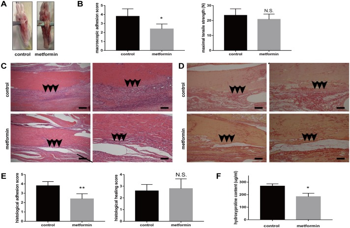
Figure 1. Metformin treatment reduces peritendinous tissue adhesion in tendon-injured rats.
(A) Macroscopic images showed less adhesive tissues in metformin-treated (200 mg/kg/d) rats. (B) Gross adhesion scores (left) were significantly lower in metformin-treated rats compared with control rats 3 weeks after tendon operation (2.8 vs 3.9, p = 0.005). Maximal tensile strengths (right) were comparable between control and metformin-treated rats (23 vs 21 N, p = 0.173) (n = 8 for each group). (C) HE staining of peritendinous tissues showed reduced cell proliferation and inflammation by metformin treatment. Arrows indicate adhesion formation. Scale bars (left) = 250 μm. Scale bars (right) = 100 μm. (D) Sirius-red staining revealed decreased collagen deposition by metformin treatment. Arrows indicate excessive collagen deposition. Scale bars (left) = 250 μm. Scale bars (right) = 100 μm. (E) Histological scores for adhesion decreased after metformin treatment (2.4 vs 3.8, p = 0.002). Histological healing scores were comparable between control and metformin-treated rats (2.6 vs 2.8, p = 0.667) (n =5 for each group). (F) Metformin treatment significantly deceased the hydroxyproline (Hyp) content of tendon tissues at 3 weeks (184 vs 268 ug/ml, p = 0.011) (n = 5 for each group). Data are expressed as means ± SEM. * P < 0.05; ** P < 0.01; N.S. not significant.

