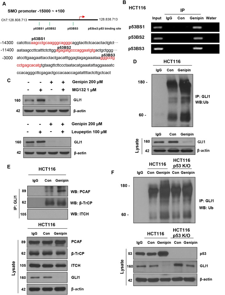
Figure 4.
(A) Illustration of the three predicted p53 binding sites in the SMO promoter. The predicted p53 binding sites are presented in the DNA sequence of the SMO promoter (-15000 to -2800). (B) HCT116 cells were treated with 200 μM genipin, and then a chromatin immunoprecipitation (ChIP) assay was performed to confirm the direct binding of p53 to the SMO promoter region. (C) HCT116 cells were treated with 1 μM MG132 for 6 h and 100 μM Leupeptin for 24 h. GLI1 protein expression of was evaluated by western blotting. (D) HCT116 cell lysates were immunoprecipitated with an anti-GLI1 antibody and then immunoblotted with an anti-ubiquitin antibody. (E) The interaction between GLI1 and three E3 ligases was measured by co-immunoprecipitation. HCT116 cell lysates were immunoprecipitated with anti-PCAF, anti-β-TrCP, and anti-ITCH antibodies, and then immunoblotted with an anti-GLI-1 antibody. (F) HCT116 or p53 knockout (KO) HCT116 cells were treated with 200 μM genipin. Lysates of HCT116 and p53 KO HCT116 cells were immunoprecipitated with an anti-GLI1 antibody and immunoblotted with an anti-ubiquitin antibody. Data are expressed as the means of three independent experiments.

