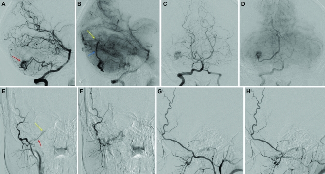
FIGURE 1.
Cerebral digital subtraction angiograms of a 19-yr-old female presenting with intractable headaches and syncopal episodes demonstrate a characteristic AVM in an HHT patient. Lateral A, B and anteroposterior C, D projections of vertebral artery injection in arterial and capillary phases showing a right superficial cerebellar 1.2 × 1.3 cm arteriovenous malformation (red arrow in A) supplied by posterior inferior cerebellar artery with superficial hemispheric drainage (blue arrow in B) to the straight sinus near the torcula (yellow arrow in B). Although there are no flow-related or intranidal aneurysms, there is a high-grade stenosis of the venous outflow at the junction between the cortical vein and the straight sinus. Anteroposterior E, F and lateral G, H projections of selective right occipital artery injection in the same patient also show a hypoglossal canal arteriovenous fistula (yellow arrow in E) fed by the hypoglossal branch of the right occipital artery (red arrow in E) and draining into condylar veins.

