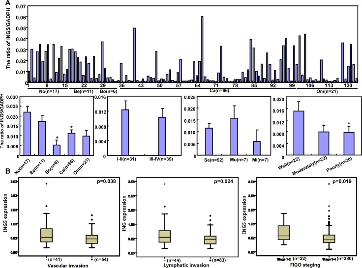
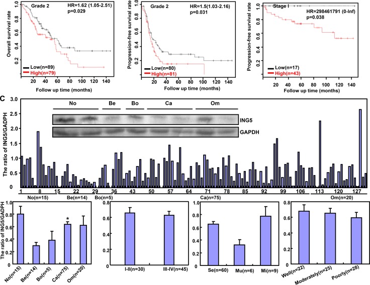
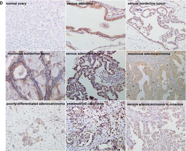
Figure 7. The clinicopathological significances of ING5 expression in ovarian cancers.
ING5 mRNA was quantified in ovarian normal tissue (No), benign (Be) and borderline (Bo) tumor, primary cancer (Ca), and metastatic cancer in omentum (Om) by real-time PCR with its correlation between its expression and clinicopathological features analyzed (A). The clinicopathological and prognostic significances of ING5 mRNA expression were analyzed using TCGA dataset and KM plotter respectively (B). Tissue lysate was loaded and probed with anti-ING5 antibody with GAPDH as an internal control by Western blot. ING5 protein was examined in No, Be, Bo, Ca and Om, and compared with aggressive parameters (C). Additionally, ING5 was not expressed in normal fiber cells, but strongly in the nucleus and cytoplasm of serous adenoma, serous and mucinous borderline tumor, serous, mucinous, and poorly-differentiated adenocarcinoma, endometriod carcinoma, serous adenocarcinoma in omentus by immunohistochemistry (D). The data is expressed as mean ± standard error. *p < 0.05.


