Abstract
Disaster victim identification (DVI) is an intensive and demanding task involving specialists from various disciplines. The forensic dentist is one of the key persons who plays an important role in the DVI human identification process. In recent years, many disaster incidents have occurred that challenged the DVI team with various kinds of difficulties related to disaster management and unique situations in each disaster. New technologies have been developed to make the working process faster and more effective and the different DVI protocols have been evaluated and improved. The aim of this article is to collate all information regarding diagnostic tools and methodologies pertaining to forensic odontological DVI, both current and future. It can be concluded that lessons learned from previous disaster incidents have helped to optimize working protocols and to develop new tools that can be applied in future DVI operation. The working procedures have been greatly improved by newly developed technologies.
KEYWORDS: forensic odontology, Disaster Victim Identification (DVI), human identification, mass casualty
Introduction
Each natural or man-made disaster presents a different set of circumstances and, as a consequence, each event results in new challenges for identification teams. Although the exact number of deceased persons to define an event as a disaster varies by jurisdiction, it is widely agreed that mass fatality incidents always exert an onerous impact on local resources.
Dental DVI team leaders conduct training exercises to familiarize their team with standard operating procedures and to be better prepared for any kind of eventuality. (1) Attempts are made during training exercises to demonstrate the complex challenges using simulations and by studying previous responses and events. Interpol and other agencies have developed standardized forms to record dental traits at the time of PM examination. Similar forms are used to translate and transcribe the original data from collected AM dental records into a common nomenclature. These PM and AM data are entered into a computer database that will ultimately search for best possible matches. Examples of the most common computer applications are DVI System International from Plass Data® (approved and used by Interpol), (2) WinID® (North America) and DAVID® (Australia).
Interpol Disaster Victim Identification philosophy on mass casualties
The first manual on Disaster Victim Identification was issued in 1984 by the Interpol DVI Standing Committee to emphasize the multidisciplinary approach of victim identification. The Disaster Victim Identification Guide (Interpol) describes the basic principles of the Interpol philosophy in relation to DVI, and aims to stimulate DVI teams to apply ‘the best practice’ to obtain maximum results in DVI operations. (3, 4)
The 2004 Asian Tsunami was a prime example of the application of Interpol’s interdisciplinary disaster victim identification philosophy and how an international effort for DVI was set up and coordinated by the Interpol Secretariat General (IPSG) and DVI Standing Committee in Lyon. DVI teams from more than 20 countries took part in the identification process which, because of the complexity of the situation and different legal systems, had to be conducted in an internationally agreed upon way. After the incident, a thorough assessment and a review of all procedures and related issues were carried out on behalf of IPSG in Lyon to set new standards for the future. (5) Consequently new guidelines were implemented in the Guide.
Disaster Victim Identification (DVI) process
Standard Operating protocols (SOP) for PM and AM procedures were established for fingerprinting, forensic pathology, forensic odontology and DNA profiling. Such protocols were found crucial in the quality of the entire DVI process, especially in case of rapidly decomposing bodies.
The overall identification process involves recovery-, AM-, PM- and identification teams. The mission and tasks of these various teams are outlined below, as well as the position of the forensic odontologist in each of these teams. It is clear that actions and operation of these teams should be interactive and well coordinated. A firm chain of command is thus essential.
Recovery team
The recovery team has the important task to collect evidence such as bodies and body parts, personal property from the disaster scene and to record the findings accurately. This requires accurate mapping – photographic aerial overview or GPS mapping - of the disaster area, which allows the team to record in which part of the site the given evidence was recovered. Usually the incident site will be organized in a grid system. (3) Body numbering is done according to Interpol guidelines and has to be applied by all teams to avoid errors and creation of even more chaos. The given body numbering system – international country telephone code, site number and body number as applied in the Tsunami disaster (ex. 32-1-00596) – is the reference for future disasters. This unique body number has to stay with the body during the subsequent stages of the identification process and will be visible on all related documentation (forms, photographs).
It is recommended that a forensic odontologist is part of the recovery team, as the trained specialist has a better eye for dental evidence. In some cases, such as with charred bodies, it might be necessary for the odontologist on the recovery team to consolidate or describe the dental evidence on site before it is removed, to avoid destruction of the brittle dental substances during transportation to the mortuary.
AM team
The work of the AM teams starts with eliciting missing persons lists from each country and entering this information into a missing person database.
There is very limited information in this database; however, it can serve as a master list that should be compared with the names of those for which AM records exist. This AM information of the reported missing people is obtained through the missing persons’ family members who will provide names of health care providers, where medical and/or dental AM information can be obtained.
After the missing persons’ dentist has been contacted by the local police, a forensic dentist should allocate the dental AM data and materials. Other information sources such as specialists, hospitals, dental insurance companies should be contacted as well to obtain additional AM information. All available material (dental records, X-rays, CT scans, dental models, full face photographs, mouth guards, etc.) should be collected, with respect for the patient’s rights to medical secrecy. The source and content of the original dental records will be carefully read, analysed and transcribed onto the AM F1/F2 Interpol forms before being transmitted to the identification centre. In case of any doubts, the forensic odontologist in the AM team should contact treating dentists to discuss the issue and clarify the problem.
The records (personal, medical, dental, DNA and fingerprints) forwarded by the specialists of countries with missing citizens will be entered into a central computer system: DVI System International (Plass Data®) or WinID® or DAVID® or other software available by trained and experienced forensic odontologists.
When AM fingerprint records are received with these records, they are scanned into a separate computer system called the Automated Fingerprint Identification System (AFIS). (2)
The quantity and quality of AM dental records is extremely variable across the world. (6) This is mainly due to differences in legislation in the way dental records are compiled and kept, their content and the legally required retention periods. Managing this AM information (searching, collecting, receiving, quality assurance, transcribing, tasking, analysing) from all countries is a crucial step in the quality system of a DVI process. (7) The importance of proper (complete and accurate) dental records should be emphasized to all dentists, dental and health organizations throughout the world.
PM or mortuary team
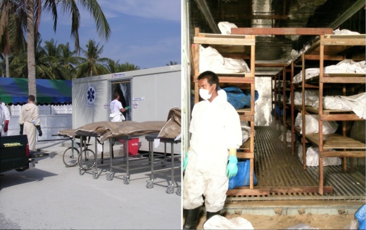
The temporary mortuary, where the PM information will be collected, needs to be constructed for body storage and examination, and established on premises affording the best possible facilities in the given circumstances (Fig. 1). (8) In the mortuary, the body will be thoroughly examined by a multidisciplinary team of specialists (fingerprint experts, policemen, pathologists, odontologists and DNA experts), who will register their findings on the pink PM Interpol forms.
Fig.1.
Temporary mortuary in Thailand after the tsunami in 2004
Each body to be moved into the examination room for physical description should be placed under custody of a PM records officer, who follows the body through all the examination stages until it is returned for storage. The PM records officer should be in possession of all PM DVI forms for each body.
The first step is finger/palm print analysis by specialists from forensic police labs. The recovered fingerprints will be entered into the AFIS system for comparison with existing AM data. In the second phase, bodies will be photographed, followed by an extensive external description of the body, clothing and personal belongings. All these items are photographed preferably in colour after being cleaned and labelled, with the reference body number clearly visible. Personal effects such as documents, jewellery, watches, clothing and pocket contents may constitute valuable circumstantial evidence of identity, but never proof. They must be combined with other evidence to conclude to a positive identification.
During the next step, the pathologist starts the external and internal examination and description of the body. It should be standard practice to perform full autopsies on all disaster victims not only for identification and cause of death purposes, but also to assist in preventing or minimizing the effects of similar incidents in the future.
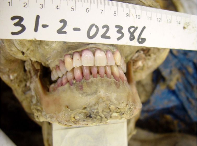
Dental examination in this phase is carried out by forensic odontologists. All dental related details will be registered on the PM F1/F2 Interpol forms. As a general rule, jaws should not be removed by dental experts unless a more specific examination is mandatory. To create more access to the dentition, a non-destructive method of mandibular dissecting technique is recommended. (9) This method allows an easy access to both maxilla and mandible and still enables a complete repositioning of the facial tissue after autopsy, so that the body can still be shown to relatives if required. All dental characteristics should be recorded by colour photography and radiography (Fig. 2). Dental age estimation is a major component of the identification process. Post- mortem dental age estimation allows forensic odontologists to focus on the search for a matching ante-mortem file on a specific age range among the possible candidates for identification from the missing persons list. Dental age estimation may be performed in different ways using morphological or radiological parameters that are all age-related. Obvious examples are tooth development in children and adolescents or morphological changes in adults (enamel wear, cementum incremental lines, root translucency or secondary dentine formation), which play an essential role in dental age assessments. In the context of identification, the most appropriate fitting age estimation method in relation to the presented evidence should be chosen out of all existing methods. This is an advantage compared with the aging of living individuals where for instance the methods of age estimation performed after tooth extraction have to be excluded. In essence, during identification, the dental age estimation protocols are divided in two groups based on the availability or absence of developing teeth. Therefore, radiological dental investigations and preferably full body CT’s are essential at the start of the identification process. They allow, if developing teeth are detected, to apply immediately the most appropriate age estimation method, (10-18) providing an instant age result. If all available teeth are fully developed, 2D radiographs can be used to apply the Kvaal technique (19) on the related monoradicular teeth. On 3D reconstructions of cone-beam computed tomography (CBCT) images, volumetric pulp tooth ratios can be calculated and implemented in an according age estimation technique. (20) After tooth development has been completed, methods on extracted and sectioned teeth will have to be considered. In this context, measurements of morphological changes related to age, need to be examined. The length of the apical translucency of the tooth roots (21) provides immediate age information on both intact and cut teeth. If possible the described methods on mature teeth are combined with methods taking into consideration attrition, periodontal attachment, cementum annulations, root resorption, and secondary dentine apposition. (22, 23)
Fig. 2.
Photographs of dental structures together with body number must be taken during the post mortem examination.
Genetic identification techniques provide a powerful tool in the identification of disaster victims. DNA analysis techniques currently in use complement other methods commonly used in disaster victim identification, especially when a body has been severely mutilated. As dental pulp material is a good source for DNA analysis, two vital teeth (canines/premolars) can be extracted and sent to the forensic DNA laboratories. (24)
Identification Centre
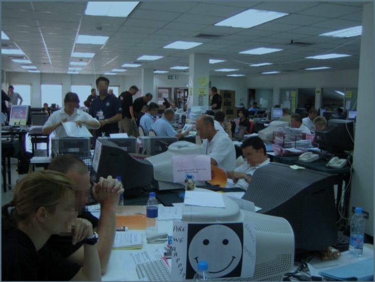
The Identification Centre handles and compares AM and PM documents forwarded from the AM and PM Units. In the different sections of the identification bureau - missing persons; ante-mortem; post-mortem; fingerprint; dental; DNA analysis and reconciliation - the quality controls and the transcription of the AM and PM documents take place. Results obtained from the specialised sections are fed back to the Identification Files Section to be combined into one master list of results (Fig.3).
Fig. 3.
The identification team, working during Tsunami identification process, putting all AM and PM data into the Plass Data® software. The system matches two datasets (AM and PM) and the matches will be discussed again in the reconciliation board.
The identification software runs an automatic comparison and matching between the ante and post mortem data but the final judgment must be made by professional experts and be based on personal evaluation of the evidence. (2) The matches will then in the next stage be verified by the different experts on the Reconciliation Board. The responsibility of this Identification Board is to check the results of comparison made by the various specialised sections. It is also responsible for scrutinising and eventual reconciliation of possible inconsistencies and will combine the results into one final list of identifications.
Other aspects assisting in The identification process
During the recent years, many disasters occurred at different intensities. DVI teams must respond systematically, using all facilities and new technologies available in the process of victim identification. The fast growing technology and ever more powerful equipment are mostly in respect of the field of imaging processes.
Dental radiology
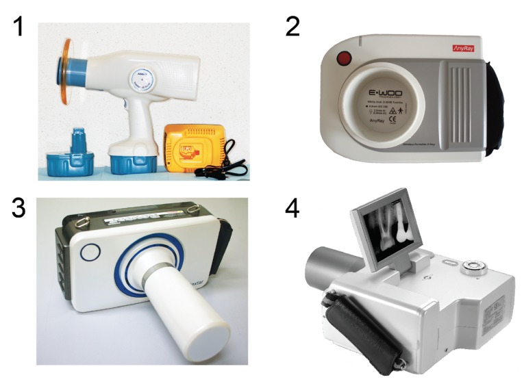
Many factors may affect PM-radiographic image collection: the presence of suitable dental X-ray equipment but also the body condition such as rigor mortis, positioning the victim’s bodies and aiming the x-ray beam, electricity supply, working areas and equipment. Therefore, during mass disasters the recovery team needs to transport the remains to compartments - equipped with fixed dental x-ray unit(s) - suitable for performing dental autopsies. Even when electric power is supplied, the fixed x-ray devices can be damaged by constant line fluctuation as was reported during the Asian tsunami disaster. (25, 26) Recently developed portable and handheld digital dental x-ray units can solve these practical forensic problems as shown during the Tsunami crisis in 2005 when Nomad® was introduced for the first time in mass disasters. These light weight and autonomic working devices can easily be brought next to the bodies, allowing an immediate forensic odontologic investigation in combination with digital imaging and management systems, enabling potentially an immediate AM-PM matching (Fig. 4). (27, 28)
Fig. 4.
Several portable dental x-ray devices are available on the market nowadays. (1) Nomad® (Aribex, Utah, USA) was first introduced in 2005 in the identification process after Tsunami disaster. (2) AnyRay® (VATECH Co., Ltd., Gyeonggi-do, Republic of Korea), (3) Rextar® (Sungwon Econet, Seoul, Republic of Korea) (4) ADX4000 (DEXCOWIN Co., Ltd. Seoul, Republic of Korea)
The dosimetry studies performed with the Nomad® portable X-ray machine, which is provided with circular lead-filled shields attached to the end of the exit tube, showed that exposure of the operator to leaking or backscatter radiation is below the maximum permissible for occupational dose. (29, 30) A study reporting dose measurements carried out with Nomad® in normal conditions (i.e. patient seated in dental chair and operator in the “safe zone” provided by the circular lead-shield) as well as in “atypical” situations (i.e. forensic sites, field work, sedated patients) has demonstrated that the whole body exposure was equivalent to less than 1% of the occupational dose limit. (30) Another report (28) shows that exposure at the operator’s hand was lowest when a protection shield was used or with the use of an exposure switch cable (distance > 1m).
In addition, new technologies provide new accessories for X-ray viewing in the field of disaster. Some companies have launched a portable X-ray machine with a viewing display attached to the machine itself. Others provide a small portable X-ray viewing gadget. When operated in a wireless environment, data and images could thus be easily transmitted in both directions. This may help to facilitate the radiographic interpretation more rapidly than in the past.
Facial reconstruction
Today, thanks to the multidisciplinary approach towards an unidentified body, a tremendous amount of information concerning the victim can be obtained. Unfortunately even the biggest and most detailed post-mortem (PM) files are useless when no match with any AM file can be made. When confronted with a corpse that is unrecognisable due to its state of decomposition, skeletisation, mutilation or calcination, a cranio-facial reconstruction (CFR) should be considered. The goal of CFR is to recreate a likeness with the face of missing individuals immediately prior to their death. Presenting this reconstructed face to the public can get the identification-process out of the impasse by triggering recognition. (31-37)
Several 3D manual methods for CFR are currently being used. One of these reconstruction methods consists of physically modelling a face on a skull replica (the target skull) with clay or plasticine; however, this method requires a high degree of anatomical and sculptural expertise and, as a result, remains difficult and subjective. The progress in computer science and the improvement of medical imaging technologies during recent years have lead to the development of alternative computer-based CFR methods. A computer, compared to a human expert, is consistent and objective. Knowing all the modelling assumptions and given the same input data, a computer always generates the same output data. Furthermore, certain procedures can be automated so that the creation of multiple reconstructions from the same skull using different modelling assumptions (age, BMI, ancestry, gender, etc) becomes possible. As a result, the CFR process becomes accessible to a wide range of people without the need for extensive expertise. (38)
Virtual autopsy
The role of 3D imaging in the forensic field is growing quickly, not only in cranio-facial reconstruction (CFR) but also in the whole autopsy process.
Some institutions have already implemented CT in post-mortem forensic investigations, such as at the Armed Forces Medical Examiners autopsy room (Armed Forces Institute of Pathology, Washington, D.C., and Dover, Del., USA), where CT scans on military personnel killed in combat are used on a routine basis. (39)
During the Victorian Bushfire in 2009, CT scanning also proved to be very useful in the victim identification process. (40)
In Switzerland, the Virtopsy project implements a variety of imaging methods: 3D photogrammetry-based optical surface scanning, MSCT (Multi-slice CT) and MRI (Magnetic Resonance Imaging). Virtopsy is a non-invasive or minimally invasive approach that has several advantages to current forensic examination techniques, as it can help to provide precise, objective and clear documentation of forensic findings for testimony in court. This technique also helps improve quality assurance through digital data archiving and transfer. Because of its minimal invasiveness to the body, it can also improve judicature in cultures with low autopsy acceptance. (41)
In some cases, physical examination should still be performed in addition to the virtopsy to provide more physical and external information of the victims.
PAST AND FUTURE DVI
World globalisation has resulted in mass disasters these days often involving victims from many different nationalities, requiring the assistance of DVI teams from various countries often with different levels of expertise. (5, 42)
After the Tsunami in 2004, all the DVI protocols were re-evaluated by an Interpol working group. (43-45) This Tsunami Evaluation report is a summary of what the DVI teams experienced during the 2004-2005 incident. In the current Interpol DVI guidelines, the methods of identification process are categorized into 2 groups: primary and secondary identification methods. (3, 42, 46) Primary identification means that the method by itself can lead to a 100% scientific identification which is able to withstand global legal scrutiny. Forensic odontology, as one of the primary identification methods (DNA, forensic odontology, finger printing), has proved to be an effective identification method especially in large scale disasters with the overall ID rate of 83.3% in South-East Asia Tsunami. (25)
The Forensic Odontology Working Group of the Interpol DVI Standing Committee has been working to develop new guidelines and adapted forms (F1/F2). The content of the modified AM and PM forms will be simplified. Unnecessary parts will be removed. All captured data will be directly linked to the Plass Data® system. (2)
In terms of AM records, as internet access is growing rapidly, the AM records should be made available online when disaster occurs. This will help speed up the identification process and minimize possible risk of losing AM evidence during transportation. The AM team, the insurance companies and personnel who are dealing with the victims’ data should follow the legal obligation of the medical confidentiality. (47, 48)
At this stage, Interpol DVI dental forms are being updated. Our suggestion is that as 3D data and virtopsy are more accessible, the DVI forms should be adapted to handle these 3D evidences. Civilians have better access to medical care. Images from multi-slice computed tomography (MSCT) and cone-beam computed tomography (CBCT) will become more widely available. It is possible that one of the potential victims of a future disaster may have these types of AM data, yet the old forms are not supporting this new type of evidence and thus, there is a need to revise the forms, considering new diagnostic material in more dimensions. More research should be performed on how to apply 3D information in the identification process more efficiently. If the PM records contain a 3D scan, the challenge will be to find a suitable match to the 2D AM datasets. To enable this, there is a need for new matching algorithms.
It is not only of crucial importance that rules and SOP’s as written out in the Interpol DVI Guidelines are followed and applied by all DVI team members, but also that all specialists involved in DVI are suitably trained and qualified and will be deployed in appropriate roles. Different levels of experience with Interpol DVI guidelines, documents and standards, made clear the need for standardization.
Internationally agreed upon common minimum standards of training for the personnel of the different specialist sections would be beneficial to the international community. This of course requires specialists accredited to provide these training programs and also the creation of a framework for quality control within each speciality in the overall DVI process.
Overall conclusions
Disaster victim identification (DVI) is a demanding task that can only be brought to a successful conclusion if properly planned, by selecting the appropriate forensic diagnostic tools and involving a team of well-trained key experts. Lessons learned since the Tsunami disaster in 2004 have changed the Interpol DVI vision and standards tremendously. The DVI community is moving forward and continuously improving the guidelines and protocols.
Forensic odontology as one of the primary identification methods is a dynamic field and has developed tremendously since the Tsunami in 2004. Recent developments in computer-aided 3D imaging have been applied for forensic odontology, forensic radiology, forensic craniofacial reconstruction and virtual autopsy. New research challenges include developing forensic diagnostic tools, with maximal use of remnants and information, increased efficacy for various forensic applications and optimized protocols for DVI operations.
ACKNOWLEDGEMENTS
The authors wish to thank Dr. Nop Porntrakulseree from Unit of Oral and Maxillofacial Surgery, Department of Dental, Lamphun Hospital, Lamphun.
Footnotes
The authors declare that they have no conflict of interest.
References
- 1.Pretty IA, Webb DA, Sweet D. Dental participants in mass disasters—A retrospective study with future implications. J Forensic Sci. 2002;47:117–20. 10.1520/JFS15210J [DOI] [PubMed] [Google Scholar]
- 2.Andersen Torpet L. DVI System International: software assisting in the Thai tsunami victim identification process. J Forensic Odontostomatol. 2005;23:19–25. [PubMed] [Google Scholar]
- 3.Disaster Victim Identification Guide [Internet]. [updated 2009; cited 2012 Jan 17]; Available from: http://www.interpol.int/INTERPOL-expertise/Forensics/DVI-Pages/DVI-guide.
- 4.De Valck E. De tandarts als lid van het DVI team: De interdisciplinaire DVI-filosofie. Belg Tijdschr voor tandheelkunde 2005;3:171-188.
- 5.James H. Thai tsunami victim identification – overview to date. J Forensic Odontostomatol. 2005;23:1–18. [PubMed] [Google Scholar]
- 6.Soomer H, Lincoln MJ, Ranta H. Dentists’ qualifications affect the accuracy of radiographic identification. J Forensic Sci. 2003;48:1121–6. 10.1520/JFS2003142 [DOI] [PubMed] [Google Scholar]
- 7.De Valck E. Major incident response: Collecting ante-mortem data. Forensic Sci Int. 2006;159:S15–9. 10.1016/j.forsciint.2006.02.004 [DOI] [PubMed] [Google Scholar]
- 8.Leditschke J, Collett S, Ellen R. Mortuary operations in the aftermath of the 2009 Victorian bushfires. Forensic Sci Int. 2011;205:8–14. 10.1016/j.forsciint.2010.11.002 [DOI] [PubMed] [Google Scholar]
- 9.Beauthier JP, De Valck E, Lefevre P, De Winne J. Mass Disaster Victim Identification: The Tsunami Experience. Open Forensic Sci J. 2009;2:54–62. 10.2174/1874402800902010054 [DOI] [Google Scholar]
- 10.Demirjian A, Goldstein H, Tanner JM. A new system of dental age assessment. Hum Biol. 1973;45:211–27. [PubMed] [Google Scholar]
- 11.Demirjian A, Goldstein H. New systems for dental maturity based on seven and four teeth. Ann Hum Biol. 1976;3:411–21. 10.1080/03014467600001671 [DOI] [PubMed] [Google Scholar]
- 12.Demirjian A, Buschang PH, Tanguay R, Patterson DK. Interrelationships among measures of somatic, skeletal, dental, and sexual maturity. Am J Orthod. 1985;88:433–8. 10.1016/0002-9416(85)90070-3 [DOI] [PubMed] [Google Scholar]
- 13.Willems G, Van Olmen A, Spiessens B, Carels C. Dental age estimation in Belgian children: Demirjian’s technique revisited. J Forensic Sci. 2001;46:893–5. 10.1520/JFS15064J [DOI] [PubMed] [Google Scholar]
- 14.Willems G, Thevissen PW, Belmans A, Liversidge HM, Willems II. Non-gender-specific dental maturity scores. Forensic Sci Int. 2010;201:84–5. 10.1016/j.forsciint.2010.04.033 [DOI] [PubMed] [Google Scholar]
- 15.Liversidge HM, Chaillet N, Mörnstad H, Nyström M, Rowlings K, Taylor J, et al. Timing of Demirjian’s tooth formation stages. Ann Hum Biol. 2006;33:454–70. 10.1080/03014460600802387 [DOI] [PubMed] [Google Scholar]
- 16.Thevissen PW, Alqerban A, Asaumi J, Kahveci F, Kaur J, Kim YK, et al. Human dental age estimation using third molar developmental stages: Accuracy of age predictions not using country specific information. Forensic Sci Int. 2010;201:106–11. 10.1016/j.forsciint.2010.04.040 [DOI] [PubMed] [Google Scholar]
- 17.Thevissen PW, Fieuws S, Willems G. Human dental age estimation using third molar developmental stages: does a Bayesian approach outperform regression models to discriminate between juveniles and adults? Int J Legal Med. 2010;124:35–42. 10.1007/s00414-009-0329-8 [DOI] [PubMed] [Google Scholar]
- 18.Thevissen PW, Kaur J, Willems G. Human age estimation combining third molar and skeletal development. Int J Legal Med. 2012;•••: [cited 17 January 2012] 10.1007/s00414-011-0639-5 [DOI] [PubMed] [Google Scholar]
- 19.Kvaal SI, Kolltveit KM, Thomsen IO, Solheim T. Age estimation of adults from dental radiographs. Forensic Sci Int. 1995;74:175–85. 10.1016/0379-0738(95)01760-G [DOI] [PubMed] [Google Scholar]
- 20.Star H, Thevissen P, Jacobs R, Fieuws S, Solheim T, Willems G. Human dental age estimation by calculation of pulp-tooth volume ratios yielded on clinically acquired cone beam computed tomography images of monoradicular teeth. J Forensic Sci. 2011;56 Suppl 1:S77–82. 10.1111/j.1556-4029.2010.01633.x [DOI] [PubMed] [Google Scholar]
- 21.Bang G, Ramm E. Determination of age in humans from root dentin transparency. Acta Odontol Scand. 1970;28:3–35. 10.3109/00016357009033130 [DOI] [PubMed] [Google Scholar]
- 22.Solheim T. A new method for dental age estimation in adults. Forensic Sci Int. 1993;59:137–47. 10.1016/0379-0738(93)90152-Z [DOI] [PubMed] [Google Scholar]
- 23.Kvaal S, Solheim T. A non-destructive dental method for age estimation. J Forensic Odontostomatol. 1994;12:6–11. [PubMed] [Google Scholar]
- 24.Hartman D, Drummer O, Eckhoff C, Scheffer JW, Stringer P. The contribution of DNA to the disaster victim identification (DVI) effort. Forensic Sci Int. 2011;205:52–8. 10.1016/j.forsciint.2010.09.024 [DOI] [PubMed] [Google Scholar]
- 25.Schuller-Götzburg P, Suchanek J. Forensic odontologists successfully identify tsunami victims in Phuket, Thailand. Forensic Sci Int. 2007;171:204–7. 10.1016/j.forsciint.2006.08.013 [DOI] [PubMed] [Google Scholar]
- 26.Dawidson I. The dental identification of the Swedish Tsunami victims in Thailand. Forensic Sci Int. 2007;169S:S47–8. 10.1016/j.forsciint.2007.04.105 [DOI] [Google Scholar]
- 27.Pittayapat P, Thevissen P, Fieuws S, Jacobs R, Willems G. Forensic oral imaging quality of hand-held dental X-ray devices: comparison of two image receptors and two devices. Forensic Sci Int. 2010;194:20–7. 10.1016/j.forsciint.2009.09.024 [DOI] [PubMed] [Google Scholar]
- 28.Pittayapat P, Oliveira-Santos C, Thevissen P, Michielsen K, Bergans N, Willems G, et al. Image quality assessment and medical physics evaluation of different portable dental X-ray units. Forensic Sci Int. 2010;201:112–7. 10.1016/j.forsciint.2010.04.041 [DOI] [PubMed] [Google Scholar]
- 29.Goren AD, Bonvento M, Biernacki J, Colosi DC. Radiation exposure with the NOMAD portable X-ray system. Dentomaxillofac Radiol. 2008;37:109–12. 10.1259/dmfr/33303181 [DOI] [PubMed] [Google Scholar]
- 30.Danforth RA, Herschaft EE, Leonowich JA. Operator exposure to scatter radiation from a portable hand-held radiation emitting device (Aribex NOMAD) while making 915 intraoral dental radiographs. J Forensic Sci. 2009;54:415–21. 10.1111/j.1556-4029.2008.00960.x [DOI] [PubMed] [Google Scholar]
- 31.Stephan CN. Beyond the sphere of the English facial approximation literature: ramifications of German papers on western method concepts. J Forensic Sci. 2006;51:736–9. 10.1111/j.1556-4029.2006.00175.x [DOI] [PubMed] [Google Scholar]
- 32.Gerasimov M. The Face Finder. Philadelphia, JB Lippencott Co.; 1971. [Google Scholar]
- 33.Lebedinskaya G, Balueva T, Veselovskaya E. Development of methodological principles for reconstruction of the face on the basis of skull material. In: Iscan MY, and Helmer RP. (ed.). Forensic Analysis of the Skull, New York, Wiley-Liss Inc.; 1993. [Google Scholar]
- 34.Snow CC, Gatliff B, Williams KM. Reconstruction of the facial features from skull: an evaluation of its usefulness in forensic anthropology. Am J Phys Anthropol. 1970;33:221–8. 10.1002/ajpa.1330330207 [DOI] [PubMed] [Google Scholar]
- 35.Prag J, Neave R. Making Faces Using Forensic and Archeological Evidence. London, British Museum Press; 1997. [Google Scholar]
- 36.Wilkinson C. Forensic Facial Reconstruction. Cambridge, Cambridge University Press; 2004. [Google Scholar]
- 37.Wilkinson C. Facial reconstruction--anatomical art or artistic anatomy? J Anat. 2010;216:235–50. 10.1111/j.1469-7580.2009.01182.x [DOI] [PMC free article] [PubMed] [Google Scholar]
- 38.Claes P, Vandermeulen D, De Greef S, Willems G, Clement JG, Suetens P. Computerized craniofacial reconstruction: Conceptual framework and review. Forensic Sci Int. 2010b;201:138–45. 10.1016/j.forsciint.2010.03.008 [DOI] [PubMed] [Google Scholar]
- 39.Levy AD, Abbott RM, Mallak CT, Getz JM, Harcke HT, Champion HR, et al. Virtual autopsy: preliminary experience in high-velocity gunshot wound victims. Radiology. 2006;240:522–8. 10.1148/radiol.2402050972 [DOI] [PubMed] [Google Scholar]
- 40.Hinchliffe J. Forensic odontology, part 3. The Australian bushfires – Victoria state, February 2009. Br Dent J. 2011;210:317–21. 10.1038/sj.bdj.2011.239 [DOI] [PubMed] [Google Scholar]
- 41.Bolliger SA, Thali MJ, Ross S, Buck U, Naether S, Vock P. Virtual autopsy using imaging: bridging radiologic and forensic sciences. A review of the Virtopsy and similar projects. Eur Radiol. 2008;18:273–82. 10.1007/s00330-007-0737-4 [DOI] [PubMed] [Google Scholar]
- 42.Sweet D. INTERPOL DVI best-practice standards--An overview. Forensic Sci Int. 2010;201:18–21. 10.1016/j.forsciint.2010.02.031 [DOI] [PubMed] [Google Scholar]
- 43.INTERPOL Tsunami Evaluation Working Group. The DVI response to the South East Asian Tsunami between December 2004 and February 2006. 2010. Available from: https://www.interpol.int/Public/DisasterVictim/TsunamiEvaluation20100330.pdf [cited 4 April 2012].
- 44.Hill AJ, Hewson I, Lain R. The role of the forensic odontologist in disaster victim identification: Lessons for management. Forensic Sci Int. 2011;205:44–7. 10.1016/j.forsciint.2010.08.013 [DOI] [PubMed] [Google Scholar]
- 45.Hinchliffe J. Forensic odontology, part 2. Major disasters. Br Dent J. 2011;210:269–74. 10.1038/sj.bdj.2011.199 [DOI] [PubMed] [Google Scholar]
- 46.Lessig R, Rothschild M. International standards in cases of mass disaster victim identification (DVI). Forensic Sci Med Pathol. 2012;•••: [cited 4 Arpil 2012] 10.1007/s12024-011-9272-3 [DOI] [PubMed] [Google Scholar]
- 47.Disaster Victim Identification (DVI). Beroepsgeheim (Dutch). Orde van geneesheren April 30 2011: 133. Available from: http://www.ordomedic.be/nl/adviezen/advies/disaster-victim-identification-team-(dvi)-beroepsgeheim2 [cited 4 Arpil 2012].
- 48.European Patients’ Forum. Patients’ Right in the European Union. 2009. Available from: http://www.eu-patient.eu/Documents/Projects/Valueplus/Patients_Rights.pdf [cited 9 April 2012].


