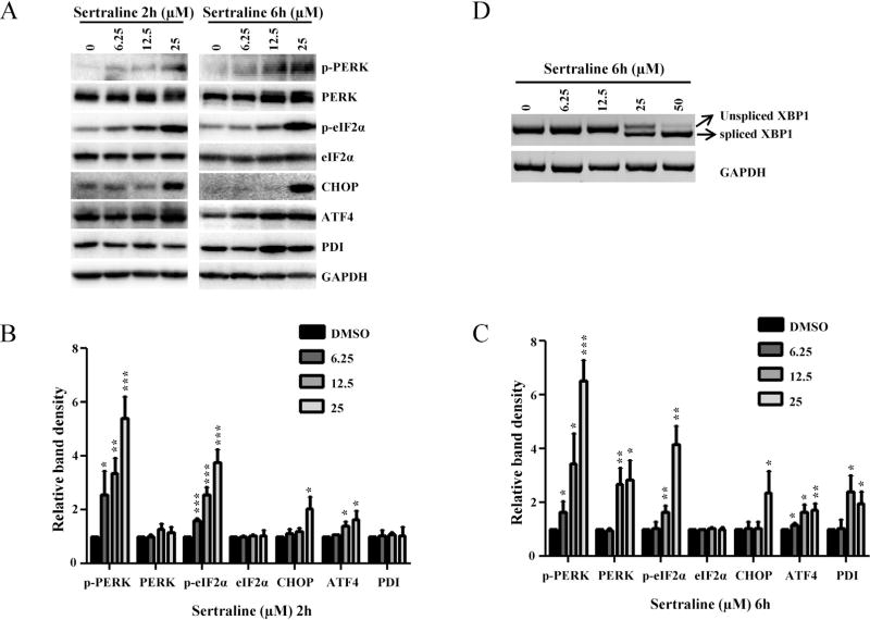
Fig. 3.
Effects of sertraline in HepG2 cells on the expression of proteins involved in the ER stress response and on splicing of XBP mRNA. (A) Total cellular proteins were extracted at 2 and 6 h after sertraline treatment. The level of ER stress related proteins including phospho-PERK, PERK, phospho-eIF2α, eIF2α, CHOP, ATF-4, and PDI was determined by Western blotting. GAPDH was used as a loading control. Data are typical of three experiments. (B and C) Relative protein level to DMSO control by densitometric analysis. (D) HepG2 cells were treated with the indicated concentration of sertraline for 6 h. The total RNA was isolated and semi-quantitative RT-PCR analysis was performed to detect both spliced (263 bp) and unspliced (289 bp) XBP mRNA. GAPDH was also amplified and used as an internal control. A representative DNA gel from three independent experiments is shown.

