Abstract
Tarsal coalitions have an incidence of 2% and are often underdiagnosed. These are considered to be one of the causes of chronic ankle and foot pain. Among all tarsal coalitions, the talonavicular type represents a rare and uncommon condition. The purpose of this article was to present the case of a 35-year-old male patient with a bilateral talonavicular coalition treated conservatively. A review of the literature was also performed to understand the management of this rare condition.
Keywords: talonavicular, tarsal, coalition, foot, treatment
Introduction
Tarsal coalitions are defined as congenital fusions of two or more tarsal bones inducing chronic ankle and foot pain. These are classified on the basis of the morphology of the bridging as fibrous (syndesmosis), chondral (synchondrosis), or bony (synostosis) bridge. 1 The incidence in the general population is approximately 2%. 2 The clinical presentation may vary from an asymptomatic form to a chronic foot and ankle pain, often leading to a complex differential diagnosis.
Among several anatomic variants, the talonavicular (TN) coalition is an infrequent hindfoot syndrome. 2 3 It represents 1% of all tarsal coalitions and considered to be linked to genetic mutations (autosomal recessive disease). 4 5 TN coalition is frequently bilateral and usually associated with several orthopaedic anomalies, such as clinodactyly, symphalangism, great toe being shorter than the second, and ball-and-socket ankle. 4 6 In recent studies, the genetic anomalies have been described as mutations of the noggin (NOG) gene. 7
Although the condition is usually asymptomatic, a small proportion of patients may have a painful bony prominence, particularly during sports or working activities. 4 8 9
The present report describes a case of a patient with a bilateral TN coalition managed by conservative treatment.
Case Report
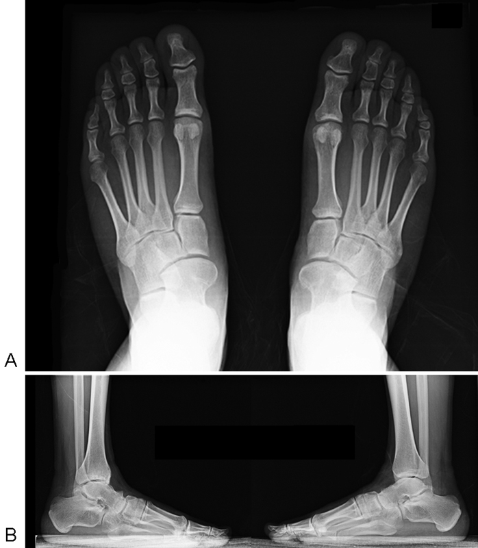
A 35-year-old patient arrived at our Department of Foot and Ankle Surgery complaining of bilateral foot and ankle pain without any history of trauma. Pain was referred from 3 years only during sports activities or after a significant effort. The patient managed such symptoms by oral analgesics. The physical examination revealed the loss of the medial longitudinal arch of both feet (flatfoot deformity), with an elective pain in the area of the navicular bone during inversion/eversion movements. He complained of pain over the calcaneocuboid joint. No swelling was referred or present at the examination, as no range of motion (ROM) limitations were assessed. A mild pain on the first metatarsophalangeal joint associated with signs of mild hallux limitus was detected, but both great toes showed a full ROM. No associated alterations, congenital disorders, or neurological impairments were recorded. Standard X-rays revealed an uncommon TN coalition, an increased talo first metatarsal angle > 5 degrees, and a metatarsus primus elevatus with clear signs of osteoarthritis of the talus, particularly in the right foot ( Fig. 1 ). No family history resulted for tarsal coalitions.
Fig. 1.

Weight-bearing X-rays of both feet in dorsoplantar ( A ) and lateral ( B ) view, showing a bilateral talonavicular coalition.
The patient, after receiving complete information on the different treatment options and prognosis of his tarsal coalition, agreed to be treated by conservative measures, such as paracetamol 1 g and ibuprofen 600 mg in case of pain, physical therapy (eccentric exercises of the calf and laser therapy) and functional foot orthoses with medial arch supports for a 12-month period. Surgical treatment was considered as the further strategy in case of failure. No genetic analysis of the NOG gene was performed because it was an isolated case. The conservative treatment was well tolerated.
Discussion
TN coalition is reported to be less common than talocalcaneal or calcaneonavicular type. Calcaneonavicular and talocalcaneal coalitions are more symptomatic than TN. These usually are incidentally discovered on plain X-rays after a minor trauma. 9 Diagnosis is made at variable age; previous publications have reported cases of 20- as well as 50-year-old patients. 2 6 8
As for other deformities, its etiology is probably a failure of differentiation and segmentation of the primitive mesenchymal tissue. 6 Moreover, the majority of such congenital alterations are reported as bilateral 4 8 9 and associated with other deformities, such as symphalangism, multiple synostosis syndrome, tarsal–carpal coalition syndrome, brachydactyly type, and stapes ankylosis with broad thumb and toes. 7
As revealed by gait analysis studies, the abnormal union of tarsal bones may lead to excessive strain on the other joints that are characterized by overuse stresses to compensate the loss of ROM due to coalition. 10 11 12 A TN coalition may have an almost complete restriction of inversion–eversion movement, thereby increasing the overload on the subtalar joint. Also, the first metatarsophalangeal joint may suffer for such increase of stress resulting in hyperkeratosis and secondary hyperpronation of the foot. 8
In the present case, there was no family history indicating a probable autosomal recessive nature of the coalition. All degenerative changes mentioned before have been found in the described case, particularly for the right foot. Furthermore, the mechanical overload of the calcaneocuboid joint referred by the patient could be observed in the right foot of the patient.
Treatment options for tarsal coalitions may vary from conservative to surgical procedures. Conservative therapy is necessarily considered first line, while surgery is performed in the case of failure. 13 14 15 In such cases, both osteotomy and joint fusion have been considered useful strategies. 11 12 13
Different types of surgeries were described in talocalcaneal coalitions with long-term results, but no such findings were reported in the literature on TN coalitions. 16 17 Migues et al 8 performed, in a symptomatic TN bilateral coalition, a calcaneocuboid joint distraction arthrodesis to relieve pain and improve alignment of both feet and a proximal plantar flexion first metatarsal osteotomy to induce pain relief of the metatarsophalangeal joint. 8 Ellington et al, 11 in patients with ball and socket ankle joint associated with a talonavicular tarsal coalition, compared the supramalleolar osteotomy with the tibiotalocalcaneal arthrodesis.
Surgery has demonstrated good short-term results but long-term follow-up on TN coalitions is not available. 6 13 On considering current life expectancy of the general population and undisclosed long-term results of the surgical techniques, it is legitimate to consider conservative treatment as the first option. Moreover, patient's age and the moderate symptoms referred led us to propose a conservative management as the first choice. The patient was informed that in the case of recurrence of symptoms, a surgical solution—arthrodesis or osteotomy—may be considered in the future.
Conclusion
Tarsal coalition is a rare condition that should be taken into consideration by unexperienced foot and ankle surgeons as a cause of bilateral chronic foot pain and midfoot osteoarthritis. The conservative treatment appears to be the gold standard, given the variable outcomes of surgery in the literature.
References
- 1.Lawrence D A, Rolen M F, Haims A H, Zayour Z, Moukaddam H A. Tarsal coalitions: radiographic, CT, and MR imaging findings. HSS J. 2014;10(02):153–166. doi: 10.1007/s11420-013-9379-z. [DOI] [PMC free article] [PubMed] [Google Scholar]
- 2.Shtofmakher G, Rozenstrauch A, Cohen R. An incidental talonavicular coalition in a diabetic patient: a podiatric perspective. BMJ Case Rep. 2014 doi: 10.1136/bcr-2014-204510. [DOI] [PMC free article] [PubMed] [Google Scholar]
- 3.Bonk J H, Tozzi M A. Congenital talonavicular synostosis. A review of the literature and a case report. J Am Podiatr Med Assoc. 1989;79(04):186–189. doi: 10.7547/87507315-79-4-186. [DOI] [PubMed] [Google Scholar]
- 4.David D R, Clark N E, Bier J A. Congenital talonavicular coalition. Review of the literature, case report, and orthotic management. J Am Podiatr Med Assoc. 1998;88(05):223–227. doi: 10.7547/87507315-88-5-223. [DOI] [PubMed] [Google Scholar]
- 5.Vincent K A. Tarsal coalition and painful flatfoot. J Am Acad Orthop Surg. 1998;6(05):274–281. doi: 10.5435/00124635-199809000-00002. [DOI] [PubMed] [Google Scholar]
- 6.Kembhavi R S, James B. A rare combination of ipsilateral partial talocalcaneal and talonavicular coalition. J Clin Diagn Res. 2015;9(12):RD07–RD08. doi: 10.7860/JCDR/2015/17426.6997. [DOI] [PMC free article] [PubMed] [Google Scholar]
- 7.Takano K, Ogasawara N, Matsunaga T et al. A novel nonsense mutation in the NOG gene causes familial NOG-related symphalangism spectrum disorder. Hum Genome Var. 2016;3:16023. doi: 10.1038/hgv.2016.23. [DOI] [PMC free article] [PubMed] [Google Scholar]
- 8.Migues A, Slullitel G A, Suárez E, Galán H L. Case reports: symptomatic bilateral talonavicular coalition. Clin Orthop Relat Res. 2009;467(01):288–292. doi: 10.1007/s11999-008-0500-4. [DOI] [PMC free article] [PubMed] [Google Scholar]
- 9.Bryson D, Uzoigwe C E, Bhagat S B, Menon D K. Complete bony coalition of the talus and navicular: decades of discomfort. BMJ Case Rep. 2011;2011 doi: 10.1136/bcr.03.2011.4031. [DOI] [PMC free article] [PubMed] [Google Scholar]
- 10.Pontious J, Hillstrom H J, Monahan T, Connelly S. Talonavicular coalition. Objective gait analysis. J Am Podiatr Med Assoc. 1993;83(07):379–385. doi: 10.7547/87507315-83-7-379. [DOI] [PubMed] [Google Scholar]
- 11.Ellington J K, Myerson M S. Surgical correction of the ball and socket ankle joint in the adult associated with a talonavicular tarsal coalition. Foot Ankle Int. 2013;34(10):1381–1388. doi: 10.1177/1071100713488762. [DOI] [PubMed] [Google Scholar]
- 12.Downey M S, Dewaters A M.Tarsal coalition New York: Lippincott Williams & Wilkins; 2012;126:598–635. [Google Scholar]
- 13.Doyle S M, Kumar S J. Symptomatic talonavicular coalition. J Pediatr Orthop. 1999;19(04):508–510. doi: 10.1097/00004694-199907000-00016. [DOI] [PubMed] [Google Scholar]
- 14.Ozyurek S, Guler F, Turan A, Kose O. Symptomatic talar beak in talocalcaneal coalition. BMJ Case Rep. 2013;2013.pii:bcr2013009309. doi: 10.1136/bcr-2013-009309. [DOI] [PMC free article] [PubMed] [Google Scholar]
- 15.Varner K E, Michelson J D. Tarsal coalition in adults. Foot Ankle Int. 2000;21(08):669–672. doi: 10.1177/107110070002100807. [DOI] [PubMed] [Google Scholar]
- 16.Swiontkowski M F, Scranton P E, Hansen S. Tarsal coalitions: long-term results of surgical treatment. J Pediatr Orthop. 1983;3(03):287–292. [PubMed] [Google Scholar]
- 17.Khoshbin A, Law P W, Caspi L, Wright J G. Long-term functional outcomes of resected tarsal coalitions. Foot Ankle Int. 2013;34(10):1370–1375. doi: 10.1177/1071100713489122. [DOI] [PubMed] [Google Scholar]


