
Figure 6. Immunohistochemical analysis of HLA-G protein expression in Reed-Sternberg cells in pediatric HL patients.
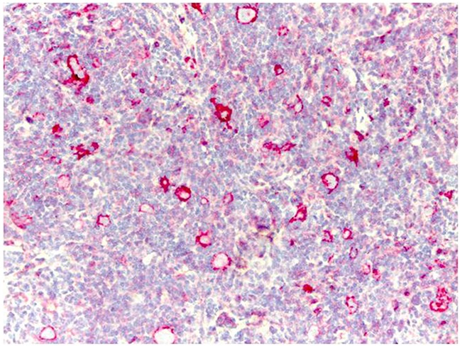
Representative image shows immunohistochemical staining for HLA-G in a HL patient carrying the wild type C/C +3027 genotype. Formalin/PFA-fixed paraffin-embedded sections were stained with primary anti-HLA-G antibody (4H84). Lymphoma cells show strong membrane staining for HLAG. Note: Only 5 out of 25 patients analyzed were positive for HLA-G in our analysis.