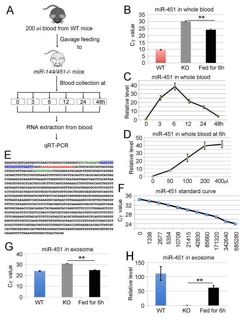
Figure 1. Ingestion of wild type blood increases the levels of miR-451 and miR-144 in peripheral blood of miR-144/451 null mice.
(A) Schematic view for the ingestion of WT blood into miR-144/451 KO mice. miR-144/451 KO mice were gavage fed with 200 μl fresh blood from WT animals. Blood was then drawn from the KO mice at different time points ranging from 0 to 48 hours. 200 μl of whole blood was used for RNA extraction and miR-451 levels in blood were assessed by two different PCR methods: TaqMan probe-based qRT-PCR assay and All-in-One miRNA detection kit. (B) Quantitative analysis of miR-451 levels in peripheral blood of miR-144/451 KO mice 6 hours after feeding WT blood. The Y-axis shows the CT value of miR-451. WT blood was used as positive control. (C) Quantitative analysis of miR-451 levels in peripheral blood of miR-144/451 KO mice after ingestion of WT blood. The Y-axis shows relative fold change of miR-451 with the miR-451 level at zero hour assigned as a relative value of 1. X-axis shows hours after feeding WT blood. Data is from three independent experiments. (D) qRT-PCR analysis of miR-451 expression 6 hours after gavage feeding different amounts of WT blood. The Y-axis shows relative levels of miR-451 in miR-144/451 KO blood. X-axis shows amount of WT blood fed to miR-144/451 KO mice. Data is from three independent experiments. (E) Sequence of PCR product. PCR products were gel-purified, cloned and sequenced. Note: the sequence highlighted with blue color is the sequence of mature miR-451 and the poly A highlighted with red color is the adapter sequence for reverse transcription. (F) Standard curve for miR-451. CT values were plotted against copy number. (G-H) Quantitative analysis of miR-451 levels in exosomes isolated from peripheral blood of miR-144/451 KO mice 6 hours after ingestion of WT blood. The Y-axis shows the CT value of miR-451 (G) and fold change of miR-451 with the miR-451 level at zero hour (KO) assigned as a relative value of 1 (H). WT blood was used as positive control. The loading control for all miRNA detection was small nuclear RNA U6.

