Abstract
Toric intraocular lenses (IOLs) are the procedure of choice to correct corneal astigmatism of 1 D or more in cases undergoing cataract surgery. Comprehensive literature search was performed in MEDLINE using “toric intraocular lenses,” “astigmatism,” and “cataract surgery” as keywords. The outcomes after toric IOL implantation are influenced by numerous factors, right from the preoperative case selection and investigations to accurate intraoperative alignment and postoperative care. Enhanced accuracy of keratometry estimation may be achieved by taking multiple measurements and employing at least two separate devices based on different principles. The importance of posterior corneal curvature is increasingly being recognized in various studies, and newer investigative modalities that account for both the anterior and posterior corneal power are becoming the standard of care. An ideal IOL power calculation formula should take into account the surgically induced astigmatism, the posterior corneal curvature as well as the effective lens position. Conventional manual marking has given way to image-guided systems and intraoperative aberrometry, which provide a mark-less IOL alignment and also aid in planning the incisions, capsulorhexis size, and optimal IOL centration. Postoperative toric IOL misalignment is the major factor responsible for suboptimal visual outcomes after toric IOL implantation. Realignment of the toric IOL is needed in 0.65%–3.3% cases, with more than 10° of rotation from the target axis. Newer toric IOLs have enhanced rotational stability and provide precise visual outcomes with minimal higher order aberrations.
Keywords: Astigmatism, cataract surgery, toric intraocular lenses
Toric intraocular lenses (IOLs) were first introduced in 1992 by Shimizu et al. as 3-piece nonfoldable polymethyl methacrylate implants to be inserted through a 5.7 mm incision.[1] Since then, the increased predictability and enhanced safety of toric IOL implantation has firmly established it as the procedure of choice to correct significant corneal astigmatism in cases undergoing cataract surgery.[2,3,4,5,6,7,8,9,10,11,12,13,14,15,16,17,18,19,20] A preoperative corneal astigmatism of 1 D or more may be present in up to one-third of the cases undergoing cataract surgery, with 22% having more than 1.5 D of astigmatism and 8% having more than 2.0 D of astigmatism.[9,21,22] In these cases, toric IOLs help to achieve postoperative spectacle independence and optimal patient satisfaction. Technological advancements in terms of IOL material as well as design have resulted in better rotational stability and precise visual outcomes.[2,7,8,9,11]
This review provides a comprehensive overview of toric IOLs along with the preoperative planning, various marking methods, intraoperative alignment, and postoperative management to achieve optimal outcomes. The literature search was performed in MEDLINE using “toric intraocular lenses,” “astigmatism,” and “cataract surgery” as keywords. The relevant references cited in those articles were also searched. Abstracts of relevant non-English articles were used. All articles were reviewed since the first use of toric IOLs in 1992. For statements that are frequently mentioned in the literature, we chose the earliest publication and other important articles.
Patient Selection
Ideal case selection is a prerequisite before surgery to ensure patient satisfaction as well as optimal outcomes. The decision to implant a toric IOL is governed by the magnitude and axis of corneal astigmatism, patient expectations, type of IOL, and the presence of other ocular comorbidities.
At present, standard toric IOLs are available in cylinder powers of 1.5 D to 6.0 D (1.03 D to 4.11 D at the corneal plane) and are intended to correct preexisting regular corneal astigmatism ranging from 0.75 D to 4.75 D.[23] Extended series and customized toric IOLs to correct higher cylinder powers are also available [Tables 1 and 2]. Toric IOLs are universally recommended in cases with significant preoperative corneal astigmatism of 1.5 D or more. Even in cases with low astigmatism with a magnitude of around 1 D, the superiority of toric IOLs over monofocal IOLs has been demonstrated in terms of better-uncorrected distance visual acuity (UDVA).[24]
Table 1.
Intraocular lens material, design and range of power of commercially available monofocal toric intraocular lens
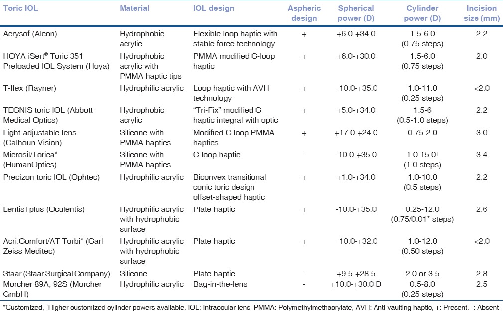
Table 2.
Intraocular lens material, design and range of power of commercially available multifocal toric intraocular lens
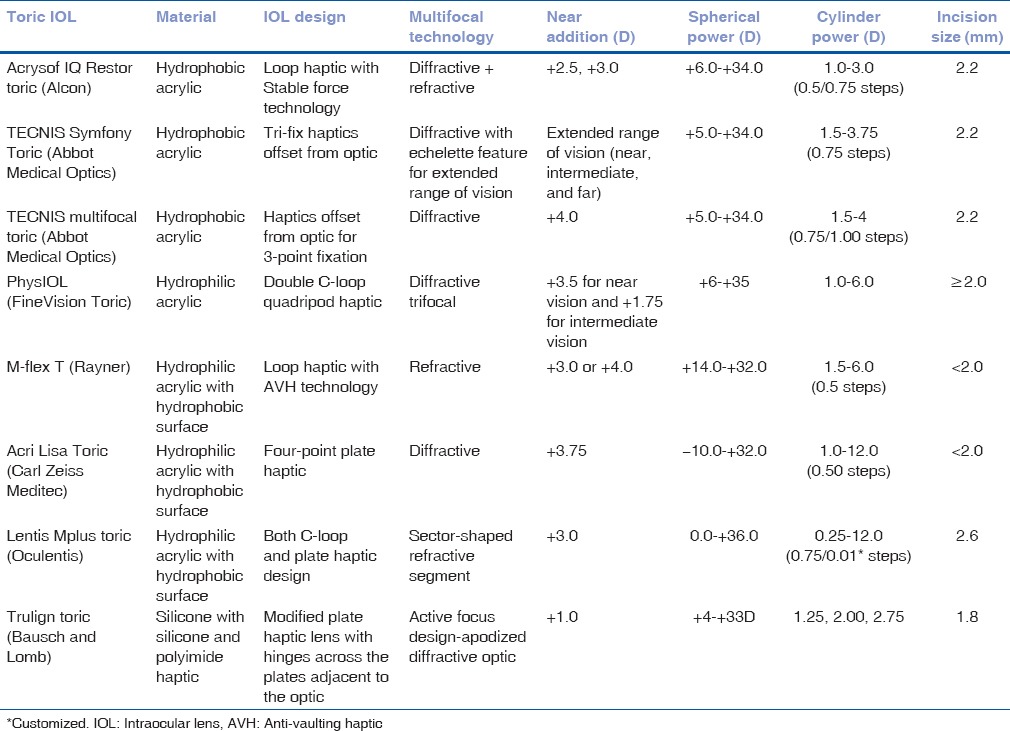
However, patients undergoing premium IOL implantation such as multifocal IOLs may not tolerate residual astigmatism of even <1 D and a toric multifocal IOL may be required in such cases.
A comprehensive ocular examination should be undertaken to rule out any ocular comorbidities that may interfere with the postoperative outcomes. Cases with irregular astigmatism resulting from corneal scars or ectatic disorders are not ideal candidates for toric IOL implantation. They are unlikely to achieve complete refractive correction with toric IOLs; however, the amount of astigmatism may be reduced with a decreased dependence on spectacles or contact lenses and such cases may be considered for surgery after adequate counseling.[25,26,27] Zonular instability and posterior capsular dehiscence are contraindications for implanting toric IOLs, as a stable capsular bag-IOL complex is essential for the rotational stability of the IOL. Poor pupillary dilatation is also a relative contraindication, as it may hamper the visualization of the alignment marks which are located in the periphery of the toric IOL. Patients that have undergone prior vitreoretinal procedures, buckling, and glaucoma drainage surgeries may not achieve the intended results with toric IOLs due to their primary pathology as well as the surgically induced changes in the anatomical configuration.
Preoperative patient counseling is of paramount importance, and it is essential to address unrealistic patient expectations at the stage of planning itself. Patients who desire good uncorrected near vision may be counseled for toric multifocal IOLs.
Preoperative Investigations
A detailed preoperative ocular examination should be undertaken in all cases to evaluate the ocular surface and tear film status, characterize the type and grade of cataract and rule out any posterior segment pathology or other ocular comorbidities.
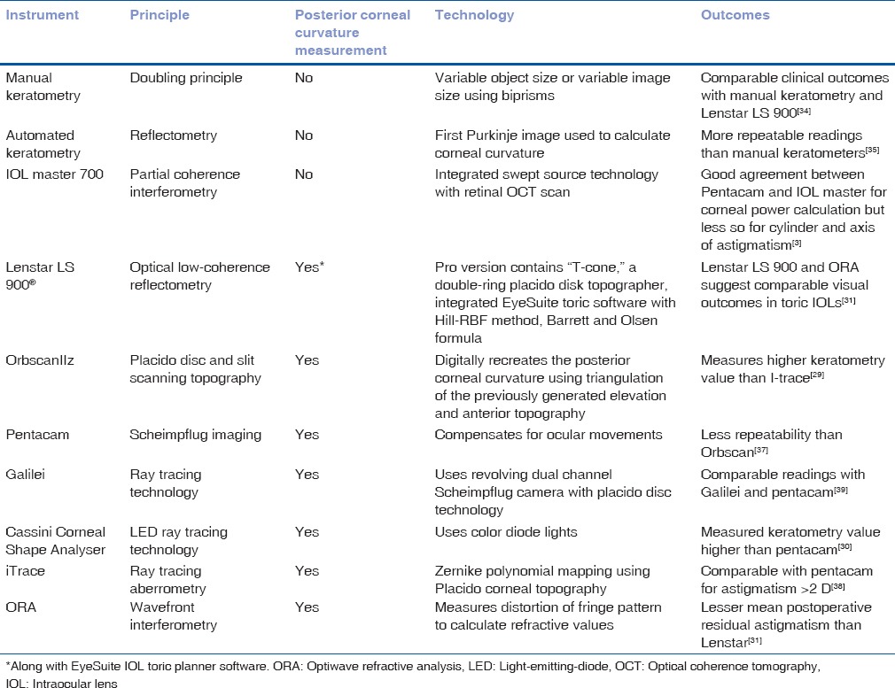
An accurate biometry is a prerequisite for precise IOL power calculation. The axial length may be estimated by either ultrasonic biometry or optical systems such as IOL Master (Carl Zeiss Meditec, Germany) and Lenstar (Haag Streit, Switzerland). Keratometry estimation is of paramount importance to determine the power as well as the axis of the toric IOL. Various instruments based on different principles may be used for keratometry estimation, such as manual and automated keratometers, placido-based corneal topographers, slit scanning systems, Scheimpflug imaging systems, aberrometers and optical coherence tomography (OCT)-based systems [Table 3].[28,29,30,31,32,33,34,35,36,37,38,39] Enhanced accuracy of keratometry estimation may be achieved by taking multiple measurements and employing at least two separate devices based on different principles.[31,32,33] Cases with similar steep corneal meridian on different devices are good candidates for toric IOL implantation. However, if significant variability in both the axis and magnitude of toric IOL is observed on different devices, the patient should be evaluated to rule out coexistent ocular comorbidities. The visual outcomes may not be satisfactory in such cases.
Table 3.
Investigative modalities to assess preoperative keratometry before toric intraocular lens implantation
It is essential to evaluate the posterior corneal astigmatism while calculating total corneal astigmatism to avoid errors in IOL power calculation. Posterior cornea acts as a minus lens, and the astigmatism is generally against-the-rule and stable over time. In contrast, the anterior corneal astigmatism shifts from with-the-rule in the younger age group to against-the-rule in the older age group.[40,41,42,43] Relying solely on the anterior corneal curvature measurements results in residual astigmatism after toric IOL implantation, overcorrecting by a factor of 1.38 in eyes having with-the-rule astigmatism and under correcting by a factor of 0.65 in the eyes with against-the-rule astigmatism.[42]
The keratometers and placido-based topography systems do not take into account the posterior corneal curvature. They assume a fixed ratio between the anterior and posterior curvature and are prone to errors in keratometry estimation.[42] The slit scanning systems, Scheimpflug imaging systems and OCT measure both the anterior and posterior corneal curvature and are deemed to be more accurate. The superiority of one single method has not been definitively established in numerous studies.[32,33] Hoffmann et al. evaluated five different systems for corneal power measurement including a swept source fourier domain OCT, an autokeratometer, a hybrid topographer, a Placido topographer and a Scheimpflug tomographer.[33] They observed more precise results with OCT and hybrid topography systems as they incorporate posterior curvature measurements. Although posterior curvature data are also measured by Scheimpflug-based systems, they have the disadvantage of high measuring noise. The highest precision for planning toric IOL power and axis was achieved by combining the keratometry and OCT data.
The image-guided systems such as VERION and CALLISTO Eye aid in preoperative planning of the location and size of the surgical incisions and capsulorhexis, as well as IOL positioning. The CALLISTO eye also assists in planning the position of limbal relaxing incisions. VERION™ Reference Unit allows comprehensive astigmatism management, wherein the surgeons can optimize the incision locations and toric IOL power as per their SIA. Moreover, the position and length of arcuate incisions can be determined in cases planned for femtosecond laser-assisted cataract surgery (FLACS).
The iTrace System (Tracey Technologies, Houston, Tx) combines Placido disc corneal topography with a ray tracing aberrometer to analyze the entire visual system and helps in the preoperative planning, decision-making as well as postoperative assessment in cases undergoing toric IOL implantation.[44] It provides the corneal curvature and power maps, measures angle alpha and calculates the corneal, internal and total higher order aberrations. Moreover, the iTRACE workstation incorporates an in-built toric IOL planner, which calculates the IOL power and also provides the axis of placement, taking into account the surgically induced astigmatism. Integrated Zaldivar toric caliper with toric calculator can be used to assess the accuracy of preoperative reference axis marking. In addition, the postoperative toric IOL enhancement software provides the degree of misalignment of the toric IOL, and the direction as well as the magnitude of the required postoperative rotation to achieve optimal results.
Intraocular Lens Power Calculation
Various formulae and toric calculators are available for IOL power calculation, which determine the axis as well as the magnitude of the toric IOL to be implanted. An ideal formula should take into account the SIA, the posterior corneal curvature as well as the effective lens position (ELP). It is essential for the operating surgeon to determine his SIA using standard astigmatism vector analysis or online tools such as http://www. doctor-hill.com.[45,46] Many of the commonly used toric IOL calculators do not take into account the contribution of the posterior cornea in IOL power calculation. Another potential source of error is the use of a single conversion factor for converting the cylindrical power from the IOL plane to the corneal plane for a particular spherical equivalent, without considering the anterior chamber depth and pachymetry. This may result in erroneous calculations, especially in eyes with extremes of axial lengths.[47]
The AcrySof online toric calculator and the iTRACE calculator employ a fixed ratio to convert power from IOL to corneal plane.[48] The TECNIS calculator incorporates the anterior chamber depth based on the axial length and keratometry values, and the Holladay formula incorporates the ELP in its calculations.[49]
Various nomograms have been described in literature to adjust for these confounding variables.[50,51] The Baylor nomogram incorporates the posterior corneal curvature in its measurements and has been observed to be more precise than Alcon and Holladay toric calculators.[51] The Barrett toric calculator takes into account both the ELP as well as the posterior corneal astigmatism and has better predictability than the Baylor nomogram as well as Holladay and Alcon toric calculators.[51]
The online calculators have been revised to incorporate corrections for posterior corneal astigmatism. The revised AcrySof toric calculator incorporates the Barrett toric algorithm, and the Tecnis calculator received FDA approval in 2016 to incorporate posterior corneal astigmatism compensation.
Intraoperative wavefront aberrometry is increasingly being used to estimate the toric IOL power and axis of placement based on the aphakic refraction, especially in postrefractive surgery cases. A retrospective analysis observed a mean prediction error of 0.43 ± 0.33 D with Optiwave Refractive Analysis (ORA) in postlaser-assisted in situ keratomileusis (LASIK) cases undergoing toric IOL implantation. The results were more accurate than those obtained by the standard SRK-T formula and the online ASCRS calculator.[52]
Intraocular Lens Selection
Various toric IOLs are available commercially, with different material, design, and range of toricity [Tables 1 and 2]. The choice of IOL depends on the surgeon comfort, patient expectations, financial considerations and availability. A monofocal or multifocal toric IOL may be selected based on patient's preference and preoperative assessment.
Marking Techniques
Accurate alignment of toric IOL is a prerequisite to achieve successful outcomes. Various methods have been described to place the preoperative reference and axis marks and may be broadly categorized as manual methods, iris fingerprinting techniques, image-guided systems, and intraoperative aberrometry-based methods.
Manual techniques
The three-step technique is commonly used for toric IOL alignment, which involves the preoperative marking of the reference axis, intraoperative alignment of the reference marks with the degree gauge of the fixation ring and intraoperative marking of the target axis.[53] The reference marks are commonly placed in the 3’o, 6’o, and 9’o clock positions to improve predictability, though some surgeons may prefer to mark only the horizontal 3’o and 9’o clock positions, or only the inferior 6’o clock position. The marking may be performed with a skin-marking pen in a free-hand manner, or with the help of various devices such as a thin slit-beam, weighted thread, pendulum marker or Nuijts-Solomon bubble marker. This is followed by the intraoperative alignment of these reference marks to the degree gauge on a fixation ring, and the target axis is then marked with a corneal meridian marker. One-step axis marking may be done with the help of various devices such as tonometer markers, electronic toric markers, Neuhann one-step toric bubble marker, and Geuder-Gerten Pendulum marker.[54,55]
A change in patient position from sitting to supine may induce significant cyclotorsion, and up to 28° of cyclotorsion has been observed in 68% cases.[56] Hence, the patient should be sitting erect with the back resting against a wall and a straight-ahead gaze while marking the reference axis to avoid inadvertent errors. The cornea should be dry, and adequate topical anesthesia should be administered to improve patient comfort during marking.
The three-step marking method is fairly accurate, and a mean error of 2.4° ± 0.8° has been observed during axis marking with a bubble marker, with a total error of 4.9° ± 2.1° in toric IOL alignment.[57] Both bubble marker and pendulum marker are easy and reproducible techniques with fairly accurate results.[58] A comparative evaluation of four different marking techniques including coaxial slit beam, bubble marker, pendular marker, and tonometer marker observed minimum rotational deviation with the pendular marker and least vertical misalignment with the slit lamp marking technique.[59] The least accurate results were observed with the tonometer marker, whereas the other three methods provided fairly accurate results. Slit-lamp assisted pendular marker has been observed to give more accurate results than using a horizontal slit-beam alone or a direct nonpendular marker.[55]
The manual marking methods have inherent sources of errors, such as smudging of the dye, irregular, and broad marks. Moreover, they are associated with a significant learning curve, and intersurgeon variability may be observed in the accuracy of marking.
Osher ThermoDot Marker (Beaver-Visitec International, BVI, Waltham, Mass.) has been developed to eliminate the ink-associated problems in reference axis marking. It employs a bipolar cautery to create an ink-free, precise reference mark during surgery. Anterior stromal puncture using a 26-gauge bent needle stained with sterile blue ink has been described for reference axis marking, to obtain precise reference marks with no smudging.[60]
Image-guided techniques
The concept of iris-fingerprinting was introduced by Osher in 2010, wherein the iris crypts, nevi, brush fields, etc., were used as landmarks to place the axis marks.[61,62] It formed the basis for the development of various image-guided systems, such as CALLISTO Eye and Z align, VERION image-guided system and the TrueVision 3D Surgical System [Table 4].[63,64,65,66]
Table 4.
Image-guided systems for toric intraocular lens alignment
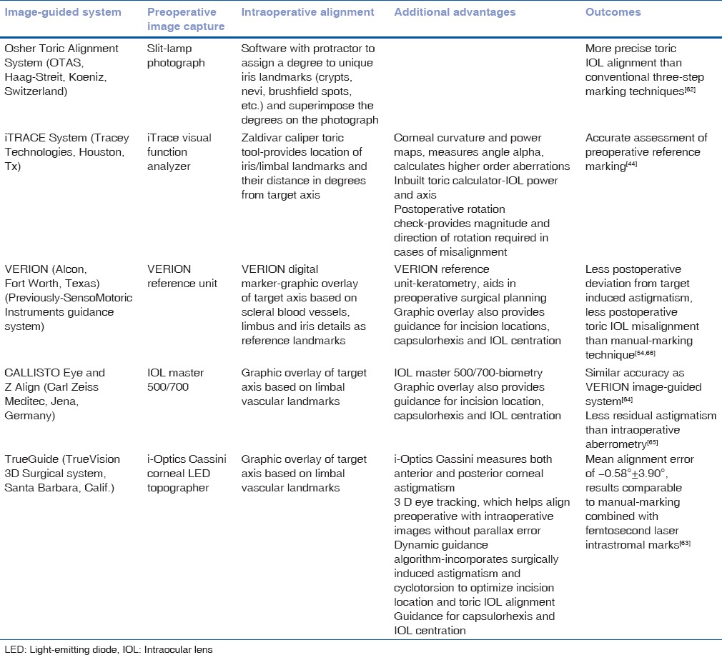
The iris and limbal landmarks may be captured by the iTRACE system, and the Zaldivar Toric Caliper tool can be used to calculate the location of these marks and their distance in degrees from the target IOL axis. A final surgical plan is generated that provides simple angular directions from each reference mark to the desired axis of IOL placement with regard to surgeon's position and view.[44]
The image-guided systems involve the capture of a preoperative reference image followed by intraoperative image registration wherein the limbal landmarks are used to match the two images with respect to each other. A graphic overlay is then superimposed on the surgical field along the target axis, which provides a guide for toric IOL alignment. The VERION image-guided system utilizes the scleral blood vessels, limbus, and iris details as reference landmarks to determine the extent of cyclotorsion.
In addition to aiding the alignment of the toric IOL, the image-guided systems also provide a step-by-step guidance during the various surgical steps including the placement of corneal incisions, the size and centration of the capsulorhexis as well as IOL centration.
Significantly more precise alignment has been observed with VERION-image guided marking as compared to manual slit lamp-assisted preoperative marking using pendulum-attached marker.[54,66] The accuracy of CALLISTO Eye and Z align is similar to VERION.[64]
The eye tracker in these systems may disengage during surgery, and a repeat registration may be required. Conjunctival chemosis, ballooning and bleeding may interfere with intraoperative registration. Registration may also not be possible in extremely uncooperative patients or difficult orbital anatomy including extremely deep-set eyes or narrow palpebral apertures. In addition to these limitations, the high financial cost involved may limit the widespread usage of this technology.
Intraoperative aberrometry
Intraoperative aberrometry devices such as ORA (ORA; WaveTec Vision Systems Inc., CA, USA) and Holos IntraOp (Clarity Medical Systems, CA, USA) perform a real-time assessment of the phakic, aphakic, or pseudophakic refraction to provide feedback for toric IOL alignment.
ORA utilizes the principle of Talbot-Moire interferometry to perform real-time calculation of IOL power as well as the axis, based on the aphakic refraction. It employs a modified refractive vergence formula for accurate IOL power calculation even in complicated postrefractive surgery cases. Moreover, it permits refinement of the axis by providing the direction as well as magnitude of rotation required to achieve minimum residual astigmatism. VerifEye has been incorporated in ORA with a fast imaging processor that confirms the stability of the system before measurements are taken.
Holos IntraOp provides continuous real-time refraction throughout the surgery. Although it does not provide the spherical IOL power to be implanted, the axis of the toric IOL can be refined based on the continuous feedback provided by this system.
Cases undergoing toric IOL implantation assisted with intraoperative aberrometry are 2.4 times more likely to have 0.50D or less residual astigmatism compared with other standard methods.[31] Wavefront aberrometry significantly affects the intraoperative decision-making, with the cylinder power changed 24% of the time, the spherical power changed 25% of the time, and three or fewer rotations needed 92% of the time.[67] Intraoperative aberrometry is also superior to conventional methods in patients with prior myopic keratorefractive surgery.[52] However, a recent study comparing Callisto eye and Z align with ORA observed more precise alignment with less residual astigmatism in cases using Callisto image-guided system.[65]
Before obtaining the readings, the anterior chamber should be uniformly filled with cohesive ocular viscoelastic devices (OVD) to maintain the intraocular pressure and ensure a uniform fundal glow. The accuracy of aberrometry readings may be affected by intraoperative corneal edema, eyelid speculum, presence of air bubbles, or clumps of dispersive OVD in the anterior chamber or inadequate intraocular pressure.[68] Multiple radial keratotomy cuts encroaching the visual axis also preclude accurate measurements. The device is mounted directly onto the bottom of the surgical microscope and takes up a significant amount of space. In addition to these limitations, the high financial cost involved may act as a deterrent for surgeons.
Intraoperative Toric Intraocular Lens Alignment
In cases with manual marking, the target axis is marked at the beginning of surgery after aligning the preoperatively placed reference marks with a degree gauge. In addition to intraoperative alignment of the toric IOL along the desired corneal meridian, the clear corneal incisions, capsulorhexis and IOL centration also play a significant role in achieving optimal outcomes. Self-sealing clear corneal incisions that are astigmatically neutral or induce minimal astigmatism should be created. Uniformity of corneal incisions in terms of location and size is essential to prevent variations in SIA. Image-guided systems compensate for cyclotorsion and assist in the precise placement of incisions.
A well centered circular continuous capsulorhexis providing adequate IOL coverage of around 0.5 mm is essential to ensure IOL stability in the postoperative period. Posterior capsular rent is a relative contraindication for in-the-bag toric IOLs, as there is a high risk of IOL tilt as well as rotation. The IOL should be centered along the coaxially sighted corneal light reflex, as represented by the first Purkinje image while the patient is fixating on the microscope light. Perfect centration is especially significant in cases undergoing toric multifocal IOLs to prevent the occurrence of dysphotic visual symptoms.
During IOL alignment, the IOL should be left about 3°–5° anticlockwise of the final desired lens position. The final alignment should be done after complete OVD removal and hydration of the wounds, as most open-loop IOLs can be rotated only clockwise and a complete re-rotation will be needed if the IOL rotates further clockwise of the target axis during these maneuvers.
The precise capsulotomy created in FLACS may further improve the outcomes of toric IOL implantation. A significant decrease in higher order aberrations has been observed with FLACS as compared to standard phacoemulsification with toric IOL implantation.[69]
Postoperative Outcomes
The postoperative outcomes after implantation of toric IOL may be assessed in terms of anatomical outcomes, such as precision and stability of IOL alignment, and functional outcomes, such as visual acuity and quality [Tables 5 and 6].
Table 5.
Visual and anatomical outcomes after toric intraocular lens implantation
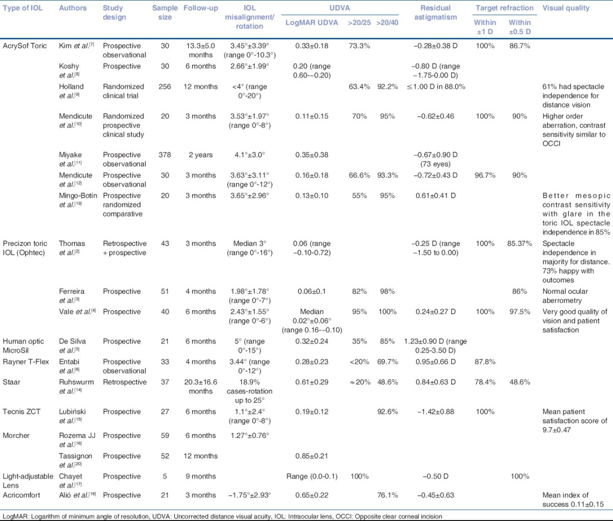
Table 6.
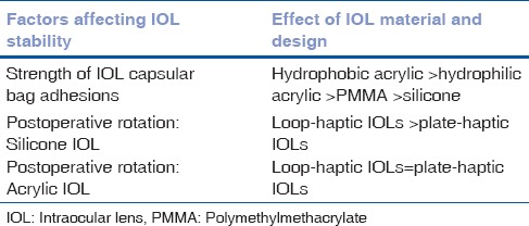
Effect of intraocular lens material and design on rotational stability of toric intraocular lenses
Anatomical outcomes
The precision of IOL alignment along the intended target axis is influenced by various factors, such as the type of marking method, IOL material and design, intraoperative factors such as capsulorhexis size, IOL coverage, sealing of corneal incisions, and surgeon experience.
Conventional three-step manual marking techniques result in fairly accurate alignment of toric IOLs, with a mean deviation from target axis of 4.9° ± 2.1°.[57] The image-guided systems and intraoperative aberrometry have improved the precision of toric IOL alignment, with <5° of deviation from the intended axis in the majority of cases.[64,65,67]
Rotational stability of the IOL varies with design and material, and maximum rotational stability has been observed with hydrophobic acrylic lenses [Tables 5 and 6]. This may be attributed to the development of strong adhesions between the IOL and lens capsule in the early postoperative period.
A long-term prospective study of AcrySof toric IOLs observed the significant postoperative rotation of more than 10° in only 1.68% eyes, and maximum rotation occurred within the initial 10 days in the postoperative period in cases with high axial length.[11] A total of 76.7% eyes were within 5 degrees of the intended target axis even 2 years after surgery.
Similar results were observed in a randomized control trial comparing AcrySof toric IOL with spherical IOLs, with a mean rotation of <4° (range, 0°–20°). A rotation of >10° was observed in only 6.7% of the cases at the end of 1 year.[9]
Implantation of the hydrophobic single-piece toric IOL (Tecnis ZCT, Abbott Medical Optics) yielded stable results with a mean absolute IOL rotation of 1.1° ± 2.4° at 6 months of follow-up, with more than 5° rotation observed in only 6.9% cases.[15] Accurate alignment of the IOL with its intended axis was obtained in 70.37% cases.
The silicone lenses have the least rotational stability, though the plate haptic design confers better stability as compared to conventional three-piece lenses with polypropylene loop haptics.[70,71] The “C-” shaped polypropylene loop haptics invariably rotate anticlockwise within 2 postoperative weeks.[70] Early postoperative rotation is also influenced by a large capsular bag size, high axial length, retained OVDs, and small diameter of the haptic.[72]
Less than 5° rotation has been observed with the Trulign (Bausch and Lomb) toric presbyopia-correcting IOL at 1 month of follow-up.[66] However, 10.6% cases with Lentis Unico L-312T aspheric toric IOL (Oculentis GmbH), underwent postoperative rotation of 30° or more, despite having a hydrophobic surface and an open loop haptic.[67]
Functional outcomes
A UDVA of 20/40 or better is achieved in 70%–100% of cases undergoing toric IOL implantation [Table 5].[2,3,4,5,6,7,8,9,10,11,12,13,14,15,16,17,18,19,20] Spectacle-independence for distance vision has been reported in 60%–97% of patients with toric IOLs. A randomized control trial compared the outcomes of AcrySof toric IOL with conventional spherical IOL in patients with preexisting corneal astigmatism of 0.75D or more, and observed a UDVA of 20/40 or better in 92.2% cases undergoing toric IOL implantation, with 63.4% having a UDVA of 20/25 or better. In contrast, only 81.4% cases undergoing nontoric IOL implantation had a UDVA of 20/40 or better, and 41.4% had a UDVA of 20/25 or better.[9]
Lower degree of mean residual astigmatism is observed with toric IOLs as compared to nontoric IOL's with or without limbal relaxing incisions.[9,10,12,73] Residual astigmatism may result from preoperative measurement errors, marking errors, posterior corneal astigmatism, ELP, and postoperative IOL rotation. A randomized control trial observed residual astigmatism of 1.0D or less in 88% cases and 0.5D or less in 53% cases undergoing toric IOL implantation.[9]
Satisfactory visual quality has been reported by patients undergoing toric IOL implantation, and a mean patient satisfaction score of 9.7 ± 0.47 has been reported in a study evaluating the outcomes of Tecnis toric IOL.[15] The higher-order aberrations and contrast sensitivity after toric IOL implantation is similar to that observed with conventional monofocal IOLs.[10] Better mesopic contrast sensitivity and less glare have been observed with AcrySof toric IOLs compared to peripheral corneal relaxing incisions.[13]
Toric multifocal IOLs demonstrate good visual outcomes with UDVA better than 20/40 in 97% to 100% of patients, uncorrected near visual acuity better than 20/40 in 100% of patients, spectacle independence in 79% to 100% of patients and residual refractive astigmatism lower than 0.50 D in 38%–79% of patients.[74,75,76,77,78,79] However, dysphotic symptoms such as glare and halos may limit the patient satisfaction achieved with these IOLs.
Intraocular Lens Misalignment
Postoperative toric IOL misalignment is the major factor responsible for suboptimal visual outcomes after toric IOL implantation. It may be attributed to three factors, namely, inaccurate preoperative prediction of the axis of IOL alignment, inaccurate intraoperative alignment, and postoperative IOL rotation. Newer investigative modalities and advanced image-guided and aberrometry systems help to minimize the incidence of pre- and intra-operative alignment errors and have been discussed in detail in the previous sections.
Misalignment of the toric IOL axis can cause reduction of the cylinder power along the desired meridian and induction of cylinder in a new meridian when misalignment exceeds 30°. The new residual cylinder may be estimated by the formula R = ∣2C sin θ∣, where C is the cylinder power of the toric lens and θ is the degree of misalignment. One degree of misalignment causes a loss of approximately 3% of the effective cylinder power, and the entire toric effect is lost in cases with 30° of misalignment.[80,81] The UDVA is significantly worse in misaligned multifocal toric IOLs as compared to monofocal toric lenses.[82]
IOL rotation may be observed as early as 1 h after surgery, and a majority of rotations occur within the initial 10 days.[11] Early IOL rotation likely results from incomplete OVD removal, whereas late postoperative rotation, is influenced by the IOL architecture, design, and axial length. The axis of IOL implantation is associated with postoperative rotation, and an increased incidence of rotation has been observed in cases with vertical axis of IOL implantation (with-the-rule astigmatism).[14] Capsulorhexis extension or inadequate IOL coverage also contribute to postoperative rotation.
The axis of implanted toric IOL may be assessed at the slit-lamp with a rotating slit and rotational gauge. This method requires adequate mydriasis to visualize the IOL optic marks. The 10° steps on the slit-lamp measuring reticule limit the accuracy of this method. A simple and inexpensive method to measure the toric IOL axis using a camera-enabled cellular phone and (ImageJ) computer software has also been described.[83] An online toric results analyzer (www.astigmatismfix.com) has been developed, which determines the ideal position of the toric IOL in cases of postoperative malrotation.[84,85] It uses the patient's postoperative manifest refraction, power and current axis of the toric IOL to predict the ideal axis of toric IOL and postrotation refraction. Wavefront aberrometers such as the iTRACE system determine the orientation of the toric IOL based on the internal ocular aberrations. The toric IOL enhancement software of iTRACE also provides the magnitude and direction of the required rotation to achieve accurate alignment with minimal residual astigmatism.
Realignment of the toric IOL is needed in cases with more than 10° of rotation from the target axis. A rotation of less than 10° changes the manifest refraction by 0.5D and usually does not warrant any additional intervention.[86] Realignment may be required in 0.65%–3.3% cases and an incidence of 1.1% has been observed with AcrySof toric IOL, 2.3% with Tecnis Toric IOL and 3.3% with silicone lenses.[11,87,88,89] In a retrospective evaluation of 6431 eyes implanted with toric IOLs, realignment was performed in 0.653% cases, and it reduced the magnitude of misalignment from 32.9° ± 15.7° to 8.8° ± 9.7°.[90] A significant negative correlation was observed between the interval from cataract surgery to repositioning procedure and the degree of residual misalignment. A repositioning performed after 1 week of primary cataract surgery had superior outcomes in terms of precision of IOL alignment and minimal residual refractive cylinder.
Intraoperative techniques to rotate toric IOLs depend on the length of time from the initial surgery and the degree of adhesions between the IOL and the capsular bag. Optimal results have been observed in cases with early rotation using a long cannula mounted on a balanced salt solution-filled syringe to rotate the IOL through the paracentesis incision.[89] The new target axis is determined relative to the current axis; therefore, intraoperative marking is only necessary relative to the implanted IOL. This reduces repositioning variability and maximizes outcomes after IOL rotation.
In cases with the large residual cylinder not amenable to correction by rotation alone, an IOL exchange, piggyback IOLs or corneal ablative procedures may be considered. LASIK has been observed to be superior to lens exchange and piggyback IOLs, with a greater reduction in spherocylinder refractive error.[91] Customized surface ablation or femtosecond laser-assisted intrastromal keratotomies may also be attempted to correct residual astigmatism.[92]
Conclusion
The outcomes after toric IOL implantation are influenced by numerous factors, right from the preoperative case selection and investigations to accurate intraoperative alignment and postoperative care. The importance of posterior corneal curvature is increasingly being recognized in various studies, and newer investigative modalities that account for both the anterior and posterior corneal power are becoming the standard of care. The conventional manual marking has given way to image-guided systems and intraoperative aberrometry, which provide a mark-less IOL alignment and also aid in planning the incisions, capsulorhexis size, and optimal IOL centration. Newer IOLs are being introduced for commercial use, with superior design, expanding the range of cylinder powers, enhanced rotational stability, and minimal induction of higher-order aberrations.
The applications of toric IOLs are expanding to include cases with high astigmatism, irregular astigmatism, corneal ectatic disorders, and postkeratoplasty cases.[25,26,27,93,94] Future technological advancements may further refine the outcomes of toric IOL, with more precise visual results and enhanced IOL stability. Newer customized IOLs are being introduced that may be implanted at the 0°–180° axis without a need for rotational alignment such as the Ultima Smart Toric Customised Hydrophilic IOL (EyePharma, Care Group, Cape Town, South Africa). Cirle surgical navigation system (Bausch and Lomb, Rochester, New York, USA) is being developed for commercial use, which will provide 3-D guidance using microscope oculars during cataract surgery. We may see the development of integrated image-guided systems that incorporate preoperative keratometry and IOL power estimation, intraoperative surgical guidance, and toric alignment as well as postoperative assessment in a single platform.
Financial support and sponsorship
Nil.
Conflicts of interest
There are no conflicts of interest.
References
- 1.Shimizu K, Misawa A, Suzuki Y. Toric intraocular lenses: Correcting astigmatism while controlling axis shift. J Cataract Refract Surg. 1994;20:523–6. doi: 10.1016/s0886-3350(13)80232-5. [DOI] [PubMed] [Google Scholar]
- 2.Thomas BC, Khoramnia R, Auffarth GU, Holzer MP. Clinical outcomes after implantation of a toric intraocular lens with a transitional conic toric surface. Br J Ophthalmol. 2017 doi: 10.1136/bjophthalmol-2017-310386. [Epub ahead of print] [DOI] [PubMed] [Google Scholar]
- 3.Ferreira TB, Berendschot TT, Ribeiro FJ. Clinical outcomes after cataract surgery with a new transitional toric intraocular lens. J Refract Surg. 2016;32:452–9. doi: 10.3928/1081597X-20160428-07. [DOI] [PubMed] [Google Scholar]
- 4.Vale C, Menezes C, Firmino-Machado J, Rodrigues P, Lume M, Tenedório P, et al. Astigmatism management in cataract surgery with precizon(®) toric intraocular lens: A prospective study. Clin Ophthalmol. 2016;10:151–9. doi: 10.2147/OPTH.S91298. [DOI] [PMC free article] [PubMed] [Google Scholar]
- 5.De Silva DJ, Ramkissoon YD, Bloom PA. Evaluation of a toric intraocular lens with a Z-haptic. J Cataract Refract Surg. 2006;32:1492–8. doi: 10.1016/j.jcrs.2006.04.022. [DOI] [PubMed] [Google Scholar]
- 6.Entabi M, Harman F, Lee N, Bloom PA. Injectable 1-piece hydrophilic acrylic toric intraocular lens for cataract surgery: Efficacy and stability. J Cataract Refract Surg. 2011;37:235–40. doi: 10.1016/j.jcrs.2010.08.040. [DOI] [PubMed] [Google Scholar]
- 7.Kim MH, Chung TY, Chung ES. Long-term efficacy and rotational stability of AcrySof toric intraocular lens implantation in cataract surgery. Korean J Ophthalmol. 2010;24:207–12. doi: 10.3341/kjo.2010.24.4.207. [DOI] [PMC free article] [PubMed] [Google Scholar]
- 8.Koshy JJ, Nishi Y, Hirnschall N, Crnej A, Gangwani V, Maurino V, et al. Rotational stability of a single-piece toric acrylic intraocular lens. J Cataract Refract Surg. 2010;36:1665–70. doi: 10.1016/j.jcrs.2010.05.018. [DOI] [PubMed] [Google Scholar]
- 9.Holland E, Lane S, Horn JD, Ernest P, Arleo R, Miller KM, et al. The AcrySof toric intraocular lens in subjects with cataracts and corneal astigmatism: A randomized, subject-masked, parallel-group, 1-year study. Ophthalmology. 2010;117:2104–11. doi: 10.1016/j.ophtha.2010.07.033. [DOI] [PubMed] [Google Scholar]
- 10.Mendicute J, Irigoyen C, Ruiz M, Illarramendi I, Ferrer-Blasco T, Montés-Micó R, et al. Toric intraocular lens versus opposite clear corneal incisions to correct astigmatism in eyes having cataract surgery. J Cataract Refract Surg. 2009;35:451–8. doi: 10.1016/j.jcrs.2008.11.043. [DOI] [PubMed] [Google Scholar]
- 11.Miyake T, Kamiya K, Amano R, Iida Y, Tsunehiro S, Shimizu K, et al. Long-term clinical outcomes of toric intraocular lens implantation in cataract cases with preexisting astigmatism. J Cataract Refract Surg. 2014;40:1654–60. doi: 10.1016/j.jcrs.2014.01.044. [DOI] [PubMed] [Google Scholar]
- 12.Mendicute J, Irigoyen C, Aramberri J, Ondarra A, Montés-Micó R. Foldable toric intraocular lens for astigmatism correction in cataract patients. J Cataract Refract Surg. 2008;34:601–7. doi: 10.1016/j.jcrs.2007.11.033. [DOI] [PubMed] [Google Scholar]
- 13.Mingo-Botín D, Muñoz-Negrete FJ, Won Kim HR, Morcillo-Laiz R, Rebolleda G, Oblanca N, et al. Comparison of toric intraocular lenses and peripheral corneal relaxing incisions to treat astigmatism during cataract surgery. J Cataract Refract Surg. 2010;36:1700–8. doi: 10.1016/j.jcrs.2010.04.043. [DOI] [PubMed] [Google Scholar]
- 14.Ruhswurm I, Scholz U, Zehetmayer M, Hanselmayer G, Vass C, Skorpik C, et al. Astigmatism correction with a foldable toric intraocular lens in cataract patients. J Cataract Refract Surg. 2000;26:1022–7. doi: 10.1016/s0886-3350(00)00317-5. [DOI] [PubMed] [Google Scholar]
- 15.Lubiński W, Kańmierczak B, Gronkowska-Serafin J, Podborączyńska-Jodko K. Clinical outcomes after uncomplicated cataract surgery with implantation of the tecnis toric intraocular lens. J Ophthalmol. 2016;2016:3257217. doi: 10.1155/2016/3257217. [DOI] [PMC free article] [PubMed] [Google Scholar]
- 16.Rozema JJ, Gobin L, Verbruggen K, Tassignon MJ. Changes in rotation after implantation of a bag-in-the-lens intraocular lens. J Cataract Refract Surg. 2009;35:1385–8. doi: 10.1016/j.jcrs.2009.03.037. [DOI] [PubMed] [Google Scholar]
- 17.Chayet A, Sandstedt C, Chang S, Rhee P, Tsuchiyama B, Grubbs R, et al. Use of the light-adjustable lens to correct astigmatism after cataract surgery. Br J Ophthalmol. 2010;94:690–2. doi: 10.1136/bjo.2009.164616. [DOI] [PubMed] [Google Scholar]
- 18.Alió JL, Agdeppa MC, Pongo VC, El Kady B. Microincision cataract surgery with toric intraocular lens implantation for correcting moderate and high astigmatism: Pilot study. J Cataract Refract Surg. 2010;36:44–52. doi: 10.1016/j.jcrs.2009.07.043. [DOI] [PubMed] [Google Scholar]
- 19.Xiao XW, Hao J, Zhang H, Tian F. Optical quality of toric intraocular lens implantation in cataract surgery. Int J Ophthalmol. 2015;8:66–71. doi: 10.3980/j.issn.2222-3959.2015.01.12. [DOI] [PMC free article] [PubMed] [Google Scholar]
- 20.Tassignon MJ, Gobin L, Mathysen D, Van Looveren J. Clinical results after spherotoric intraocular lens implantation using the bag-in-the-lens technique. J Cataract Refract Surg. 2011;37:830–4. doi: 10.1016/j.jcrs.2010.12.042. [DOI] [PubMed] [Google Scholar]
- 21.Hoffmann PC, Hütz WW. Analysis of biometry and prevalence data for corneal astigmatism in 23,239 eyes. J Cataract Refract Surg. 2010;36:1479–85. doi: 10.1016/j.jcrs.2010.02.025. [DOI] [PubMed] [Google Scholar]
- 22.Ferrer-Blasco T, Montés-Micó R, Peixoto-de-Matos SC, González-Méijome JM, Cerviño A. Prevalence of corneal astigmatism before cataract surgery. J Cataract Refract Surg. 2009;35:70–5. doi: 10.1016/j.jcrs.2008.09.027. [DOI] [PubMed] [Google Scholar]
- 23.Khan MI, Ch’ng SW, Muhtaseb M. The use of toric intraocular lens to correct astigmatism at the time of cataract surgery. Oman J Ophthalmol. 2015;8:38–43. doi: 10.4103/0974-620X.149865. [DOI] [PMC free article] [PubMed] [Google Scholar]
- 24.Statham M, Apel A, Stephensen D. Comparison of the AcrySof SA60 spherical intraocular lens and the AcrySof toric SN60T3 intraocular lens outcomes in patients with low amounts of corneal astigmatism. Clin Exp Ophthalmol. 2009;37:775–9. doi: 10.1111/j.1442-9071.2009.02154.x. [DOI] [PubMed] [Google Scholar]
- 25.Luck J. Customized ultra-high-power toric intraocular lens implantation for pellucid marginal degeneration and cataract. J Cataract Refract Surg. 2010;36:1235–8. doi: 10.1016/j.jcrs.2010.04.009. [DOI] [PubMed] [Google Scholar]
- 26.Kersey JP, O’Donnell A, Illingworth CD. Cataract surgery with toric intraocular lenses can optimize uncorrected postoperative visual acuity in patients with marked corneal astigmatism. Cornea. 2007;26:133–5. doi: 10.1097/ICO.0b013e31802be5cc. [DOI] [PubMed] [Google Scholar]
- 27.Stewart CM, McAlister JC. Comparison of grafted and non-grafted patients with corneal astigmatism undergoing cataract extraction with a toric intraocular lens implant. Clin Exp Ophthalmol. 2010;38:747–57. doi: 10.1111/j.1442-9071.2010.02336.x. [DOI] [PubMed] [Google Scholar]
- 28.Shajari M, Cremonese C, Petermann K, Singh P, Müller M, Kohnen T, et al. Comparison of axial length, corneal curvature, and anterior chamber depth measurements of 2 recently introduced devices to a known biometer. Am J Ophthalmol. 2017;178:58–64. doi: 10.1016/j.ajo.2017.02.027. [DOI] [PubMed] [Google Scholar]
- 29.Chen Y, Xia X. Comparison of the Orbscan II topographer and the iTrace aberrometer for the measurements of keratometry and corneal diameter in myopic patients. BMC Ophthalmol. 2016;16:33. doi: 10.1186/s12886-016-0210-8. [DOI] [PMC free article] [PubMed] [Google Scholar]
- 30.Hidalgo IR, Rozema JJ, Dhubhghaill SN, Zakaria N, Koppen C, Tassignon MJ, et al. Repeatability and inter-device agreement for three different methods of keratometry: Placido, scheimpflug, and color LED corneal topography. J Refract Surg. 2015;31:176–81. doi: 10.3928/1081597X-20150224-01. [DOI] [PubMed] [Google Scholar]
- 31.Woodcock MG, Lehmann R, Cionni RJ, Breen M, Scott MC. Intraoperative aberrometry versus standard preoperative biometry and a toric IOL calculator for bilateral toric IOL implantation with a femtosecond laser: One-month results. J Cataract Refract Surg. 2016;42:817–25. doi: 10.1016/j.jcrs.2016.02.048. [DOI] [PubMed] [Google Scholar]
- 32.Browne AW, Osher RH. Optimizing precision in toric lens selection by combining keratometry techniques. J Refract Surg. 2014;30:67–72. doi: 10.3928/1081597X-20131217-07. [DOI] [PubMed] [Google Scholar]
- 33.Hoffmann PC, Abraham M, Hirnschall N, Findl O. Prediction of residual astigmatism after cataract surgery using swept source fourier domain optical coherence tomography. Curr Eye Res. 2014;39:1178–86. doi: 10.3109/02713683.2014.898376. [DOI] [PubMed] [Google Scholar]
- 34.Gundersen KG, Potvin R. Prospective study of toric IOL outcomes based on the Lenstar LS 900® dual zone automated keratometer. BMC Ophthalmol. 2012;12:21. doi: 10.1186/1471-2415-12-21. [DOI] [PMC free article] [PubMed] [Google Scholar]
- 35.Manning CA, Kloess PM. Comparison of portable automated keratometry and manual keratometry for IOL calculation. J Cataract Refract Surg. 1997;23:1213–6. doi: 10.1016/s0886-3350(97)80318-5. [DOI] [PubMed] [Google Scholar]
- 36.Lee BW, Galor A, Feuer WJ, Pouyeh B, Pelletier JS, Vaddavalli PK, et al. Agreement between Pentacam and IOL master in patients undergoing toric IOL implantation. J Refract Surg. 2013;29:114–20. doi: 10.3928/1081597X-20130117-06. [DOI] [PubMed] [Google Scholar]
- 37.Kumar M, Shetty R, Jayadev C, Rao HL, Dutta D. Repeatability and agreement of five imaging systems for measuring anterior segment parameters in healthy eyes. Indian J Ophthalmol. 2017;65:288–94. doi: 10.4103/ijo.IJO_729_16. [DOI] [PMC free article] [PubMed] [Google Scholar]
- 38.Delrivo M, Ruiseñor Vázquez PR, Galletti JD, Garibotto M, Fuentes Bonthoux F, Pförtner T, et al. Agreement between placido topography and scheimpflug tomography for corneal astigmatism assessment. J Refract Surg. 2014;30:49–53. doi: 10.3928/1081597X-20131217-06. [DOI] [PubMed] [Google Scholar]
- 39.Baradaran-Rafii A, Motevasseli T, Yazdizadeh F, Karimian F, Fekri S, Baradaran-Rafii A, et al. Comparison between two scheimpflug anterior segment analyzers. J Ophthalmic Vis Res. 2017;12:23–9. doi: 10.4103/jovr.jovr_104_16. [DOI] [PMC free article] [PubMed] [Google Scholar]
- 40.Ho JD, Liou SW, Tsai RJ, Tsai CY. Effects of aging on anterior and posterior corneal astigmatism. Cornea. 2010;29:632–7. doi: 10.1097/ICO.0b013e3181c2965f. [DOI] [PubMed] [Google Scholar]
- 41.Koch DD, Jenkins RB, Weikert MP, Yeu E, Wang L. Correcting astigmatism with toric intraocular lenses: Effect of posterior corneal astigmatism. J Cataract Refract Surg. 2013;39:1803–9. doi: 10.1016/j.jcrs.2013.06.027. [DOI] [PubMed] [Google Scholar]
- 42.Goggin M, Zamora-Alejo K, Esterman A, van Zyl L. Adjustment of anterior corneal astigmatism values to incorporate the likely effect of posterior corneal curvature for toric intraocular lens calculation. J Refract Surg. 2015;31:98–102. doi: 10.3928/1081597X-20150122-04. [DOI] [PubMed] [Google Scholar]
- 43.Koch DD. The posterior cornea: Hiding in plain sight. Ophthalmology. 2015;122:1070–1. doi: 10.1016/j.ophtha.2015.01.022. [DOI] [PubMed] [Google Scholar]
- 44.Farooqui JH, Sharma M, Koul A, Dutta R, Shroff NM. Evaluation of a new electronic preoperative reference marker for toric intraocular lens implantation by two different methods of analysis: Adobe Photoshop versus iTrace. Oman J Ophthalmol. 2017;10:96–9. doi: 10.4103/ojo.OJO_163_2013. [DOI] [PMC free article] [PubMed] [Google Scholar]
- 45.Holladay JT, Moran JR, Kezirian GM. Analysis of aggregate surgically induced refractive change, prediction error, and intraocular astigmatism. J Cataract Refract Surg. 2001;27:61–79. doi: 10.1016/s0886-3350(00)00796-3. [DOI] [PubMed] [Google Scholar]
- 46.East Valley Ophthalmology. IOL Power Calculations: Surgically Induced Astigmatism Calculator. [Last accessed on 2017 Aug 25]. Available from: http://www.doctor-hill.com/
- 47.Savini G, Hoffer KJ, Carbonelli M, Ducoli P, Barboni P. Influence of axial length and corneal power on the astigmatic power of toric intraocular lenses. J Cataract Refract Surg. 2013;39:1900–3. doi: 10.1016/j.jcrs.2013.04.047. [DOI] [PubMed] [Google Scholar]
- 48.Savini G, Hoffer KJ, Ducoli P. A new slant on toric intraocular lens power calculation. J Refract Surg. 2013;29:348–54. doi: 10.3928/1081597X-20130415-06. [DOI] [PubMed] [Google Scholar]
- 49.Park HJ, Lee H, Woo YJ, Kim EK, Seo KY, Kim HY, et al. Comparison of the astigmatic power of toric intraocular lenses using three toric calculators. Yonsei Med J. 2015;56:1097–105. doi: 10.3349/ymj.2015.56.4.1097. [DOI] [PMC free article] [PubMed] [Google Scholar]
- 50.Goggin M, Moore S, Esterman A. Toric intraocular lens outcome using the manufacturer's prediction of corneal plane equivalent intraocular lens cylinder power. Arch Ophthalmol. 2011;129:1004–8. doi: 10.1001/archophthalmol.2011.178. [DOI] [PubMed] [Google Scholar]
- 51.Abulafia A, Barrett GD, Kleinmann G, Ofir S, Levy A, Marcovich AL, et al. Prediction of refractive outcomes with toric intraocular lens implantation. J Cataract Refract Surg. 2015;41:936–44. doi: 10.1016/j.jcrs.2014.08.036. [DOI] [PubMed] [Google Scholar]
- 52.Yesilirmak N, Palioura S, Culbertson W, Yoo SH, Donaldson K. Intraoperative wavefront aberrometry for toric intraocular lens placement in eyes with a history of refractive surgery. J Refract Surg. 2016;32:69–70. doi: 10.3928/1081597X-20151210-02. [DOI] [PMC free article] [PubMed] [Google Scholar]
- 53.Ventura BV, Wang L, Weikert MP, Robinson SB, Koch DD. Surgical management of astigmatism with toric intraocular lenses. Arq Bras Oftalmol. 2014;77:125–31. doi: 10.5935/0004-2749.20140032. [DOI] [PubMed] [Google Scholar]
- 54.Elhofi AH, Helaly HA. Comparison between digital and manual marking for toric intraocular lenses: A randomized trial. Medicine (Baltimore) 2015;94:e1618. doi: 10.1097/MD.0000000000001618. [DOI] [PMC free article] [PubMed] [Google Scholar]
- 55.Woo YJ, Lee H, Kim HS, Kim EK, Seo KY, Kim TI, et al. Comparison of 3 marking techniques in preoperative assessment of toric intraocular lenses using a wavefront aberrometer. J Cataract Refract Surg. 2015;41:1232–40. doi: 10.1016/j.jcrs.2014.09.045. [DOI] [PubMed] [Google Scholar]
- 56.Ciccio AE, Durrie DS, Stahl JE, Schwendeman F. Ocular cyclotorsion during customized laser ablation. J Refract Surg. 2005;21:S772–4. doi: 10.3928/1081-597X-20051101-25. [DOI] [PubMed] [Google Scholar]
- 57.Visser N, Berendschot TT, Bauer NJ, Jurich J, Kersting O, Nuijts RM, et al. Accuracy of toric intraocular lens implantation in cataract and refractive surgery. J Cataract Refract Surg. 2011;37:1394–402. doi: 10.1016/j.jcrs.2011.02.024. [DOI] [PubMed] [Google Scholar]
- 58.Farooqui JH, Koul A, Dutta R, Shroff NM. Comparison of two different methods of preoperative marking for toric intraocular lens implantation: Bubble marker versus pendulum marker. Int J Ophthalmol. 2016;9:703–6. doi: 10.18240/ijo.2016.05.11. [DOI] [PMC free article] [PubMed] [Google Scholar]
- 59.Popp N, Hirnschall N, Maedel S, Findl O. Evaluation of 4 corneal astigmatic marking methods. J Cataract Refract Surg. 2012;38:2094–9. doi: 10.1016/j.jcrs.2012.07.039. [DOI] [PubMed] [Google Scholar]
- 60.Bhandari S, Nath M. Anterior stromal puncture with staining: A modified technique for preoperative reference corneal marking for toric lenses and its retrospective analyses. Indian J Ophthalmol. 2016;64:559–62. doi: 10.4103/0301-4738.191486. [DOI] [PMC free article] [PubMed] [Google Scholar]
- 61.Osher RH. Iris fingerprinting: New method for improving accuracy in toric lens orientation. J Cataract Refract Surg. 2010;36:351–2. doi: 10.1016/j.jcrs.2009.09.021. [DOI] [PubMed] [Google Scholar]
- 62.Onishi H, Torii H, Watanabe K, Tsubota K, Negishi K. Comparison of clinical outcomes among 3 marking methods for toric intraocular lens implantation. Jpn J Ophthalmol. 2016;60:142–9. doi: 10.1007/s10384-016-0432-6. [DOI] [PubMed] [Google Scholar]
- 63.Montes de Oca I, Kim EJ, Wang L, Weikert MP, Khandelwal SS, Al-Mohtaseb Z, et al. Accuracy of toric intraocular lens axis alignment using a 3-dimensional computer-guided visualization system. J Cataract Refract Surg. 2016;42:550–5. doi: 10.1016/j.jcrs.2015.12.052. [DOI] [PubMed] [Google Scholar]
- 64.Hura AS, Osher RH. Comparing the zeiss callisto eye and the alcon verion image guided system toric lens alignment technologies. J Refract Surg. 2017;33:482–7. doi: 10.3928/1081597X-20170504-02. [DOI] [PubMed] [Google Scholar]
- 65.Solomon JD, Ladas J. Toric outcomes: Computer-assisted registration versus intraoperative aberrometry. J Cataract Refract Surg. 2017;43:498–504. doi: 10.1016/j.jcrs.2017.01.012. [DOI] [PubMed] [Google Scholar]
- 66.Webers VS, Bauer NJ, Visser N, Berendschot TT, van den Biggelaar FJ, Nuijts RM, et al. Image-guided system versus manual marking for toric intraocular lens alignment in cataract surgery. J Cataract Refract Surg. 2017;43:781–8. doi: 10.1016/j.jcrs.2017.03.041. [DOI] [PubMed] [Google Scholar]
- 67.Hatch KM, Woodcock EC, Talamo JH. Intraocular lens power selection and positioning with and without intraoperative aberrometry. J Refract Surg. 2015;31:237–42. doi: 10.3928/1081597X-20150319-03. [DOI] [PubMed] [Google Scholar]
- 68.Stringham J, Pettey J, Olson RJ. Evaluation of variables affecting intraoperative aberrometry. J Cataract Refract Surg. 2012;38:470–4. doi: 10.1016/j.jcrs.2011.09.039. [DOI] [PubMed] [Google Scholar]
- 69.Espaillat A, Pérez O, Potvin R. Clinical outcomes using standard phacoemulsification and femtosecond laser-assisted surgery with toric intraocular lenses. Clin Ophthalmol. 2016;10:555–63. doi: 10.2147/OPTH.S102083. [DOI] [PMC free article] [PubMed] [Google Scholar]
- 70.Patel CK, Ormonde S, Rosen PH, Bron AJ. Postoperative intraocular lens rotation: A randomized comparison of plate and loop haptic implants. Ophthalmology. 1999;106:2190–5. doi: 10.1016/S0161-6420(99)90504-3. [DOI] [PubMed] [Google Scholar]
- 71.Prinz A, Neumayer T, Buehl W, Vock L, Menapace R, Findl O, et al. Rotational stability and posterior capsule opacification of a plate-haptic and an open-loop-haptic intraocular lens. J Cataract Refract Surg. 2011;37:251–7. doi: 10.1016/j.jcrs.2010.08.049. [DOI] [PubMed] [Google Scholar]
- 72.Shah GD, Praveen MR, Vasavada AR, Vasavada VA, Rampal G, Shastry LR, et al. Rotational stability of a toric intraocular lens: Influence of axial length and alignment in the capsular bag. J Cataract Refract Surg. 2012;38:54–9. doi: 10.1016/j.jcrs.2011.08.028. [DOI] [PubMed] [Google Scholar]
- 73.Titiyal JS, Khatik M, Sharma N, Sehra SV, Maharana PK, Ghatak U, et al. Toric intraocular lens implantation versus astigmatic keratotomy to correct astigmatism during phacoemulsification. J Cataract Refract Surg. 2014;40:741–7. doi: 10.1016/j.jcrs.2013.10.036. [DOI] [PubMed] [Google Scholar]
- 74.Venter J, Pelouskova M. Outcomes and complications of a multifocal toric intraocular lens with a surface-embedded near section. J Cataract Refract Surg. 2013;39:859–66. doi: 10.1016/j.jcrs.2013.01.033. [DOI] [PubMed] [Google Scholar]
- 75.Chen X, Zhao M, Shi Y, Yang L, Lu Y, Huang Z, et al. Visual outcomes and optical quality after implantation of a diffractive multifocal toric intraocular lens. Indian J Ophthalmol. 2016;64:285–91. doi: 10.4103/0301-4738.182939. [DOI] [PMC free article] [PubMed] [Google Scholar]
- 76.Bellucci R, Bauer NJ, Daya SM, Visser N, Santin G, Cargnoni M, et al. Visual acuity and refraction with a diffractive multifocal toric intraocular lens. J Cataract Refract Surg. 2013;39:1507–18. doi: 10.1016/j.jcrs.2013.04.036. [DOI] [PubMed] [Google Scholar]
- 77.Ferreira TB, Marques EF, Rodrigues A, Montés-Micó R. Visual and optical outcomes of a diffractive multifocal toric intraocular lens. J Cataract Refract Surg. 2013;39:1029–35. doi: 10.1016/j.jcrs.2013.02.037. [DOI] [PubMed] [Google Scholar]
- 78.Gangwani V, Hirnschall N, Findl O, Maurino V. Multifocal toric intraocular lenses versus multifocal intraocular lenses combined with peripheral corneal relaxing incisions to correct moderate astigmatism. J Cataract Refract Surg. 2014;40:1625–32. doi: 10.1016/j.jcrs.2014.01.037. [DOI] [PubMed] [Google Scholar]
- 79.Lehmann R, Modi S, Fisher B, Michna M, Snyder M. Bilateral implantation of +3.0 D multifocal toric intraocular lenses: Results of a US food and drug administration clinical trial. Clin Ophthalmol. 2017;11:1321–31. doi: 10.2147/OPTH.S137413. [DOI] [PMC free article] [PubMed] [Google Scholar]
- 80.Ma JJ, Tseng SS. Simple method for accurate alignment in toric phakic and aphakic intraocular lens implantation. J Cataract Refract Surg. 2008;34:1631–6. doi: 10.1016/j.jcrs.2008.04.041. [DOI] [PubMed] [Google Scholar]
- 81.Till JS, Yoder PR Jr , Wilcox TK, Spielman JL. Toric intraocular lens implantation: 100 consecutive cases. J Cataract Refract Surg. 2002;28:295–301. doi: 10.1016/s0886-3350(01)01035-5. [DOI] [PubMed] [Google Scholar]
- 82.Garzón N, Poyales F, de Zárate BO, Ruiz-García JL, Quiroga JA. Evaluation of rotation and visual outcomes after implantation of monofocal and multifocal toric intraocular lenses. J Refract Surg. 2015;31:90–7. doi: 10.3928/1081597X-20150122-03. [DOI] [PubMed] [Google Scholar]
- 83.Teichman JC, Baig K, Ahmed II. Simple technique to measure toric intraocular lens alignment and stability using a smartphone. J Cataract Refract Surg. 2014;40:1949–52. doi: 10.1016/j.jcrs.2014.09.029. [DOI] [PubMed] [Google Scholar]
- 84.Berdahl JP, Hardten DR. Residual astigmatism after toric intraocular lens implantation. J Cataract Refract Surg. 2012;38:730–1. doi: 10.1016/j.jcrs.2012.01.018. [DOI] [PubMed] [Google Scholar]
- 85.Lockwood JC, Randleman JB. Toric intraocular lens rotation to optimize refractive outcome despite appropriate intraoperative positioning. J Cataract Refract Surg. 2015;41:878–83. doi: 10.1016/j.jcrs.2015.02.007. [DOI] [PubMed] [Google Scholar]
- 86.Felipe A, Artigas JM, Díez-Ajenjo A, García-Domene C, Alcocer P. Residual astigmatism produced by toric intraocular lens rotation. J Cataract Refract Surg. 2011;37:1895–901. doi: 10.1016/j.jcrs.2011.04.036. [DOI] [PubMed] [Google Scholar]
- 87.Waltz KL, Featherstone K, Tsai L, Trentacost D. Clinical outcomes of TECNIS toric intraocular lens implantation after cataract removal in patients with corneal astigmatism. Ophthalmology. 2015;122:39–47. doi: 10.1016/j.ophtha.2014.06.027. [DOI] [PubMed] [Google Scholar]
- 88.Chang DF. Comparative rotational stability of single-piece open-loop acrylic and plate-haptic silicone toric intraocular lenses. J Cataract Refract Surg. 2008;34:1842–7. doi: 10.1016/j.jcrs.2008.07.012. [DOI] [PubMed] [Google Scholar]
- 89.Chang DF. Repositioning technique and rate for toric intraocular lenses. J Cataract Refract Surg. 2009;35:1315–6. doi: 10.1016/j.jcrs.2009.02.035. [DOI] [PubMed] [Google Scholar]
- 90.Oshika T, Inamura M, Inoue Y, Ohashi T, Sugita T, Fujita Y, et al. Incidence and outcomes of repositioning surgery to correct misalignment of toric intraocular lenses. Ophthalmology. 2017 doi: 10.1016/j.ophtha.2017.07.004. [Epub ahead of print] [DOI] [PubMed] [Google Scholar]
- 91.Fernández-Buenaga R, Alió JL, Pérez Ardoy AL, Quesada AL, Pinilla-Cortés L, Barraquer RI, et al. Resolving refractive error after cataract surgery: IOL exchange, piggyback lens, or LASIK. J Refract Surg. 2013;29:676–83. doi: 10.3928/1081597X-20130826-01. [DOI] [PubMed] [Google Scholar]
- 92.Rückl T, Dexl AK, Bachernegg A, Reischl V, Riha W, Ruckhofer J, et al. Femtosecond laser-assisted intrastromal arcuate keratotomy to reduce corneal astigmatism. J Cataract Refract Surg. 2013;39:528–38. doi: 10.1016/j.jcrs.2012.10.043. [DOI] [PubMed] [Google Scholar]
- 93.Wade M, Steinert RF, Garg S, Farid M, Gaster R. Results of toric intraocular lenses for post-penetrating keratoplasty astigmatism. Ophthalmology. 2014;121:771–7. doi: 10.1016/j.ophtha.2013.10.011. [DOI] [PubMed] [Google Scholar]
- 94.Srinivasan S, Ting DS, Lyall DA. Implantation of a customized toric intraocular lens for correction of post-keratoplasty astigmatism. Eye (Lond) 2013;27:531–7. doi: 10.1038/eye.2012.300. [DOI] [PMC free article] [PubMed] [Google Scholar]


