Abstract
Splenic hamartoma (or splenoma) is a rare, benign, vascular tumor, often incidentally found at imaging, surgery or autopsy. Albeit usually asymptomatic and rare in children, when it occurs in the pediatric population it is more commonly symptomatic. We report a case of a 15-year-old girl with iron-deficiency anemia and beta-thalassemia, who had a large (10 × 8 × 7 cm) splenic lesion with calcifications, incidentally found during follow-up for splenomegaly and histologically characterized as hamartoma with calcified areas. The association of hamartoma and hematological disorders is a very unusual condition in children.
Keywords: Splenic hamartoma, splenoma, splenomegaly, iron-deficiency anemia, thalassemia, children, hematologic disorder
CASE REPORT
A 15-years-old girl had complained of left hypochondriac swelling for two years. She had been diagnosed 3 years earlier with underlying iron-deficiency anemia and beta-thalassemia and for this reason she was submitted to transfusions every 4 weeks. Her hemoglobin level dropped to 8.7 g/dl.
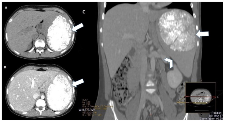
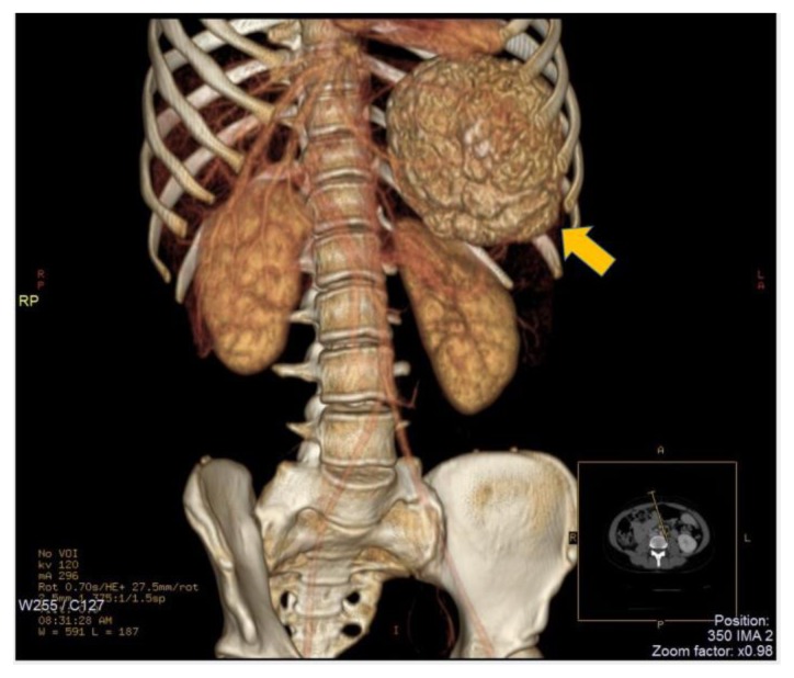
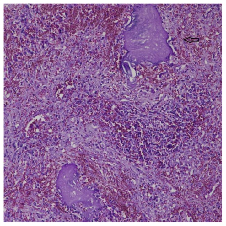
Computed tomography (CT) imaging displayed a large (maximum diameters 10 × 8 × 7 cm), inhomogeneous, solid mass in the upper pole of the spleen with calcifications inside (Fig. 1 A). On the arterial and portal venous phases, despite the evaluation of different Region of Interests within the lesion, no significant enhancement of the splenic mass has been observed (Fig. 1 B, C and Fig. 2).
Figure 1.
15-years-old girl with beta-thalassemia, splenomegaly and a calcified splenic hamartoma. - Technique: Abdominal CT scans (120 kV, modulated mA, 3 mm slice thickness, 80 ml of Iomeron 400) - Findings: A: Axial non-contrast CT image shows a large (10 × 8 × 7 cm) splenic lesion (arrow) with many calcifications widely scattered inside. B: Axial contrast enhanced CT image on portal venous phase demonstrates the calcified non-enhancing splenoma (arrow), with well-defined margins. C: Coronal reformatted contrast enhanced CT of the abdomen (bone window) demonstrates the significantly enlarged spleen (arrowhead) with a calcified mass in the splenic parenchyma, related to splenoma (arrow).
Figure 2.
15-years-old girl with beta-thalassemia, splenomegaly and a calcified splenic hamartoma. - Technique: Abdominal CT scan after intravenous administration of 80 ml of Iomeron 400 (120 kV, modulated mA, 3 mm slice thickness). 3D Volume Rendering of spiral CT data. - Findings: this image depicts the large splenic hamartoma (10 × 8 × 7 cm) with calcium density (arrow).
MANAGEMENT
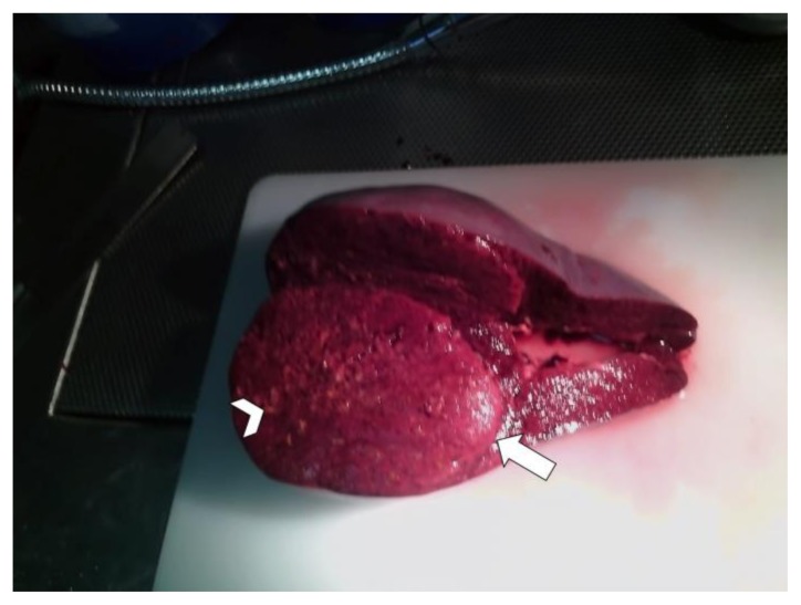
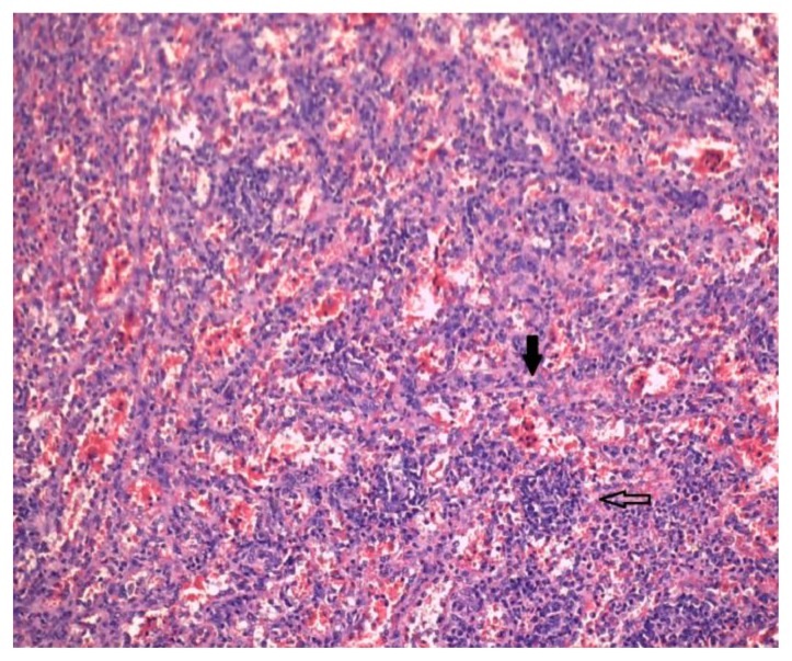
The patient was subsequently treated with splenectomy and the spleen was pathologically examined (Fig. 3). Histologically, the lesion consisted of lymphoid aggregates, with small-medium sized lymphocytes surrounded by mature plasma cells, interspersed with vascular areas of capillaries and sinusoids (Fig. 4). Moreover, there were multiple calcified areas within the lesion (Fig. 5). Atypical cells were not observed. The remaining splenic parenchyma showed mild hyperplasia of the white pulp, with expansion of the marginal zone.
Figure 3.
15-years-old girl with beta-thalassemia, splenomegaly and a calcified splenic hamartoma. - Technique: standard macroscopy image after splenectomy - Findings: Gross photograph of the resected spleen illustrating a solid mass (arrow), measuring 10 × 8 × 7 cm, arising in the upper pole of the spleen (weighted 870 g). The lesion was well circumscribed from the surrounding splenic parenchyma but without a capsule and was dark red in appearance, mottled with numerous yellowish focal areas (arrowhead).
Figure 4.
15-years-old girl with beta-thalassemia, splenomegaly and a calcified splenic hamartoma. - Technique: standard hematoxylin-eosin stain, original magnification ×400 - Findings: High magnification of the lesion shows various lymphoid aggregates (empty arrow), interspersed with vascular structures (capillaries and sinusoids of splenic red pulp-like area) lined by endothelial cells with no cytologic atypia (black arrow).
Figure 5.
15-years-old girl with beta-thalassemia, splenomegaly and a calcified splenic hamartoma. - Technique: standard hematoxylin-eosin stain, original magnification ×200 - Findings: Histological section of the hamartomatous lesion reveals deposit of amorphous, calcified material (empty arrow).
Immunohistochemical studies, with antibodies to T- and B-cells and immunoglobulin light chains, showed a prevalence of B-cells, associated with a lesser amount of T-cells (positive for CD 20 and CD 5) and plasma cells, in the lymphoid aggregates, with no suggestion of a clonal population. The vascular areas interposed between the lymphoid aggregates were characterized by lining cells of capillaries (positive for endothelial marker CD 34) and sinusoids (positive for endothelial marker CD 8); they were also present scattered histiocytes positive for CD 68 (PGM1). Antibodies to the proliferation antigen Ki-67 (MIB-1) by immunohistochemistry showed that all cells within the lesion had a low proliferationindex of 5%. The reaction for myeloperoxidase detected the presence of some localizations of heterotopic hemopoiesis in proximity to the calcified areas. These features were consistent with the diagnosis of a hamartomatous lesion.
To our knowledge, this is the first described case of a symptomatic, almost completely calcified splenic hamartoma in a pediatric patient associated with beta-thalassemia. The authors believe that such calcifications and avascularity are late manifestations of a slowly-growing splenic tumor, which may be well hyper-vascular if examined earlier.
DISCUSSION
ETIOLOGY & DEMOGRAPHICS
Splenic hamartomas (or splenomas) are very rare benign vascular tumors of the spleen, with a wide incidence range reported in a review of autopsies from 0.024% to 0.13% and equal sex preponderance [1,2]. It was firstly reported in 1861 by Rokitansky [3] with approximately 150 cases documented in the literature to date [1]. The pathogenesis of hamartoma is controversial and includes congenital malformation of the splenic red pulp, neoplasia, excessive and disorganized growth of abnormally formed red pulp or a reactive lesion to prior trauma [1].
Splenomas are usually asymptomatic and discovered incidentally in adult patients at imaging studies performed for other reasons or at autopsy; however, in literature there are also some documented cases of symptomatic hamartomas in children, associated with hematologic disorders, such as pancytopenia, anemia, and thrombocytopenia [4,5]. Splenic hamartomas have also been reported in association with hypersplenism, tuberous sclerosis and Wiskott-Aldrich syndrome [1,6].
Our case was accidentally discovered on CT examination of the abdomen in a patient in follow-up for splenomegaly.
CLINICAL & IMAGING FINDINGS
Although imaging features of splenic hamartoma can be nonspecific, hamartomas usually appear on CT as homogeneous iso-hypodense solid ball-like masses on non-enhanced scans, with progressive heterogeneous contrast enhancement, relatively to adjacent normal splenic parenchyma, after contrast agent injection [7].
In literature, there are few case reports describing splenic hamartomas with focal coarse calcifications [8,9]. Furthermore, most of the previously reported examples of splenoma were asymptomatic.
Although not performed in our patient, magnetic resonance imaging (MRI) can be useful for distinction of splenic hamartoma from hemangiomas: most lesions are isointense on T1-weighted images and hyperintense on T2-weighted images; heterogeneous enhancement on post-contrast late arterial phase is demonstrated, while a relative uniform and intense enhancement is found on delayed post-contrast images. Most splenic hamartomas are hyperechoic masses on sonography, with or without cystic changes and are hypervascular on color-Doppler ultrasound (US) [7].
TREATMENT & PROGNOSIS
As mentioned previously, splenic hamartoma usually remains asymptomatic, but the presence of the clinical symptoms occurs from mass effect if they grow larger.
Our case was an example of a patient with a large, atypical splenic lesion, therefore she was subsequently treated with splenectomy and the spleen was pathologically examined. The multiple calcified areas of the splenic hamartoma, detected on CT imaging and later confirmed on cytological examination, are unusual features of hamartoma, which may be due to hemorrhage within a large lesion or secondary to ischemia [8].
DIFFERENTIAL DIAGNOSES
The main pathologic differential diagnoses for splenic hamartoma are atypical vascular tumors, including hemangioma, littoral cell angioma, lymphangioma, hemangioendothelioma and angiosarcoma. These conditions may have similar clinical and radiological features, although these latter are various and nonspecific (Tab. 2) [10]. Therefore, immunohistochemical staining is mandatory to distinguish a splenic hamartoma from the other conditions [1]. Hemangioma appears on US as hyperechoic mass without any intrinsic color flow on color-Doppler, on unenhanced CT as a hypoattenuating mass and on MRI as an isointense lesion on T1 weighted image and hyperintense on T2 weighted image; on contrast enhancement evaluation it shows a homogeneous enhancement with centripetal filling. Littoral cell angioma appears on US as multiple isoechoic, hypoechoic or hyperechoic lesions within a heterogeneous splenic echotexture, on CT scans with or without contrast agent as multiple hypoattenuating lesions, becoming isodense only on delayed contrast-enhanced image and on MRI as hypointense nodular lesions on both T1 and T2 weighted pulse sequences. Lymphangioma appears on US as a cystic lesion with occasional internal septation or calcification, on unenhanced CT as a hypoattenuating mass with sharp margins and on MRI as a hypointense lesion on T1 weighted image and multiloculated and hyperintense on T2 weighted image; no significant contrast enhancement is typically seen. Hemangioendothelioma appears on US as a hypoechoic mass with disordered vascularization on color-Doppler, on unenhanced CT scans as a hypoattenuating mass and on MRI as a heterogeneous solid mass, with low signal intensity on both T1 and T2 weighted images in presence of hemosiderin; on contrast enhancement evaluation it shows enhancement of the solid portions of the tumor that may appear hypovascular relative to the normal splenic parenchyma. Angiosarcoma appears on US as a complex mass with heterogeneous echotexture and increased Doppler flow in the more solid portions, on CT scans as an ill-defined splenic mass with heterogeneous contrast enhancement; MRI demonstrates the hemorrhagic nature of the tumor, with areas of increased and decreased signal intensity which may be seen on images obtained with both T1 and T2 weighted pulse sequences [10].
Table 2.
Differential diagnosis table for splenic hamartoma.
| CT | Ultrasound | MRI | Pattern of contrast enhancement | PET | |
| Hamartoma | Hypodense | Hyperechoic | T1 isointense T2 hyperintense | Heterogeneous enhancement | moderate uptake |
| Hemangioma | Hypodense | Hyperechoic | T1 isointense T2 hyperintense | Centripetal fill | moderate uptake |
| Littoral cell angioma | Isodense | Iso/hypo/hyper-echoic | T1 and T2 hypointense | Hypodense; isodense in delayed phase | moderate uptake |
| Lymphangioma | Hypodense | Hypoechoic | T1 hypointense T2 hyperintense | No enhancement | moderate uptake |
| Hemangio-endothelioma | Hypodense | Hypoechoic | T1 and T2 heterogeneous | Hypovascular | intense uptake |
| Angiosarcoma | Heterogeneous | Heterogeneous | T1 and T2 heterogeneous | Heterogeneous enhancement | intense uptake |
The role of Positron Emission Tomography/Computed Tomography (PET/CT) in defining splenic lesions remains to be defined and such investigations should be approached with caution, so that an indeterminate or even positive PET scan cannot be assumed to represent malignancy [11]. Usually, PET-CT shows moderate uptake of radiolabeled 18F-fluoro-deoxy-D-glucose (FDG) in benign splenic lesions like hamartoma, hemangioma, littoral cell angioma and lymphangioma, while an intense uptake of FDG is suspicious for malignant tumors like hemangioendothelioma and angiosarcoma [11,12].
Histologic findings of hamartoma reveal disorganized vascular channels lined by slightly plump endothelial cells without atypia, mixed with intervening splenic red pulp–like stroma with or without white pulp; splenic hemangiomas are the most common benign neoplasm arising from sinusoidal epithelial cells, histologically composed of proliferating vascular channels; littoral cell angioma is a rare vascular tumor, arising from littoral cells originating from the splenic sinuses; lymphangioma is another rare benign spleen tumor, which manifests as a subcapsular nodule or as diffuse lymphangiomatosis in young patients, characterized by cystic spaces filled with proteinaceous fluids; hemangioendothelioma is a quite rare entity of the spleen, with an intermediate histology between that of hemangioma and angiosarcoma, with lining cells of vascular channels showing an intermediate degree of atypia; angiosarcoma is a malignant primary tumor of non-lymphoid origin, characterized by irregular, anastomosing vessels associated with cellular atypia and invasion of adjacent organs [1].
Calcified splenic hamartoma is an unusual type of splenic lesion. Spleen calcifications have been reported most frequently in tumors or in infective and in accumulation diseases (for example splenic hemangioma, lymphangioma, tuberculosis, histoplasmosis, brucellosis, amyloidosis, phlebolith, hydatid cyst, epidermoid cyst and abscess) [13]. All of these conditions are rare entities that may be clinically controversial and diagnosis is possible only in presence of an appropriate clinical assessment.
TEACHING POINT
Imaging features of splenic hamartoma are variegated, although it usually appears on CT as homogeneous iso-hypodense solid ball-like mass with progressive heterogeneous contrast enhancement after contrast agent injection.
However, atypical findings may be encountered, such as calcifications and lack of contrast enhancement.
Table 1.
Summary table for splenic hamartoma.
| Etiology | Controversial: congenital malformation of the splenic red pulp, neoplasia, excessive and disorganized growth of abnormally formed red pulp, reactive lesion to prior trauma. |
| Incidence | Wide incidence range (from 0.024% to 0.13% in an autopsies review). |
| Gender ratio | Equal sex preponderance. |
| Age predilection | All age groups, although most of the reports in the literature consist of with adult patients. |
| Risk factors | Tuberous sclerosis, Wiskott-Aldrich like syndrome, hypersplenism, hematologic conditions. |
| Treatment | Splenectomy. |
| Prognosis | Usually asymptomatic; clinical symptoms occur from mass effect if they grow larger. |
| Findings on imaging |
US: Hyperechoic masses, with or without cystic changes; hyper-vascular on color-Doppler ultrasound. CT: homogeneous iso-hypodense solid ball-like masses on non-enhanced scans; calcifications can be found. MRI: isointense on T1-weighted images; hyperintense on T2-weighted images. Pattern of contrast enhancement: progressive heterogeneous enhancement on post-contrast phases is emonstrated, with a relative uniform and intense enhancement on delayed post-contrast images. |
ABBREVIATIONS
- CT
Computed tomography
- FDG
18F-fluoro-deoxy-D-glucose
- MRI
Magnetic Resonance Imaging
- PET
Positron Emission Tomography
- US
Ultrasound
REFERENCES
- 1.Lee H, Maeda K. Hamartoma of the Spleen. Archives of Pathology & Laboratory Medicine. 2009 Jan;133(1):147–151. doi: 10.5858/133.1.147. [DOI] [PubMed] [Google Scholar]
- 2.Tavangar SM, Abdollahi A. Splenic Hamartoma: Immunohistochemical Profile. Acta Med Iran. 2017 Jan;55(1):77–78. [PubMed] [Google Scholar]
- 3.Rokitansky K. Lehrbuch der Pathologischen Anatomie. Vol. 3. 1861. Über splenome; p. 9. [Google Scholar]
- 4.Abramowsky C, Alvarado C, Wyly JB, Ricketts R. “Hamartoma” of the spleen (splenoma) in children. Pediatr Dev Pathol. 2004;(7):231–236. doi: 10.1007/s10024-003-9097-5. [DOI] [PubMed] [Google Scholar]
- 5.Wirbel RJ, Uhlig U, Futterer KM. Case report: splenic hamartoma with hematologic disorders. American Journal of the Medical Sciences. 1996;311(5):243–246. doi: 10.1097/00000441-199605000-00009. [DOI] [PubMed] [Google Scholar]
- 6.Tsitouridis I, Michailides M, Tsitouridis K, Davidis I, Efstratiou I. Symptomatic splenoma (hamartoma) of the spleen: a case report. Hippokratia. 2010;14:54–6. [PMC free article] [PubMed] [Google Scholar]
- 7.Yu RS, Zhang SZ, Hua JM. Imaging findings of splenic hamartoma. World J Gastroenterol. 2004;10(17):2613–2615. doi: 10.3748/wjg.v10.i17.2613. [DOI] [PMC free article] [PubMed] [Google Scholar]
- 8.Zissin R, Lishner M, Rathaus V. Case report: unusual presentation of splenic hamartoma; computed tomography and ultrasonic findings. Clin Radiol. 1992;45:410–411. doi: 10.1016/s0009-9260(05)81003-7. [DOI] [PubMed] [Google Scholar]
- 9.Komaki S, Gombas OF. Angiographic demonstration of a calcified splenic hamartoma. Radiology. 1976 Oct;121(1):77–8. doi: 10.1148/121.1.77. [DOI] [PubMed] [Google Scholar]
- 10.Abbott RM, Levy AD, Aguilera NS, Gorospe L, Thompson WM. From the archives of the AFIP: Primary vascular neoplasms of the spleen: radiologic-pathologic correlation. Radiographics. 2004;24:1137–1163. doi: 10.1148/rg.244045006. [DOI] [PubMed] [Google Scholar]
- 11.Avila L, Sivaprakasam P, Viero S. Splenic hamartoma in a child in the era of PET-CT. Pediatr Blood Cancer. 2009;53:114–116. doi: 10.1002/pbc.21962. [DOI] [PubMed] [Google Scholar]
- 12.Metser U, Even-Sapir E. The role of 18F-FDG PET/CT in the evaluation of solid splenic masses. Semin Ultrasound CT MR. 2006;27:420–425. doi: 10.1053/j.sult.2006.06.005. [DOI] [PubMed] [Google Scholar]
- 13.Burgener FA, Kormano M, Pudas T. Differential Diagnosis in Conventional Radiology. Thieme Medical Pub; 2007. pp. 631–653. [Google Scholar]


